Cysts within Otherwise Probably Benign Solid
Breast Masses and the Risk of Malignancy
Cistos no interior de nódulo mamários benignos: risco de
malignidade
Rodrigo Menezes Jales
1Karla Galvão Araujo
2Luis Otávio Zanata Sarian
1Kátia Piton Serra
3Helena Keppke
3Juliana Francisco
3Sophie Françoise Mauricette Derchain
11Hospital da Mulher Prof. Dr. Jose Aristodemo Pinotti (Women’s Hospital), CAISM - Department of Obstetrics and Gynecology of the School of Medical Sciences, Universidade Estadual de Campinas– Unicamp, Campinas, SP, Brazil
2Department of Obstetrics and Gynecology of the School of Medical Sciences, Unicamp, Campinas, SP, Brazil
3Breast Imaging Extension Course of the School of Medical Sciences, Unicamp, Campinas, SP, Brazil
Rev Bras Ginecol Obstet 2016;38:170–176.
Address for correspondence Rodrigo Jales, Seção de Imagens, Departamento de Obstetrícia e Ginecologia, Faculdade de Ciências Médicas, Universidade Estadual de Campinas, PO Box 6111, Campinas, São Paulo 13083-970, Brazil (e-mail: [email protected]).
Keywords
►
breast cancer
►
breast imaging
reporting and data
system
►
breast sonography
►
circumscribed masses
►
complex masses
Abstract
Objective
The objective of this study is to assess whether the largest cyst diameter is
useful for BI-RADS ultrasonography classification of predominantly solid breast masses
with an oval shape, circumscribed margins, and largest axis parallel to the skin, which,
except for the cystic component, would be likely classi
fi
ed as benign.
Methods
This study received approval from the local institutional review board. From
March 2009 to August 2014, we prospectively biopsied 170 breast masses from 164
women. We grouped the largest cyst and mass diameters according to
histopatholog-ical diagnoses. We used Student
’
s
t
-test, linear regression, and the area under the
receiver operating characteristic curve (AUC) for statistical assessment.
Results
Histopathological examination revealed 143 (84%) benign and 27 (16%) malignant
masses. The mean largest mass diameter was larger among malignant (mean
standard
deviation, 34.1
16.6 mm) than benign masses (24.7
16.7 mm) (
P
<
0.008). The mean
largest cyst diameter was also larger among malignant (9.9
7.1 mm) than benign masses
(4.6
3.6 mm) (
P
<
0.001). Agreement between measurements of the largest mass and
cyst diameters was low (R
2¼
0.26). AUC for the largest cyst diameter (0.78) was similar to
the AUC for the largest mass diameter (0.69) (
p
¼
0.2). A largest cyst diameter
<
3,
3
to
<
11, and
11 mm had a positive predictive value of 0, 15, and 52%, respectively.
Conclusion
A largest cystic component
<
3 mm identified within breast masses that
show favorable characteristics may be considered clinically inconsequential in
ultrasonog-raphy characterization. Conversely, masses with a largest cystic component
3 mm
should be classified as BI-RADS-US category 4.
received
December 15, 2015 accepted
January 18, 2016 published online April 19, 2016
DOI http://dx.doi.org/ 10.1055/s-0036-1582398. ISSN 0100-7203.
Copyright © 2016 by Thieme Publicações Ltda, Rio de Janeiro, Brazil
Introduction
Breast lesions may be sonographically characterized with the Breast Imaging Reporting and Data System
(BI-RADS-US) according to the malignancy risk.1–3Breast masses with
solid and cystic echo pattern should be described as
com-plex solid and cystic and classified as BI-RADS-US category
4.3–9 In this category, the risk of malignancy is>2% to
95% and such lesions should be investigated by biopsy.3–9
In contrast, breast masses with an oval shape, circum-scribed margins, orientation parallel to the skin, and hypo-echoic echo pattern are coded as probably benign and
classified as BI-RADS-US category 3.3,10The risk of
malig-nancy in this category is<2%, and biopsy may be avoided if
the mass maintains the same morphology and stable
di-mensions during follow-up.3,10 Therefore, sonographic
identification of a cyst or several cysts within a
predomi-nantly solid breast mass, in which the other morphologic features are consistent with BI-RADS-US category 3, still
justifies a description of the mass as complex. Hence, a
predominantly solid mass with an oval shape, circum-scribed margins, and largest axis parallel to the skin should
be classified as BI-RADS-US category 4.11However,
recom-mending biopsy may seem exaggerated in such cases,
particularly if there is only one discrete cystic focus.11
This issue led us to carry out a prospective study of predominantly solid breast masses with an oval shape, circumscribed margins, and largest axis parallel to the skin, which, except for the cystic component, would
other-wise be classified as probably benign. A previous pilot study
presented the results of a sample of 48 breast masses. These masses accounted for 3% of breast ultrasound studies in a breast imaging section located in a reference university
hospital for breast cancer treatment.11 The prevalence of
malignancy in that sample was 25%, confirming that such
masses should be described as BI-RADS-US category 4.
Moreover, the largest cyst diameter of each mass was signifi
-cantly related to the histopathological diagnosis of
malig-nancy. All masses with a largest cyst diameter<3 mm were
benign, and all masses with a largest cyst diameter>13 mm
were malignant.11
The purpose of the current study was to assess whether the largest cyst diameter is a useful ultrasonography variable
for BI-RADS-US classification of predominantly solid breast
masses with an oval shape, circumscribed margins, and largest axis parallel to the skin, which, except for the cystic
Resumo
Objetivo
Avaliar se o maior diâmetro do cisto é útil para a classificação
ultrassono-gráfica BI-RADS de nódulos mamários predominantemente sólidos, com forma oval,
margens circunscritas e maior eixo paralelo à pele que, exceto pela presença do
componente cístico, seriam classi
fi
cados como provavelmente benignos.
Métodos
Este estudo foi aprovado pelo Comitê de Ética local. De março de 2009 a
agosto de 2014, 170 nódulos mamários de 164 mulheres foram prospectivamente
biópsiados. As medidas do maior diâmetro do maior cisto e do maior diâmetro do
nódulo foram agrupados de acordo com os diagnósticos histopatológicos. O teste t de
Student, a regressão linear e a área sob a curva ROC (AUC) foram utilizados para a
avaliação estatística.
Resultados
O exame histopatológico revelou 143 (84%) nódulos benignos e 27
(16%) nódulos malignos. A média da medida do maior diâmetro dos nódulos foi
maior entre os nódulos malignos (média
desvio padrão, 34,1
16,6 mm) do que
nos nódulos benignos (24,7
16,7 mm) (
p
<
0,008). A média do maior diâmetro do
maior cisto também foi maior entre os nódulos malignos (9,9
7,1 mm) do que nos
nódulos benignos (4,6
3,6 mm) (
p
<
0,001). A concordância entre as medidas dos
maiores diâmetros dos nódulos e do maior diâmetro do maior cisto foi baixa
(R
2¼
0,26). A AUC do maior diâmetro do maior cisto (0,78) foi semelhante à
AUC do maior diâmetro do nódulo (0,69) (
p
¼
0,2). Os maiores diâmetros dos
maiores cistos medindo
<
3;
3 e
<
11; e
11 mm tiveram um valor preditivo
positivo de 0, 15 e 52%, respectivamente.
Conclusão
Componentes císticos
<
3 mm identificados dentro de nódulos
mamá-rios que apresentam as demais características provavelmente benignas podem ser
considerados clinicamente irrelevantes na caracterização ultrassonográ
fi
ca. Por outro
lado, nódulos que apresentam um componente cístico medindo
3 mm devem ser
classificadas na categoria BI-RADS-US 4.
Palavras-chave
►
câncer de mama
►
BI-RADS
►
ultrassonografia
mamária
►
nódulos
circunscritos
component, would otherwise be classified as probably benign.
Methods
This cross-sectional study with prospective collection data was approved by our Institutional Review Board under number 031/2009. All participants signed an informed con-sent term. The research was performed according to the Declaration of Helsinki, which was reviewed in 2008.
In a previous pilot study published in 2012, we presented
the results of 48 breast masses.11This prior article suggested
that the largest cyst diameter of each mass was significantly
related to the histopathological diagnoses of malignancy. In
this article, we report thisfinding in a more representative
sample of 170 masses and assess the association between largest cyst diameter and largest mass diameter.
Among all women who underwent breast ultrasound, we selected those with predominantly solid breast masses with an oval shape, circumscribed margins, and largest axis parallel to the skin, which, except for the cystic component,
would otherwise be classified as probably benign. The
medi-cal indications for the examinations were sonographic eval-uation of masses categorized as BI-RADS-MG category 0, sonographic follow-up of breast masses previously coded as BI-RADS-US category 3, sonographic screening in high-risk patients with mammographically dense breasts, and sono-graphic assessment of palpable breast masses.
In total, 170 masses from 164 women were included in this study. Prior mammograms were available for 95/170 (56%) of the breast masses. Most women undergoing mam-mography (70/95; 74%) were categorized as having BI-RADS category 0 masses because of circumscribed/obscured mass margins or focal asymmetry. In 20/95 (21%) women, the mammogram was negative (BI-RADS-MG category 1 or 2).
Only 3/95 (3%) mammograms identified suspicious lesions
(BI-RADS-US-MG category 4), including microlobulated margins (2 masses) or architectural distortion (1 mass).
All ultrasonographic examinations were performed from March 2009 to August 2014 by the same clinician (R.M.J.) with 12 years of experience in breast imaging. No patient that met the ultrasonographic criteria was excluded from the study. The Voluson 730 Expert (GE Healthcare, Little Chal-font, Buckinghamshire, United Kingdom) and Accuvix V10 (Medison Co., Ltd., Seoul, Korea) ultrasound machines were the diagnostic equipment used.
We assessed predominantly solid breast masses with an oval shape, circumscribed margins, and largest axis parallel to the skin, which except for the cystic component would
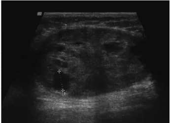
Fig. 1 First illustrative example of a mass in this study. There were two cystic foci that alone or combined did not occupy more than half of the mass. (A) The largest cystic diameter of the largest cyst measured 13 mm. (B) The largest mass diameter measured 49 mm. In our routine breast ultrasound practice, this mass would be described as complex solid and cystic, classified as BI-RADS-US category 4, and biopsied. This patient was 28 years old, and the mass was palpable. Pathological analysis confirmed pseudoangiomatous stromal hyperplasia.
Fig. 2 Second illustrative example of a mass in this study. Only one discreet cystic focus was present. (A) The largest cystic diameter measured 2 mm. (B) The largest mass diameter measured 31 mm. In our routine breast ultrasound practice, this mass would be described as complex solid and cystic, classified as BI-RADS-US category 4, and biopsied. The patient was 47 years old, and the mass was not palpable. Pathological analysis confirmed a complexfibroadenoma. In this study, we demonstrated that a largest cystic component<3 mm may be ignored in the description and characterization of breast masses.
otherwise be classified as probably benign. We included in our sample only masses in which the largest cystic component
measured2 mm because cystic foci<2 mm may not be
properly characterized by ultrasound. The cystic focus or foci did not occupy more than half of the breast mass, and the masses were therefore characterized as predominantly solid. In breast masses with more than one cystic focus, only the largest focus was measured and considered for
analysis.►Figs. 1 to3show examples of these breast masses.
In our routine breast ultrasound practice, these masses are
described as complex solid and cystic, classified as
BI-RADS-US category 4, and biopsied.11
We performed histopathological assessment of the ma-jority of the masses (157/170; 92% of lesions) by ultrasound-guided core needle biopsy using an automated biopsy gun with a 14-gauge needle (Magnum; Bard Biopsy Systems, Tempe, AZ). In 33/157 (21%) of the masses initially evaluated by core needle biopsy, analysis was supplemented by exci-sional biopsy. In 13/170 (8%) of the cases, we directly evaluated the lesions by excisional biopsy.
Statistical Analysis
We included age, menstrual status, and physical examination
findings of the breasts as control variables. Ultrasound
variables assessed in the masses were the largest mass diameter and the largest cyst diameter, both in millimeters
(►Fig. 1). We used a two-sample Student’st-test or a
chi-squared test to compare data according to the benign or malignant histopathological diagnosis.
The association between the largest cyst diameter and largest mass diameter was evaluated by linear regression,
including measurement of correlation coefficient (R2) and
analysis of a scatterplot graph. The R2is a numerical measure
of the strength of the relationship between two quantitative variables.
The performance of the largest cyst diameter and largest mass diameter in terms of malignancy prediction were calculated by the area under the receiver operating char-acteristic (ROC) curve. Through the coordinates of the ROC
curve, we selected ranges of the largest cyst diameter measures related to the positive predictive values (PPVs) suitable for BI-RADS categories. We also drew a pairwise comparison of the areas under the ROC curve (AUC) for
these variables using Venkatraman’s
projection-permuta-tion test, using the software R Environment for Statistical
Computing (R Project).12We performed all other statistical
calculations with SPSS software version 15 (SPSS Inc.,
Chicago, IL). APvalue<0.05 was considered as indicating
a significant difference.
Results
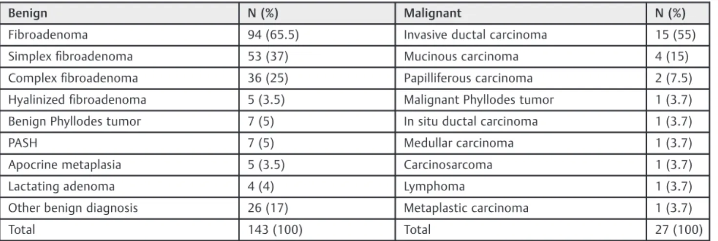
In total, 170 breast masses from 164 women were available for analysis. Histopathological examination revealed 143/170 (84%) benign and 27/170 (16%) malignant pathological diagnoses.
These masses had a high prevalence of complexfibroadenomas
(36/143, 25%) and benign phyllodes tumors (7/143, 5%), but the
major benign pathological diagnosis was simplexfibroadenoma
(53/143, 37%). In contrast, the most frequent malignant patho-logical diagnoses were invasive ductal carcinoma (15/27, 55%)
and mucinous carcinoma (4/27, 15%) (►Table 1).
Women presenting with malignant histopathologicalfi
nd-ings were older (meanstandard deviation, 55.118.2
years; range, 32–77 years) than women with benign results
(39.511.6 years; range, 14–66 years) (P<0.001).
Malig-nancy rates were higher for postmenopausal women (15/39, 38%) than for premenopausal women (12/131, 9%)
(P<0.001). Palpable masses were more frequently malignant
(22/112, 19.6%) than were nonpalpable masses (5/58, 8.6%)
(p¼0.046) (►Table 2).
The largest mass diameter was larger in malignant masses
(34.116.6 mm; range, 11–66 mm) than in benign masses
(24.716.7 mm; range, 8–135 mm) (p¼0.008). The largest
cyst diameter was also larger in malignant masses
(9.97.1; range, 3–31 mm) than in benign masses
(4.63.6 mm; range, 2–25 mm) (P<0.001) (►Table 3).
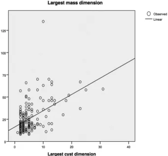
The scattergraph calculated by linear regression indicated that there was a positive relationship between the largest
Table 1 Pathological diagnosis
Benign N (%) Malignant N (%)
Fibroadenoma 94 (65.5) Invasive ductal carcinoma 15 (55)
Simplexfibroadenoma 53 (37) Mucinous carcinoma 4 (15)
Complexfibroadenoma 36 (25) Papilliferous carcinoma 2 (7.5)
Hyalinizedfibroadenoma 5 (3.5) Malignant Phyllodes tumor 1 (3.7)
Benign Phyllodes tumor 7 (5) In situ ductal carcinoma 1 (3.7)
PASH 7 (5) Medullar carcinoma 1 (3.7)
Apocrine metaplasia 5 (3.5) Carcinosarcoma 1 (3.7)
Lactating adenoma 4 (4) Lymphoma 1 (3.7)
Other benign diagnosis 26 (17) Metaplastic carcinoma 1 (3.7)
Total 143 (100) Total 27 (100)
cyst diameter and largest mass diameter. However, agree-ment between measureagree-ments of these variables was low
(R2¼0.26) (
►Fig. 4).
The AUC for the largest cyst diameter was similar to that for the largest mass diameter (0.78 and 0.69, respectively;
p¼0.2). The AUC for the largest cyst diameter was also
similar (0.73) when only considering women<40 years old,
regardless of their clinical breast examinationfindings and
women40 years of age presenting with nonpalpable
breast masses. Palpable breast masses in40-year-old
women could be considered BI-RADS-US category 4, regard-less of the cystic foci. Among all women, a largest cyst
diameter<3,3 to<11, and11 mm had a PPV of 0%,
15%, and 52%, respectively (►Fig. 5). These values are
con-sistent with BI-RADS-US categories 3, 4b, and 4c, respectively.
Discussion
In this study, we demonstrated that the largest cyst diameter
was useful for BI-RADS ultrasonography classification of
predominantly solid breast masses with an oval shape, circumscribed margins, and largest axis parallel to the skin, which, except for the cystic component, would
other-wise be classified as probably benign. The performance of the
largest cyst diameter for malignancy prediction was main-tained regardless of whether the clinical breast examination
findings were considered.
Although the largest cyst diameter tended to be larger in larger breast masses, the majority of cases plotted in the
graph did not demonstrate this trend (R2¼0.26).
Measure-ment of the largest mass dimension is performed in routine
practice during breast imaging studies.12 The low
Table 3 Distribution of sonographic variables according to thefinal pathological diagnosis
Pathological diagnosis P
Benign Malignant
Sonographic Variables
Mean/SD/Range Mean/SD/Range
Largest mass diameter (mm) 24.7/16.7/8–135 34.1/16.6/11–66 0.008
Largest cystic diameter (mm) 4.6/3.6/2–25 9.9/7.1/3–31 <0.0001
N N PPV (%)
Largest cystic diameter<3 mm 43 0 0/43 (0)
Largest cystic diameter3 mm and<11 mm 90 16 16/106 (15)
Largest cystic diameter11 mm 10 11 11/21 (52)
Total 143 (100) 27 (100)
Abbreviations: PPV, positive predictive value; SD, standard deviation.
Student
’st-test.
Table 2 Distribution of control variables according to thefinal pathological diagnosis
Pathological diagnosis
Variables Benign Malignant P
Mean/SD/Range Mean/SD/Range
Age 39.5 /11.6/14–66 55.1/18.2/32–77 <0.001
N (%) N (%) Total
Menopause <0.001#
Yes 24 (16.8) 15 (55.6) 39
No 119 (83.2) 12 (44.4) 131
Total 143 (100) 27 (100) 170
Palpable lesion 0.046#
Yes 90 (62.9) 22 (81.5) 112
No 53 (34.1) 5 (18.5) 58
Total 143 (100) 27 (100) 170
Abbreviation: SD, standard deviation.
Student
concordance rate between the largest mass dimension and the largest cyst dimension may justify including this mea-surement in the ultrasonography evaluation of complex solid and cystic masses, which, except for the presence of a cystic
focus, would otherwise be classified as BI-RADS 3.
Another important contribution of our study was the determination of the prevalence of malignancy in predomi-nantly solid breast masses with an oval shape, circumscribed margins, and largest axis parallel to the skin, which, except
for the cystic component, would otherwise be classified as
probably benign. The prevalence of malignancy in the masses included in the current study (16%) was lower than that in a
previous pilot study (25%).11This can be explained by the fact
that the current sample had a higher rate of masses in which
the cystic foci measured<3 mm. Thus, the prevalence of
16% for the total sample is slightly lower than the rate
described for complex masses (23–31%), but it is still relates
to BI-RADS category 4.3,13
With respect to the histopathological results of the breast masses studied, most masses had a histopathological result
of fibroadenoma (94/170 masses). Many thesefi
broadeno-mas were classified as complex (36/94; 38% of benign
masses). The expected prevalence rate of complexfi
broade-nomas is lower, ranging from 16% to 22% in provenfi
broa-denomas.14,15 The high prevalence of complex
fibroadenomas in our sample was not surprising because
the presence of simple cysts>3 mm withinfibroadenomas
is one histopathological criterion that defines complexfi
-broadenoma. The remaining criteria include sclerosing
ad-enosis, epithelial calcifications, and papillary apocrine
changes.15
The significance of complexfibroadenoma is that women
presenting with these lesions have a 3.1-fold higher relative risk of developing breast cancer compared with the general
population.15Thefindings of the current study suggest that
the presence of small cysts within circumscribed masses may
be related to complexfibroadenomas.
Another relevant factor in the analysis of the benign histologic results was the prevalence of benign phyllodes tumors. These tumors comprised 7/170 (4.0%) of the total
sample and 7/105 (6.7%) offibroepithelial neoplasms. These
rates are higher than twice the expected rate of benign
Fig. 5 Receiver operating characteristics curves. ROC 1: Complete sample, regardless of age and clinical breast examinationfindings (170 masses). Area under the curve (AUC) for largest cyst diameter¼0.78, and AUC for largest mass diameter¼0.69. ROC 2: Sample limited to<40-year-old women regardless of clinical breast examinationfindings and40-year-old women with nonpalpable breast masses (122 masses). AUC for largest cystic diameter¼0.73.
phyllodes tumors, which account for<1% of all breast
tumors and<3% offibroepithelial breast lesions.16
Phyllo-des tumors are associated with a risk of recurrence and
distant metastases.17Preoperative identification of
phyllo-des tumors is crucial for appropriate surgical planning and prevention of surgical complications resulting from inade-quate excision. Mammography and ultrasound examination
cannot adequately distinguish betweenfibroadenomas and
phyllodes tumors.18 The presence of a cystic foci has been
described as important in the diagnosis of this type of
tumor.19–22 Most of the masses in the present study had
histopathological results consistent withfibroadenomas, not
phyllodes tumors. Conversely, the size of the largest cystic component may prove useful in identifying malignant phyll-odes tumors.
The strengths of our study are its large sample size, which required over four years of data gathering in a breast imaging reference center; the homogeneous sample of patients, all of whomwere selected bya single experienced observer; and, most importantly, its conclusion is easily applicable in patient care.
A limitation of our study was our failure to clarify the precise histopathology of the cystic foci. Nevertheless, we can assume that the cystic foci in the four cases diagnosed as mucinous carcinoma represented mucin. Furthermore, in the remaining malignant histologic types included in our sample, the cystic foci seen on ultrasonography were likely to
correspond to small areas of necrosis associated with ineffi
-cient vascularity related to rapid tumor growth. Our sample was restricted to circumscribed masses, which are usually
related to rapidly growing lesions.23–25
In conclusion, a largest cystic component<3 mm
identi-fied within breast masses that show favorable characteristics
may be considered clinically inconsequential. Conversely,
masses with a largest cystic component3 mm should be
classified as BI-RADS-US category 4 and biopsied.
Conflicts of Interests
The authors have no conflicts of interests to declare.
Acknowledgment
This study was partiallyfinanced by the Research Support
Foundation of the State of São Paulo–Fapesp (number
2012/15059–8).
References
1 Raza S, Chikarmane SA, Neilsen SS, Zorn LM, Birdwell RL. BI-RADS 3, 4, and 5 lesions: value of US in management—follow-up and outcome. Radiology 2008;248(3):773–781
2 Jales RM, Sarian LO, Torresan R, Marussi EF, Alvares BR, Derchain S. Simple rules for ultrasonographic subcategoriza-tion of BI-RADS®-US 4 breast masses. Eur J Radiol 2013;82(8): 1231–1235
3 Mendelson EB, Böhm-Vélez M, Berg WA, Whitman GJ, Feldman MI, Madjar H. ACR BI-RADS® Ultrasound [Internet]. In: D’Orsi CJ,
Sickles EA, Mendelson EB, Morris EA. ACR BI-RADS® Atlas: breast imaging reporting and data system. 5th ed. Reston: American
College of Radiology; 2013 [cited 2015 Nov 12]. Available from: http://www.ashevilleradiology.com/physicians/ACR_BIRAD-S_ATLAS.pdf
4 Berg WA, Campassi CI, Ioffe OB. Cystic lesions of the breast: sonographic-pathologic correlation. Radiology 2003;227(1): 183–191
5 Chang YW, Kwon KH, Goo DE, Choi DL, Lee HK, Yang SB. Sono-graphic differentiation of benign and malignant cystic lesions of the breast. J Ultrasound Med 2007;26(1):47–53
6 Hsu HH, Yu JC, Lee HS, et al. Complex cystic lesions of the breast on ultrasonography: feature analysis and BI-RADS assessment. Eur J Radiol 2011;79(1):73–79
7 Huff JG. The sonographicfindings and differing clinical implica-tions of simple, complicated, and complex breast cysts. J Natl Compr Canc Netw 2009;7(10):1101–1104, quiz 1105
8 Doshi DJ, March DE, Crisi GM, Coughlin BF. Complex cystic breast masses: diagnostic approach and imaging-pathologic correlation. Radiographics 2007;27(Suppl 1):S53–S64
9 Chen M, Zhan WW, Wang WP. Cystic breast lesions by conven-tional ultrasonography: sonographic subtype-pathologic corre-lation and BI-RADS Assessment. Arch Med Sci 2014;10(1):76–83 10 Berg WA, Zhang Z, Cormack JB, Mendelson EB. Multiple bilateral circumscribed masses at screening breast US: consider annual follow-up. Radiology 2013;268(3):673–683
11 Jales RM, Sarian LO, Peralta CF, et al. Complex breast masses: assessment of malignant potential based on cyst diameter. J Ultrasound Med 2012;31(4):581–587
12 R Core Team. R: a language and environment for statistical computing: reference index [Internet]. Vienna: R Foundation for Statistical Computing; 2015 [cited Dez 20]. Available from: https://cran.r-project.org/doc/manuals/r-release/fullrefman.pdf 13 Athanasiou A, Aubert E, Vincent Salomon A, Tardivon A. Complex
cystic breast masses in ultrasound examination. Diagn Interv Imaging 2014;95(2):169–179
14 Pinto J, Aguiar AT, Duarte H, Vilaverde F, Rodrigues Â, Krug JL. Simple and complexfibroadenomas: are there any distinguishing sonographic features? J Ultrasound Med 2014;33(3):415–419 15 Dupont WD, Page DL, Parl FF, et al. Long-term risk of breast cancer
in women withfibroadenoma. N Engl J Med 1994;331(1):10–15 16 Rowell MD, Perry RR, Hsiu JG, Barranco SC. Phyllodes tumors. Am J
Surg 1993;165(3):376–379
17 Bhargav PR, Mishra A, Agarwal G, Agarwal A, Verma AK, Mishra SK. Phyllodes tumour of the breast: clinicopathological analysis of recurrent vs. non-recurrent cases. Asian J Surg 2009;32(4):224–228
18 Gatta G, Iaselli F, Parlato V, Di Grezia G, Grassi R, Rotondo A. Differential diagnosis between fibroadenoma, giant fi broade-noma and phyllodes tumour: sonographic features and core needle biopsy. Radiol Med (Torino) 2011;116(6):905–918
19 Liberman L, Bonaccio E, Hamele-Bena D, Abramson AF, Cohen MA, Dershaw DD. Benign and malignant phyllodes tumors: mammo-graphic and sonomammo-graphicfindings. Radiology 1996;198(1):121–124
20 Yilmaz E, Sal S, Lebe B. Differentiation of phyllodes tumors versus
fibroadenomas. Acta Radiol 2002;43(1):34–39
21 ChaoTC, Lo YF, Chen SC, Chen MF. Sonographic features of phyllodes tumors of the breast. Ultrasound Obstet Gynecol 2002;20(1):64–71 22 Bode MK, Rissanen T, Apaja-Sarkkinen M. Ultrasonography and core needle biopsy in the differential diagnosis offibroadenoma and tumor phyllodes. Acta Radiol 2007;48(7):708–713 23 Evans AJ, Pinder SE, James JJ, Ellis IO, Cornford E. Is
mammograph-ic spmammograph-iculation an independent, good prognostmammograph-ic factor in screen-ing-detected invasive breast cancer? AJR Am J Roentgenol 2006; 187(5):1377–1380
24 Luck AA, Evans AJ, James JJ, et al. Breast carcinoma with basal phenotype: mammographicfindings. AJR Am J Roentgenol 2008; 191(2):346–351
25 Ko ES, Lee BH, Kim HA, Noh WC, Kim MS, Lee SA. Triple-negative breast cancer: correlation between imaging and pathological