Juliana Fortes Vilarinho Braga
ETIOLOGY OF VERTEBRAL OSTEOMYELITIS IN BROILERS
Belo Horizonte
Juliana Fortes Vilarinho Braga
ETIOLOGY OF VERTEBRAL OSTEOMYELITIS IN BROILERS
Belo Horizonte
Escola de Veterinária of UFMG
2016
Final work of thesis presented to the Programa de Pós-graduação em Ciência Animal of the Universidade Federal de Minas Gerais as a requirement for obtaining the title of Doctor in Animal Science.
Area of concentration: Animal Pathology Advisor: Prof. Dr. Roselene Ecco
ACKNOWLEDGEMENTS
I thank God for his precious and immeasurable love. For his care and patience. For our conversations that renew my strength and for surrounding me with angels in every single step of my journey. Thank you, Father.
I thank my family, my root, for the example of love, trust and support they give me every day. Among many other things, I learned from you that physical distance is no barrier when souls walk side by side.
I thank my husband, who has been by my side in so many defining moments of my life, like this one. Thank you, my love, for being this exceptional man that makes me want to be better and for teaching me, in many ways, the real meaning of "love."
To my advisor, I thank for being my guide in this journey and make it lighter and pleasurable. Thank you for your restless disposition for learning and teaching and for showing me that a good professional is, above all, a good human being.
I thank my co-advisor for always being so solicitous and for contributing with his knowledge and kindness, so I could conclude this work.
I thank the members of the doctoral thesis examination for the availability to participate and contribute scientifically to the improvement of this work.
I thank the professors of the sector of Animal Pathology for all the knowledge transmitted and for the many lessons taught during these years.
To the "Ecco" family, which has been renewed over the years, I thank for the good moments of learning and collaboration, full of joy.
To all my friends of the sector of Animal pathology, I thank for helping to write one of the most special chapters of this thesis. I am glad to be part of this family, my beloved and eternal "povo da patologia". Thank you for the friendship and for, each one in your own way, make my days lighter and happier. I will carry each one of you in my heart, forever.
I thank my friends in Piauí for showing me with words and attitudes that time or distance are not able to destroy the ties that are built with love.
To CNPq, Capes and the Programa de Pós-graduação em Ciência Animal of UFMG, thanks for providing me the opportunity to perform this research, obtain this title and live this experience.
To INRA Val de Loire, especially the “Pathogénie de la colibacillose aviaire” and “Plasticité génomique, biodiversité et antibiorésistance” teams, I thank for making my experience in this research center and in France one of the most enriching of my professional and personal life.
I thank the broilers producers, veterinarians, agricultural technicians, rural workers, and veterinary school drivers for contributing with the collection of samples for this study.
“Tu escolhes, recolhes, eleges, atrais, buscas, expulsas e modificas tudo aquilo que te rodeia a existência. Teus pensamentos e vontades são a chave de teus atos e atitudes”
SUMMARY
LIST OF TABLES ... 13
LIST DE FIGURES ... 14
LIST OF APPENDIX ... 17
LIST OF ABBREVIATION ... 18
ABSTRACT ... 20
RESUMO ... 21
INTRODUCTION ... 22
OBJECTIVES ... 23
CHAPTER I: Vertebral osteomyelitis in broilers: a review …...... 24
Abstract ... 24
Introduction ... 24
Epidemiology of the disease ... 25
Etiologic agents ... 26
The genus Enterococcus sp. ... 26
Escherichia coli ... 28
Other agents involved in vertebral osteomyelitis ……... 29
Pathogenesis ... 29
Clinicopathological changes ... 32
Clinical signs ... 32
Gross changes ………... 34
Histopathology ... 34
Diagnosis ... 35
Prevention, treatment and control ... 36
Antimicrobial resistance and public health ... 36
Conclusions ... 39
References ... 39
CHAPTER II: Vertebral osteomyelitis associated with single and mixed bacterial infection in broilers ... 49
Abstract ... 49
Introduction ... 50
Materials and methods ... 51
Results ... 53
History ... 53
Frequency of the disease ... 53
Clinical signs ... 54
Necropsy ... 55
Histopathology ... 55
Bacterial isolation and identification ... 56
PCR ... 57
Discussion ... 58
References ... 60
Abstract ... 64
Background ... 65
Methods ... 66
Results ... 69
Epidemiological features of E. coli strains and PFGE ... 69
Clinical and pathological findings of the diseases ... 71
Vertebral osteomyelitis ... 71
Arthritis ... 72
APEC diagnosis ... 74
Group O serotyping and flagella ... 74
MLST and ECOR phylogroups ... 74
Virulence genes profile ... 74
Bactericidal effect of serum ... 74
Antibiotic resistance profile ... 74
Discussion ... 75
Conclusion ... 77
References ... 78
CHAPTER IV: Genetic diversity and antibiotic susceptibility of Enterococcus faecalis isolated from vertebral osteomyelitis in broilers ... 82
Abstract ... 82
Introduction ... 82
Materials and methods ... 84
Results ... 85
Discussion ... 89
Conclusion ... 92
References ... 92
GENERAL CONCLUSIONS ... 98
REFERENCES OF INTRODUCTION ... 99
LIST OF TABLES
Chapter II
Table 1. The oligonucleotide sequences and amplified product sizes used for the detection of selected etiological agents involved in cases of vertebral osteomyelitis ... 53
LIST OF FIGURES
Chapter I
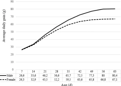
Figure 1. Average daily weight gain (g) of male and female broilers from seven to 63 days-old. Adapted from Cobb manual (2015) ... 25
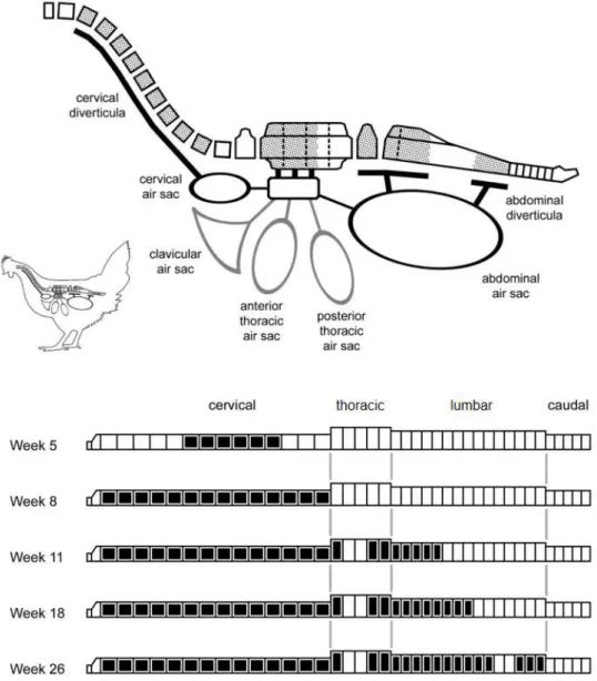
Figure 2. Pneumatization of the vertebral column in the chicken (Gallus gallus). Pneumatic vertebrae are represented in dotted (upper diagram) or black (lower diagram). The vertebral column is pneumatized by diverticula of cervical and abdominal air sacs and lungs. Adapted from King (1957) and Hogg (1984) apud Wedel (2008) ... 30
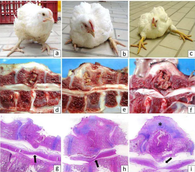
Figure 3. Clinicopathological changes of vertebral osteomyelitis and differential diagnosis in broilers. (a) Broiler showing the classical clinical sign of vertebral osteomyelitis. (b) Gross changes of vertebral osteomyelitis revealing enlargement of affected vertebral body (T4). Inset: sagittal section with caseonecrotic material in the T4 vertebra and spinal cord compression. (c) Vertebral body displacement of T4 vertebra characteristic of spondylolisthesis with spinal cord compression. (d) Scoliosis characterized by lateral deviation of vertebral column. (e, f) Histological changes of vertebral osteomyelitis. There are necrotic tissue, cell debris, heterophils, hemorrhage and fibrin. HE, 400x. Inset: Gram positive bacteria associated to vertebral lesion. Goodpasture, 400x. ... 33
Figure 4. Antibiotic targets and mechanisms of resistance in bacteria (Adapted from
Wright, 2010) ... 37
Chapter II
Figure 1. Broilers with different degrees of vertebral osteomyelitis. In broiler with (a) mild, (b) moderate and (c) marked signs. Sagittal section of the vertebral column with variable amounts of caseonecrotic material in the T4 vertebral body (d), (e) and (f). The necrotic tissue in the region of the vertebral bodies is projecting into the spinal canal leading to spinal cord compression. In the submacroscopic images of vertebral lesions (d), (e) and (f), note the increased volume of vertebral body projecting into the vertebral canal and compressing the spinal cord (arrow) to different degrees (g), (h) and (i). A thick layer of fibrous tissue and disorganized neocartilage (arrows) connecting both vertebral bodies are observed on the necrotic area. HE.. ... 54
(i.e. heterophils, erythrocytes and fibrin). HE, 400x. (d) Gram-negative and Gram-positive bacteria (*) were observed in the vertebral lesions. Goodpasture, 200x. ... 56
Chapter III
Figure 1. Molecular and phenotypic characterization of 15 Escherichia coli strains isolated from broilers with osteomyelitis and arthritis. Black and white boxes represent positive and negative results, respectively. Flock ID, number of the flock of origin; Lesion, VO: vertebral osteomyelitis, Art: arthritis; Serotype, ns: non-serotyped; Flagella, nm: non-motile, nc: non-correspondent to any flagellar type tested; ST, Sequence type; ECOR: ECOR phylogenetic group; APEC (Johnson et al.): APEC diagnosis according to Johnson et al. (2008); APEC (Schouler et al.); APEC diagnosis according to Schouler et al. (2012); Yes: APEC strain, No: non-APEC strain; pVAGs, pattern of virulence genes described by Schouler et al. (2012), nc: non-correspondent to the described patterns; Iron acquisition, genes encoding iron acquisition system; Adhesin, genes encoding adhesins; Toxin, genes encoding toxins; Protectin, genes encoding protectins; Invasin, genes encoding invasins; Miscellaneous, genes encoding different kinds of virulence; VAGs (%), percentage of APEC-associated virulence genes; Lethality score, number of chicks that died at the fourth day post-infection with E. coli; Serum resistance, R: serum resistant strain, I: intermediate resistant strain, S: serum sensitive strain; Nº resistant AB: number of antibiotics to which the strain was resistant; Antibiotic resistance profile: gentamicin, Gen; neomycin, Neo; apramycin, Apr; amoxicillin, Amx; amoxicillin + clavulanic acid, Amc; cephalotin, Cef; cefoxitin, Fox; ceftiofur, Xnl; florfenicol, Ffc; colistin, Cst; nalidixic acid, Nal; flumequine, UB; enrofloxacin, Enr; trimethoprim, Tmp; Tmp + sulfamethoxazole, TmpStx; tetracycline, Tet; pansusceptible, PanSus ... 70
Figure 2. Clinical signs and gross pathology of vertebral osteomyelitis (a, b, c) and arthritis (d, e, f) in broilers. (a) Broiler showing the classical clinical sign of severe cases of vertebral osteomyelitis. (b) Note the enlargement of affected vertebral body (T4), (c) which revels caseonecrotic material and spinal cord compression on longitudinal section. (d) Broiler with bilateral arthritis showing ventral recumbency and retracted legs. (e) Suppurative exudate in articular cavity in acute arthritis, (f) which extended to proximal tibiotarsus causing tibial osteomyelitis... 72
Figure 3. Histopathology of osteomyelitis and arthritis in broilers. (a) Vertebral osteomyelitis showing enlargment of vertebral body (T4) by caseonecrotic material (remanescent, arrow), which compresses spinal cord (*); HE. (b) Caseonecrotic hererophilic and histiocytic exudate (*) in the articular space with intralesional bacterial colonies (arrow); HE. Inset: Gram-negative bacteria stained by Goodpasture. (c) Necrotic synovitis (arrow) associated with caseonecrotic exudate within the articular space (*); HE. (d) Proximal growth plate (physis) of tibiotarsus showing extensive necrosis (*) with heterophilic exudate in a case of tibial osteomyelitis; HE. ... 73
osteomyelitis and arthritis in broilers by antibiotic class: (a) quinolones; (b) beta-lactams; (c) cephalosporins; (d) sulfonamides; (e) tetracyclines; (f) aminoglycosides; (g) phenicols; and (h) polypeptides ... 75
Chapter IV
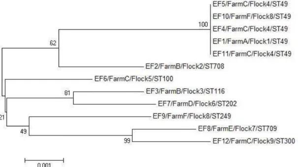
Figure 1. Evolutionary relationships among the concatenated sequences of the identified sequence types of E. faecalis isolated from vertebral osteomyelitis in broilers in Minas Gerais state, southeast Brazil, in 2012. The strain identification and its farm of origin, flock number and ST number are shown. Construction of Neighbour-joining tree was performed using Kimura 2-parameter with bootstrap values of 1000 replicates ... 86
Figure 2. Geographical distribution of E. faecalis isolated from vertebral osteomyelitis in broilers in Minas Gerais state, southeast Brazil, in 2012. The letters (A, B, C, D, E and F) represent the different municipalities included in the study, which are linked to its respective boxes with details of the strains isolated in the place (“Strain ID/Number of the flock/Sequence Type number”). Distance among farms: A to F (130 km); F to B (47 km); B to E (45 km); E to C (42 km); C to D (54 km); and D to A (161 km), comprising a total area of 10,434 km2. ... 87
Figure 3. Population snapshot of STs included in the MLST database for E. faecalis isolated from vertebral osteomyelitis in broilers in Minas Gerais state, southeast Brazil, in 2012. Each ST is represented as a node with the ST number. Clusters of linked STs correspond to clonal complexes. Black lines connect single locus variants. Primary founders are represented in blue in the cluster, and subgroup founders in yellow. Pink arrows indicates STs available in E. faecalis database that were also identified among the isolates described in this study. STs pointed by green arrows are firstly described in this study... 88
LIST OF APPENDIX
Appendix I. Approval certificate of CETEA ... 102
Chapter II
Appendix II. Confirmation of article acceptance for publication in Avian Pathology ... 103
Chapter III
Appendix III. Confirmation of article submission for publication in BMC Veterinary Research ... 104
LIST OF ABBREVIATIONS
AA amyloidosis Amyloid A protein amyloidosis
AMC Amoxicillin + clavulanic acid
AMX Amoxicillin
APEC Avian Pathogenic Escherichia coli
APR Apramycin
ATCC American Type Culture Collection
BA Blood agar
BCO Bacterial condronecrosis with osteomyelitis
BHI Brain and heart infusion
BP Base pairs
CC Clonal complex
CCCD Culture Collection of CEFAR Diagnóstica
CEF Cephalotin
CETEA Committee for Ethics in Animal Experimentation
CFU Colony forming unit
CLSI/NCCLS Clinical and Laboratory Standards Institute (Former NCCLS)
CST Colistin
DNA Deoxyribonucleic acid
E. cecorum Enterococcus cecorum
E. coli Escherichia coli
E. durans Enterococcus durans
E. faecalis Enterococcus faecalis
E. faecium Enterococcus faecium
E. hirae Enterococcus hirae
ECOR Escherichia coli Reference Collection
EDTA Ethylenediaminetetraacetic acid
ENR Enrofloxacin
ESBL Extended spectrum β-lactamases
ExPEC Extraintestinal pathogenic Escherichia coli
FFC Florfenicol
FOX Cefoxitin
GEN Gentamicin
HE Hematoxylin-eosin
HLAR High-level aminoglycoside resistance
HLGR High-level gentamicin-resistance
INRA Institut National de la Recherche Agronomique
LB Luria-Bertani
MCK MacConkey
MH Mueller-Hinton
MLST Multilocus Sequence Typing
NAL Nalidixic acid
NEO Neomycin
OD Optical density
PCR Polymerase Chain Reaction
PFGE Pulsed-field gel electrophoresis
PMSF Phenylmethylsulfonyl fluoride
RNA Ribonucleic acid
RPM Rotations per minute
S. aureus Staphylococcus aureus
SDS Sodium dodecyl sulfate
SPF Specific pathogen free
ST Sequence type
T4 4th thoracic vertebra of chicken vertebral column
TE Tris-EDTA
TET Tetracycline
TMP Trimethoprim
TmpStx Trimethoprim + sulfamethoxazole
TOC Turkey osteomyelitis complex
UB Flumequine
UFMG Universidade Federal de Minas Gerais
VO Vertebral osteomyelitis
VRE Vancomycin-resistant enterococci
20
ABSTRACT
Locomotor disorders represent a major challenge in modern poultry production worldwide and they may be related to non-infectious and infectious etiologies. Vertebral osteomyelitis is a bacterial disease described in outbreaks in many countries, characterized by infection of the mobile thoracic vertebra (T4), which results in the compression of the spine, reduced mobility and death of affected broilers. The objective of this study was to determine the frequency of vertebral osteomyelitis in broilers in the state of Minas Gerais, and to determine the bacterial etiologies involved in disease and their molecular characteristics. For this, we analyzed 608 broilers with locomotor disorders, which had their clinical signs recorded and then necropsied. Vertebral column samples and joints with gross changes were collected for bacterial isolation, molecular and histopathological analysis. Vertebral osteomyelitis was found in 5.1% (31/608) of the birds, which had different degrees of limited mobility, related to the level of spinal cord compression. The bacteria most frequently isolated from lesions were: Enterococcus spp. (53.6%), E. faecalis (32.1%) and E. hirae (7.1%); Escherichia coli (42.8%) in co-infection with E. faecalis in two cases; Staphylococcus aureus (14.3%) in co-infection with Enterococcus spp. or E. hirae in two cases. E. coli strains harbored different genetic pattern as assessed by PFGE, regardless of flock origin and lesion site (vertebral osteomyelitis or arthritis). The E. coli strains belonged to seven sequence types (STs) described previously (ST117, ST101, ST131, ST371 and ST3107) or newly described in this study (ST5766 and ST5856). Most strains belonged to ECOR phylogenetic group D (66.7%) and diverse serogroups (O88, O25, O12 and O45), some of worldwide importance. The antimicrobial susceptibility profile also showed the diversity of the strains and revealed a high proportion of multidrug-resistant strains (73%), mainly to quinolones and beta-lactams. Multilocus sequence typing (MLST) analysis of E. faecalis revealed that the strains belonged to eight different STs, being six (ST49, ST100, ST116, ST202, ST249, and ST300) previously described and ST708 and ST709 first identified in this study. ST49 was the most frequently isolated from vertebral osteomyelitis lesions. E. faecalis strains showed the highest resistance to aminoglycoside antibiotics, mainly to gentamicin (40.0%), and low resistance to vancomycin (10%). The results indicated that, in Brazil, vertebral osteomyelitis in broilers may not be caused by a single infectious agent and suggested geographical differences concerning the frequency and etiology of the disease, as comparing our region in Brazil with reports in other countries. Furthermore, our results showed the diversity of E. faecalis STs involved with this disease and high frequency of aminoglycoside resistance and low frequency of vancomycin-resistance. Also, vertebral osteomyelitis and arthritis could be associated with highly diverse E. coli, which were often multidrug-resistant. Some E. coli strains belonged to STs described also in humans, which may represent a concern to public and animal health.
21
RESUMO
As alterações locomotoras representam um desafio na produção avícola moderna em todo o mundo e podem ter origem não infecciosa e infecciosa. A osteomielite vertebral é uma doença bacteriana descrita em surtos em diversos países caracterizada por infecção da vértebra torácica móvel (T4) que resulta em compressão da medula espinhal, dificuldade de locomoção e morte das aves acometidas. O objetivo deste trabalho foi determinar a frequência da doença em frangos de corte do estado de Minas Gerais, além de conhecer e caracterizar molecularmente os agentes etiológicos envolvidos na doença. Para isso, foram analisados 608 frangos de corte com problemas locomotores que tiveram os sinais clínicos registrados e foram submetidos à necropsia. Amostras de corpo vertebral e articulações com alterações macroscópicas foram coletadas para isolamento bacteriano, histopatologia e análise molecular. Osteomielite vertebral foi encontrada em 5,1% (31/608) das aves, as quais apresentaram diferentes graus de dificuldade locomotora relacionados ao nível de compressão da medula espinal. As bactérias mais frequentemente isoladas das lesões foram: Enterococcus spp. (53,6%), E. faecalis (32,1%) e E. hirae (7,1%); Escherichia coli (42,8%) em co-infecção E. faecalis em dois casos; Staphylococcus aureus (14,3%) em dois casos em co-infecção com Enterococcus spp. ou E. hirae. Os isolados de E. coli apresentaram diferentes padrões genéticos por PFGE, independentemente do lote estudado e tipo de lesão (osteomielite vertebral ou artrite). Os isolados pertenciam a sete sequence types (STs) descritos anteriormente (ST117, ST101, ST131, ST371 e ST3107) ou descritos pela primeira vez neste estudo (ST5766 e ST5856). A maioria dos isolados pertenciam ao grupo filogenético D (66,7%) e diversos sorogrupos (O88, O25, O12 e O45), alguns de importância mundial. O perfil de susceptibilidade antimicrobiana também refletiu a diversidade dos isolados e revelou alta frequência de cepas multirresistentes (73%), principalmente às quinolonas e beta-lactâmicos. A análise do Multilocus sequence typing (MLST) revelou que os isolados de E. faecalis pertenciam a oito STs distintos. Desses, seis (ST49, ST100, ST116, ST202, ST249 e ST300) foram previamente descritos, enquanto ST708 e ST709 foram descritos pela primeira vez nesse estudo. E. faecalis ST49 foi o mais frequentemente isolado das lesões vertebrais. Os isolados da bactéria apresentaram maior percentual de resistência antimicrobiana aos aminoglicosídeos, principalmente à gentamicina (40.0%), e baixa resistência à vancomicina (10%). Os resultados desse estudo demonstram que, no Brasil, osteomielite vertebral em frangos de corte pode não ser causada por um único agente infeccioso e sugere diferenças geográficas relativas à frequência e etiologia da doença entre esta região do Brasil e outros países. Além disso, nossos resultados demonstraram a diversidade de STs de E. faecalis envolvidos na doença com alta frequência de isolados resistentes a aminoglicosídeos e baixa frequência de E. faecalis resistentes à vancomicina. Nossos resultados demonstram, ainda, que osteomielite vertebral e artrite podem estar associadas à E. coli altamente diversas, as quais são frequentemente resistentes a múltiplas drogas antimicrobianas. Alguns isolados de E. coli pertencem a STs descritos também em seres humanos, o que representa uma preocupação para a saúde pública e animal.
22
INTRODUCTION
Poultry products, meat and eggs, are a major source of animal protein available on the market due to its excellent quality, easy access to all sections of society and great variety of products at lower cost to the consumer (Amaral, 2003). Due to its popularity as a food and short production cycle, the poultry represent one of the animals most selected for production worldwide (Emmans and Kyriazakis, 2000) and the numbers of Brazilian poultry demonstrate the excellent performance of the sector in the country. Currently, Brazil is the largest exporter and second largest producer of poultry meat in world rankings (ABPA, 2015a). In 2014, the state of Minas Gerais accounted for 7.1% of the slaughtered poultry, occupying the fifth position among the Brazilian states (ABPA, 2015b).
Most broilers are created using modern intensive production systems worldwide, where birds are confined in high-density warehouses (FAO, 2007) and raised from birth to slaughter in approximately 40 days. However, there is evidence that the optimization of the production for these systems, though producing meat at low cost, results in birds with reduced viability and welfare with limited locomotion capacity (Kestin et al., 1992; Bessei, 2006).
Locomotor pathologies or "leg problems" represent a major concern in commercial flocks of broilers, particularly those that lead to limitations in mobility or lameness (Scahaw, 2000). They are responsible for significant economic losses and decrease in animal welfare in the poultry industry (Araújo et al., 2011). These losses occur for carcasses condemnations in slaughterhouse due to fractures, hematoma and lesions on the skin, as well as by the decrease in the growth and performance of affected broilers. Once these birds can not have adequate access to food and water, they become weak and lighter, presenting worst zootechnical results (Silva et al., 2001; Almeida-Paz, 2010).
The development of many locomotor diseases are related to genetic selection and the rapid growth of broilers, which is demonstrated by their frequency in broilers, broiler breeders, ducks and turkeys raised in confinement (Kestin et al., 1992). These diseases have been a problem since the beginning of intensive poultry production and has been linked to numerous causes as nutrition (poisoning, deficiencies or imbalances), genetics, management practices and others that can affect directly the growth and development of the locomotor system (Silva et al., 2001), such as infections and trauma (Julian, 1998).
23
OBJECTIVES
General
This study aimed to determine the frequency of vertebral osteomyelitis, and provides data on the etiology of the disease in poultry with locomotor disorders in the state of Minas Gerais.
Specific
1. To establish the frequency of vertebral osteomyelitis in broilers with locomotor disorders in the state of Minas Gerais;
2. To describe the clinical and pathological changes in broiler with vertebral osteomyelitis in the state of Minas Gerais;
3. To identify the etiologic agents involved in the cases of vertebral osteomyelitis in broilers with locomotor disorders in the state of Minas Gerais;
4. To investigate the antibiotic susceptibility and genetic relationships among Enterococcus faecalis isolates from vertebral osteomyelitis in broilers with locomotor disorders in the state of Minas Gerais;
5. To determine the molecular and phenotypic characteristics of Escherichia coli isolated from broiler lesions with locomotor disorders in the state of Minas Gerais; and
24
CHAPTER 1
Vertebral osteomyelitis in broilers: a review
J. F. V. Braga1, N. R. S. Martins2, R. Ecco1*
1Departamento de Clínica e Cirurgia Veterinária, Escola de Veterinária, Universidade Federal
de Minas Gerais, Av. Antônio Carlos, 6627, Campus Pampulha, 30161-970, Belo Horizonte, Minas Gerais, Brazil.
3Departamento de Medicina Veterinária Preventiva, Escola de Veterinária, Universidade
Federal de Minas Gerais, Av. Antônio Carlos, 6627, Campus Pampulha, 30161-970, Belo Horizonte, Minas Gerais, Brazil.
*To whom correspondence should be addressed. Tel: +55 31 3409 2261. E-mail: [email protected]
Abstract:
Vertebral osteomyelitis is an emerging disease in broilers worldwide. The inflammatory process in the affected thoracic vertebra (T4) and spinal cord compression leads to clinical signs related to locomotor impairment and death of birds. The pathogenesis of the disease is poorly understood and Enterococcus cecorum is the bacterium frequently associated with the disease. However, E. faecalis, E. durans, Escherichia coli and Staphylococcus aureus were recently detected in cases of the disease, raising questions about its etiopathogenesis. An important aspect related to these bacteria is their role as source virulence and antibiotic resistance genes and its possible dissemination to other bacteria, animals and humans. Since there are still many questions about vertebral osteomyelitis in broilers, the knowledge on its prevention, control and treatment are limited. In this review, we compile and discuss the current knowledge on vertebral osteomyelitis in broilers and raise relevant aspects concerning the disease.Keywords: locomotor diseases, bacterial infections, Enterococcus spp., Enterococcus cecorum, Escherichia coli.
Introduction
25
Epidemiology of the disease
Vertebral osteomyelitis has been reported in poultry in different countries of Europe, such as United Kingdon (Wood et al., 2002), Netherlands (Devriese et al., 2002; Kense and Landman, 2011), Belgium (Herdt et al., 2009), Hungary (Makrai et al., 2011), Norway (Kolbjørnsen et al., 2011), and Bulgaria (Dinev, 2013). The disease was also described in North and South America, such as in Canada (Stalker et al., 2010), several US states (Pennsylvania, Washington, North Carolina, South Carolina, Arkansas, Mississippi, Alabama, and California) (Aziz and Barnes, 2009; Gingerich, 2009) and Brazil (Braga et al., 2016c).
The disease occurs more frequently in males and several lineages can be affected (Wood et al., 2002; Gingerich, 2009). It is interesting to note that the higher body weight normally observed in males (Fig. 1) implies an increase in the weight supported by the joints and a greater chance of trauma, which is suggested for another locomotor condition known as bacterial condronecrosis with osteomyelitis (BCO), more frequent in male broilers (Wideman and Prisby, 2013).
Figure 1. Average daily weight gain (g) of male and female broilers from seven to 63 days-old. Adapted from Cobb manual (2015).
Affected broilers are usually older than 30 days-old, with reported outbreaks of the disease ranging from three to 18 week-old (Herdt et al. 2009; Armour et al., 2011; Robbins et al., 2012). However, there is a report in a flock older than 15 days. Initially, the percentage of affected birds in a flock was relatively high, ranging from 5% to 10%, and then following reports of 2% to 4% (Gingerich, 2009).
26 intestinal commensal E. cecorum, raising the question whether the emergence of clones is most likely the cause for the increased occurrence of infections (Boerlin et al., 2012).
Some evidences suggest that the higher incidence of Enterococci-associated diseases in poultry may be due to horizontal spread of dominant clones of E. cecorum which exhibit increased pathogenicity (Kense and Landman, 2011; Boerlin et al., 2012). Strains with genotypes similar to those isolated from vertebral osteomyelitis cases were rarely recovered from the cecum of birds with vertebral osteomyelitis and the presence of these isolates was not statistically associated with a higher risk of disease (Borst et al., 2012). These findings suggest that long-term cecal transport of pathogenic clones may not be necessary in the pathogenesis of vertebral osteomyelitis caused by E. cecorum. However, as the disease has a chronic character, requiring weeks from the time of infection to the onset of clinical signs, pathogenic strains may be transient in the gastrointestinal tract and therefore not recoverable in the moment of clinical presentation (Borst et al., 2012). Field observations showed that the disease occurred in successive flocks, suggesting persistence of E. cecorum on the farm (Herdt et al., 2009; Kense and Landman, 2011).
Despite the worldwide distribution, the way in which pathogenic clones of E. cecorum spread remains undetermined. Epidemiological studies on distinct outbreaks of vertebral osteomyelitis suggest that mechanical spreading by biological vectors or inadequate biosafety can contribute to disease transmission, although horizontal transmission between geographically distant locations was considered unlikely (Borst et al., 2012).
Kense and Landman (2011) demonstrated that vertical transmission does not occur. Recently, Borst et al. (2014) showed that SPF and non-SPF chicken embryos inoculated with E. cecorum isolated from vertebral lesions had lower survival rate when compared to embryos inoculated with E. cecorum isolated from the intestines of healthy birds. The embryos infected with pathogenic strains had lesions of septicemia, such as hemorrhage and edema. In embryos inoculated with non-pathogenic strains, these lesions were observed only 48 hours later.
Etiologic agents
Most inflammatory diseases of bones are caused by bacterial infections, although other agents can also infect bones (Craig et al., 2016). The bacterial agents of greatest importance in the etiology of vertebral osteomyelitis in poultry are described.
The genus Enterococcus
Enterococcus spp. are gram-positive and spherical bacteria, which occur alone, in pairs or short chains. They are non-motile, non-spore-forming, facultative anaerobic with diverse biochemical properties (Wages, 1998). However, the relationship between biochemical characteristics and pathogenicity of the species remains unknown (Thayer et al., 2008). Enterococcus spp. are ubiquitous in nature with worldwide distribution in avian species. They are considered part of the normal intestinal microbiota of chickens and commonly found in poultry environments. The frequency that different species of Enterococcus spp. are isolated from the intestinal tract of healthy birds can vary according to the age, but only a limited number of species is isolated more often. E. faecium, E. cecorum, E. faecalis, E. hirae and E. durans were the species regularly isolated in at least one of three different age groups (1 day-old, 3 to 4 week-day-old, and more than 12 week-old) examined by Devriese et al. (1991).
27 al., 2003; Perez, 2004). However, since 2002, E. cecorum was more frequently recognized as cause of outbreaks of non-vertebral and vertebral osteomyelitis (the last one also known as spondylitis) and arthritis in broiler and broiler breeders (Aziz and Barnes, 2007; Herdt et al., 2009; Aziz and Barnes, 2009; Gingerich, 2009; Stalker et al., 2010; Martin et al., 2011; Boerlin et al., 2012; Aitchison et al., 2014). Jung and Rautenschlein (2014) described Enterococcus cecorum isolation from a broiler flock with pericarditis, hepatitis, femoral head necrosis and/or vertebral osteomyelitis and concluded that bacteremia and generalized infection seem to be important steps in the pathogenesis of infection caused by this bacterium in broilers.
E. cecorum occurs more frequently in intestines of chickens older than 12 weeks of age (Devriese et al., 1991) and was rarely associated with clinical disease in these birds (Devriese et al., 2002; Wood et al., 2002; Chadfield et al., 2004; Thayer et al., 2008). This reflects on the limited number of publications regarding its role in disease and its pathogenicity (Makrai et al., 2011). Two main hypotheses were proposed to explain the recent increase in the incidence of infections with E. cecorum: 1) changes in the host or environmental factors; and 2) emergence of individual clones with increased pathogenicity (Boerlin et al., 2012). To prove the second hypothesis, the authors analyzed E. cecorum isolates recovered from the cecum of healthy birds and of birds with vertebral osteomyelitis by pulsed-field gel electrophoresis (PFGE). Genotypes of E. cecorum isolated from vertebral lesions were significantly more similar to each other than the E. cecorum isolated from the cecum of healthy birds and of birds with vertebral osteomyelitis, regardless the affected flock.
Infections by E. hirae are relatively frequent in broilers, but its importance is not as understood as the infections caused by other bacteria, such as Escherichia coli and Staphylococcus aureus. Diseases caused by E. hirae have increasing incidence in some countries, such as Norway, where the bacterium was isolated from cases of osteomyelitis in broilers (Kolbjørnsen et al., 2011). In addition, there are reports of focal cerebral necrosis in chicks (Devriese et al., 1991; Randall et al., 1993) and cases of diarrhea in one week-old chicks (Kondo et al., 1997). Velker et al. (2011) described endocarditis associated with E. hirae in different broiler flocks, with co-isolation of E. faecalis, E. coli, and a mixture of several other opportunistic bacteria from lesions. Although the meaning of this finding was considered unknown by the authors, they assumed these as opportunistic infections or resulting from tissue autolysis. Recently, Braga et al. (2016c) reported E. hirae, E. faecalis, E. coli and S. aureus in single or mixed culture from vertebral osteomyelitis cases in broilers. The molecular analysis and histopathology with special stains allowed the confirmation of concomitant agents in the lesion and discarded the possibility of contamination.
In addition to vertebral osteomyelitis, Enterococci are often associated with other diseases in poultry. In day-old chicks, Enterococci are generally responsible for infection in the yolk sac (Deeming, 2005). E. faecalis has been associated with hepatic granulomas in turkeys (Hernandez et al., 1972) and pulmonary hypertension syndrome in broilers (Tankson et al., 2001). Cases of arthropathy associated to AA amyloid and concomitant systemic amyloidosis caused by arthropathic and amyloidogenic E. faecalis was described in laying hens (Landman et al., 1994) and broiler breeders (Steentjes et al., 2002). In domestic ducks, E. faecalis have been isolated from cases of arthritis (Bisgaard, 1981), whereas E. faecium (Sandhu, 1988) and E. cecorum (Jung et al., 2013) have been associated with acute septicemia in Pekin ducks. E. durans was isolated from young chickens with bacteremia and encephalomalacia (Cardona et al., 1993; Abe et al., 2006).
28 such as adhesins, invasins, hemolysin, and pili. Specific genetic lineages of hospital-adapted strains emerged and some E. faecalis are considered high-risk Enterococci, such as the clonal complexes CC2, CC9, CC28 and CC40. These are characterized by the presence of antibiotic resistance determinants and/or virulence factors usually located on pathogenicity islands or plasmids, highlighting a major role for bacteria mobile genetic elements in establishing problematic strains (Franz et al., 2011).
Some studies showed little phylogenetic diversity of E. faecalis isolates, with nucleotide identity of 97.8% to 99.5%. However, the sequence identity of shared genetic content among isolates ranged from 70.9% to 96.5%. In general, most of E. faecalis diversity can be attributed to the inclusion of mobile genetic elements into a widely conserved genome, with these mobile elements supporting the exchange of chromosomally encoded characteristics (Palmer et al., 2012). Studies that compared E. faecalis isolates obtained from different lesions in eight broiler breeder flocks and E. faecalis isolated from healthy birds revealed 12 different sequence types (STs) and lack of correlation between ST and lesion type, although ST82, ST174 and ST177 represented 81% of the strains associated with lesions (Gregersen et al., 2010).
Escherichia coli
E. coli has been also isolated from cases of vertebral osteomyelitis in poultry (Dinev, 2013; Braga et al., 2016c), which is part of normal intestinal microbiota of humans and many animal species. E. coli are Gram-negative non-spore-forming bacillus, with 2-3 x 0.6 µm in size, and most strains are motile with peritrichous flagella (Barnes et al., 2008).
Several E. coli strains are able to express virulence factors and cause intestinal or extra-intestinal diseases (Ambrozic et al., 1998). Currently, E. coli is considered the most important Gram-negative bacterium due to its different mechanisms of pathogenicity and described diseases (Nakazato et al., 2009). In avian species, pathogenic strains are named Avian Pathogenic Escherichia coli (APEC) (Ewers et al., 2004), which are responsible for extra-intestinal diseases known generically as colibacillosis.
Colibacillosis has a worldwide occurrence and leads to significant economic losses in all types of poultry (Dziva and Stevens, 2008). The most common lesions associated with colibacillosis are perihepatitis, pericarditis and airsacculitis, although other syndromes such as osteomyelitis/arthritis, yolk sac peritonitis, salpingitis, coligranuloma, omphalitis and cellulitis can also be found (Barnes et al., 2008). According to Barnes et al. (2008), the presence of E. coli in bone and synovial tissues is a common sequel of colisepticemia and the affected birds could probably not completely eliminate the bacterial infection.
Several virulence factors are associated with APEC, such as: F1 and P fimbrial adhesins, aerobactin iron acquisition system, k1 capsular antigen, complement resistance and many proteins, such as Tsh autotransporter (Dho-Moulin and Fairbrother, 1999). Although APEC strains are the major pathogens for commercial poultry, the knowledge on virulence factors are still incomplete. Considering that avian colibacillosis occurs worldwide in its various forms, it is believed that the phylogenetic analysis of clonal relationships among E. coli isolates from different countries and regions may provide a greater understanding about its pathogenesis (Schouler et al., 2004).
29 Schouler et al. (2012) showed that four combinations of genes allowed the diagnosis of more than 70% of the APEC strains.
Phylogenetic studies based on E. coli Reference Collection (ECOR), a set of 72 E. coli strains isolated from various animal hosts and different geographic origins (Ochman and Selander, 1984), showed that there are four main phylogenetic groups for E. coli designated A, B1, B2 and D (Selander et al., 1987; Herzer et al., 1990). However, no avian strain was included in the ECOR collection and no APEC strains were placed in the E. coli phylogenetic tree. Epidemiological molecular studies showed that most APEC strains can be grouped into a limited number of clones (Ngeleka et al., 1996; Da Silveira et al., 2002; La Ragione and Woodward, 2002; Ewers et al., 2004). The clonal nature of APEC has been demonstrated by phylogenetic analysis (Whittam and Wilson, 1988; White et al., 1990; White et al., 1993; Da Silveira et al., 2002). Many studies have also revealed the prevalence of several serogroups and particular combinations of genes associated with virulent strains of APEC. These observations suggested that only a limited number of virulent genotypes exist (Blanco et al., 1998; Ngeleka et al., 2002; Ewers et al., 2004; Rodriguez-Siek et al., 2005a; Rodriguez-Siek et al., 2005b).
Some sequences of APEC strains genome was determined, still requiring extensive comparative genomic analysis of APEC strains of different serogroups (Johnson et al., 2007; Dziva et al., 2013; Mangiamele et al., 2013; Huja et al., 2015). The comparative genomic study of APEC serogroup O78 revealed that genetic variability occurs even within a single serogroup (Huja et al., 2015). According to Rasko et al. (2008), that studied different pathogenic E. coli, the pangenome of the bacterium has a reservoir consisting of more than 13,000 genes. This has great implication on diversity and pathogenesis of E. coli strains and their ability to colonize and cause disease in the human host. Approximately half of the genome content of any E. coli represents the core-conserved genome and the open pangenome of E. coli species indicates that continuous genetic sequencing should result in the identification of approximately 300 new genes per genome.
Other agents involved in vertebral osteomyelitis
Staphylococcus pyogenes was isolated from vertebral osteomyelitis cases in seven to 16 week-old chickens (Carnaghan, 1966). Nairn (1973) reported the isolation of Staphylococcus aureus of vertebral lesions in turkeys naturally affected with locomotor disorder. The experimental inoculation in turkeys resulted in osteomyelitis in the vertebral body and long bones. Van Veen (1999) reported the involvement of Aspergillus fumigatus in vertebral osteomyelitis outbreaks in two flocks of 17-19 week-old broilers.
Pathogenesis
Bacterial inflammatory processes of bones can be originated from hematogenous, local extension, and implantation routes, being the first the most common in animals. When the inflammation is originated from vascular areas of the medullary cavity or periosteum is referred as osteomyelitis or periostitis, respectively. A more general but less frequently used term for inflammation of bones is osteitis (Craig et al., 2016).
30 The disease was reproduced experimentally by Martin et al. (2011), through the inoculation of E. cecorum by oral and intravenous routes. Gross lesions were observed five weeks after the experimental infection in 6.1% and 2.9% of broilers inoculated orally or intravenously, respectively. However, histologic lesions were observed in 30.3% of broilers inoculated orally, and the macroscopic evidence of disease was suggested to be higher if the broilers were older.
The free thoracic (T4) vertebral body is singly affected in vertebral osteomyelitis and the reasons for this predilection are unknown. The single free thoracic vertebral articulation is located between the immediately anterior fused thoracic vertebrae and the posterior synsacrum (Fig. 2), enabling body position adjustments and flexibility during walk and flight, and is subject to greater biomechanical stress and microtraumas than any other vertebra. Excessive stress may lead to changes in vascular flow with development of micro-thrombi, sequestrum and multiplication of bacteria, if present in blood (Aziz and Barnes, 2007; Stalker et al., 2010; Wideman and Prisby, 2013; Aitchison et al., 2014).
31 diagram). The vertebral column is pneumatized by diverticula of cervical and abdominal
air sacs and lungs. Adapted from King (1957) and Hogg (1984) apud Wedel (2008).
According to Stashak and Mayhew (1984), vertebral osteomyelitis is usually secondary to hematogenous dissemination of a microorganism. However, other theories have been proposed to explain how the bacteria reach the mobile thoracic vertebra. Currently, the most accepted theory suggests that the agent has access to the bones via bloodstream due to rupture of the intestinal mucosal barrier (Stalker et al., 2010; Martin et al., 2011), as in coccidiosis or bacterial enteritis (Gingerich, 2009). According to Armour et al. (2011) and Martin et al. (2011), any factor that interferes negatively with intestinal health or disturbs the balance of intestinal microbiota could possibly predispose to systemic dissemination of E. cecorum.
A possible link of the vertebral osteomyelitis with air sacs and pneumatic vertebra could exist (Aziz and Barnes, 2007), as shown in Fig. 2. However, it is interesting to note that the pneumatization of the vertebra where the disease occurs (T4) begins only after eight weeks of age. The experimental inoculation of E. cecorum in two week-old broilers by air sac route did not result in vertebral osteomyelitis (Martin et al., 2011), suggesting that for experimental studies older broilers should be inoculated. It is worth mentioning that, in a study conducted by Tankson et al. (2002), E. faecalis, E. durans, and E. coli were isolated from the heart and lung of healthy birds in 15% of cases, however, there are no studies that provide this information for E. cecorum.
It is interesting to note some aspects in the pathogenesis of BCO that could assist in the understanding of the pathogenesis of vertebral osteomyelitis. This disease affects more often the femur and tibiotarsus, but it can also occur in the free thoracic vertebra. BCO initiates with the degeneration and necrosis of the cartilage followed by bacterial invasion, mainly associated to S. aureus, E. coli and E. cecorum, often in mixed culture, and with other bacteria (Wideman and Prisby, 2013).
It is believed that the BCO begins with mechanical damage to the columns of chondrocytes poorly mineralized present mainly in the proximal growth plate of fast-growing bones, such as the femur and tibia, followed by colonization of the chondronecrotic clefts by opportunistic bacteria spread through the blood. Terminal BCO presents itself as degeneration, necrosis and bacterial infection at the proximal ends (epiphyseal and metaphyseal growth plates) of the femur and tibiotarsus. A similar process may occur in the growth plates of other bones that are subject to severe torque and shear stresses, as occur in the fourth thoracic vertebra, which functions as a flexible pivot between the cranially fused vertebrae of notarium and caudally fused vertebrae of synsacrum (Carnaghan, 1966; McNamee and Smyth, 2000; Dinev, 2009; Wideman et al., 2012).
32 may possibly serve as conduits for the process of bacterial spread to the joint and surrounding soft tissues.
Clinicopathological changes
Clinical signs
The clinical signs are similar in all the vertebral osteomyelitis reports (Gingerich, 2009), although with variable onset age of clinical presentation. In cases of osteomyelitis and arthritis caused by E. cecorum, Herdt et al. (2009) reported that the clinical signs started during the first and second weeks of age with mortality rate of 7%. In the osteomyelitis cases studied by Makrai et al. (2011), the clinical signs started between the 5th and 9th week of age up to the 10th to 13th week, with a mortality rate ranging from 8% to 30%, which was higher than previously reported (Wood et al., 2002; Herdt et al., 2009).
33 Figure 3. Clinicopathological changes of vertebral osteomyelitis and differential diagnosis in broilers. (a) Broiler showing the classical clinical sign of vertebral osteomyelitis. (b) Gross changes of vertebral osteomyelitis revealing enlargement of affected vertebral body (T4). Inset: sagittal section with caseonecrotic material in the T4 vertebra and spinal cord compression. (c) Vertebral body displacement of T4 vertebra characteristic of spondylolisthesis with spinal cord compression. (d) Scoliosis characterized by lateral deviation of vertebral column. (e, f) Histological changes of vertebral osteomyelitis. There are necrotic tissue, cell debris, heterophils, hemorrhage and fibrin. HE, 400x. Inset: Gram positive bacteria associated to vertebral lesion. Goodpasture, 400x.
34 hematoma in the wings (Makrai et al., 2011). One of the consequences of impaired locomotion is the difficulty to have access to water and food, resulting in lower growth rate and death due to dehydration or starvation. In addition, sick broilers are more likely to cannibalism (Barnes et al., 2008).
Gross changes
The macroscopic examination of the vertebral column thoracolumbar region of affected broilers reveals gross changes in the free thoracic vertebra (T4), which shows a palpable whitish to yellowish enlargement (Fig. 3b). The sagittal section of these lesion shows caseonecrotic material inside the vertebral body characterized by yellow to gray exudate, granular and friable, which is surrounded by a thick whitish capsule of fibrous connective tissue (Fig. 3b) (Gingerich, 2009; Martin et al., 2011; Robbins et al., 2012, Braga et al., 2016c). Marked lesion characterized by increased volume of the vertebral body due to the infection results in narrowing of the overlying spinal canal, which would cause compression of the spinal cord (Makrai et al., 2011; Aitchison et al., 2014, Braga et al., 2016c). For early stages of disease, there is no large increase of the vertebral body and mild or no spinal compression that can be seen in the sagittal section. Body condition of affected birds is variable, from good nutritional to cachectic condition. According to Makrai et al. (2011), some broilers may have subcutaneous edematous and green-brownish lesions in the region of tibiotarsus-metatarsal joint.
In some cases, the involvement of bone and joint can occur, process named osteoarthritis. In these cases, the most commonly affected bones are tibiotarsus, femur, thoracolumbar vertebral column and humerus (Muttalib et al., 1996). In long bones, the lesion occurs more frequently in the proximal growth plate. The injuries usually occur where endochondral ossification is developing and extends to the cartilage of adjacent growth plate (McNamee and Smyth, 2000). Stalker et al. (2010) described an outbreak of typically unilateral lesions of osteomyelitis and arthritis associated with E. cecorum. These were characterized by fibrinous exudate into the articular space of tibiotarsus-metatarsal or coxo-femoral joints, extending to the tendon sheath. Rasheed (2011) reported that the joints with arthritis were increased in volume, swollen and hyperemic with purulent yellowish-white exudate inside the joint space.
Histopathology
35 degeneration, and the neuropil was disorganized and vacuolated, indicating a compressive effect.
Martin et al. (2011) reported histologic changes in broilers in the absence of macroscopic lesions, with mild histologic lesions in the subchondral vertebral areas, with no extension to the articular cartilage or adjacent vertebrae. A moderate to severe infiltration of lymphocytes and diffuse fibroplasia in the affected vertebra with intralesional bacteria was confirmed in half (4/8) of the cases. Braga et al. (2016c) also observed lesions in adjacent vertebrae, which were characterized by the degeneration and necrosis of the articular cartilage (T4/T5), and occasional presence of clefts associated or not with hemorrhages and bacterial colonies. In the study performed by Martin et al. (2011), osteochondrosis was observed in all birds, some of them with different degrees of subluxation on the free thoracic vertebra.
Stalker et al. (2010) reported the occurrence of concomitant osteomyelitis and arthritis, The arthritis was characterized by severe inflammation with heterophilic infiltration into the synovium and tendon sheaths of tibiotarsus-metatarsus joints. According to Craig et al. (2016), suppurative arthritis is characterized by greater amount of heterophils in the synovial fluid and membrane, and occasionally in adjacent structures. When the etiologic agent is a bacterium, heterophils are usually abundant and may be degenerated, which is frequently considered a septic arthritis. Most of young broilers with septic arthritis of hematogenous origin may also have osteomyelitis, possibly to the concomitant localization of the microorganism in the bone and synovial membrane, or a result of the close vascular relationship between epiphyseal bone and synovial membrane in young animals, with the spread of infection from one location to another. Foci of osteomyelitis originating in endochondral ossification sites of epiphysis below the articular cartilage may penetrate the cartilage, spreading the infection directly into the synovial fluid. In the joints that the capsule is inserted beyond the growth plate, inflammatory foci in the metaphysis can contaminate the synovial fluid by penetration in the cortical region near to the growth plate order. This region is relatively porous in young animals due to the intensive structural remodeling that occurs in the cortex of the metaphysis during rapid growth.
Diagnosis
Vertebral osteomyelitis may be suspected in birds presenting signs of sitting on the hocks (McNamee and Smyth, 2000). For the macroscopic diagnosis, lungs and kidneys must be removed to provide the visualization and careful examination of the vertebral column. A sagittal section of vertebral column should be performed in order to allow the evaluation of the vertebral body and the degree of spinal cord compression (Gingerich, 2009, Braga et al., 2016c).
36 diagnoses. In Marek’s disease, no changes in the vertebral column are observed, but in the peripheral nerves, which become yellow-gray with loss of striations, acquiring an edematous appearance in some cases (Schat and Nair, 2008).
Prevention, treatment and control
Information on prevention, treatment and control of the disease are limited, in view that the origin and pathogenesis of vertebral osteomyelitis remain unclear, and most studies are related to infections caused by E. cecorum (Kense and Landman, 2011). For the prevention of vertebral osteomyelitis, recommendations on management practices have been made to reduce the risk of developing the disease, such as: 1) avoiding excessive food restriction; 2) following the suggested weight gain patterns and nutritional recommendations; 3) promoting adequate control of coccidiosis; 4) avoiding high density of poultry; 5) ensuring adequate access to feeders; and 6) preventing respiratory diseases. All practices to prevent bacterial infections that could produce bacteremia would probably help to avoid bone and articular inflammation.
Antibiotics have been used to treat the bacterial infection in vertebral osteomyelitis. Although several antibiotics have shown efficacy against the commonly described bacteria, the difficulty is to achieve adequate concentrations of the antibiotics in the vertebral column. In the reported outbreaks of the disease, antibiotics have been ineffective in reducing mortality possibly due to antimicrobial resistance of E. cecorum or the inability of the antibiotic to effectively penetrate the anatomical areas where the bacteria is located (Kense and Landman, 2011). The antimicrobial susceptibility profiles of E. cecorum isolated from outbreaks in different countries were similar (Herdt et al., 2009; Aitchison et al., 2014). Aitchison et al. (2014) reported that, after isolation and identification of E. cecorum, it was difficult to perform the antibiotic susceptibility test due to the growth conditions. The test was performed on tryptose blood agar and nevertheless the bacteria showed poor growth. Makrai et al. (2011) reported that, after the onset of the outbreak, broilers showing clinical signs were separated from those clinically normal and the clinically normal were treated with different antibiotics (amoxycillin, amoxycillin with clavulanic acid, lincomycin or doxycycline), resulting in no new clinical case of the disease in the flock.
After the occurrence of the disease, the elimination of subsequent cases will require repeated cycles of disinfection and usually would not occur after a single cleaning and disinfection. Increased efforts in subsequent flocks are required to eliminate the disease. Some practices that can reduce the risk of vertebral osteomyelitis in the subsequent flocks include: 1) emptying and completely disinfecting the aviary; 2) changing or composting the litter bed; 3) adequate cleaning of water lines; and 4) continuously sanitizing the water (Gingerich, 2009; Stalker et al., 2010; Armour et al., 2011; Martin et al., 2011).
Antimicrobial resistance and public health
37 resistance genotypes, with special concern on vancomycin-resistant Enterococci (VRE) (Cetinkaya et al., 2000; Willems and Bonten, 2007). The VRE have become a major problem in nosocomial infections. A retrospective study of 10 human patients with osteomyelitis showed that eight of these cases were due to infection by Enterococcus faecalis resistant to vancomycin with one death reported due to bacteremia (Holtom et al., 2002).
It is interesting to note that, generally, antibiotics target basic bacterial physiology and biochemistry, causing cell death or inhibiting its growth. Bacterial targets that are different or nonexistent in eukaryotic cells (including human) are: bacterial cell wall; cell membrane; protein synthesis; DNA and RNA synthesis; and metabolism of folic acid (vitamin B9) (Fig. 4). Some examples are β-lactams, such as penicillins, cephalosporins and carbapenases that block the synthesis of the cell wall that is essential for bacterial survival. Furthermore, bacterial ribosomes are the target of tetracyclines, aminoglycosides, macrolides and other antibiotics (Wright, 2010).
Figure 4. Antibiotic targets and mechanisms of resistance in bacteria (Adapted from Wright, 2010).
38 enzymes that recognize and modify antibiotics, resulting in the elimination of the functional characteristics that allow the interaction with their targets (e.g., β-lactamase cleaves the central β-lactam ring, which is characteristic of the class and essential for antibiotic activity) (Wright, 2010).
Resistant bacteria in animals and their by-products and the possible transmission to humans through contamination of carcasses represent a concern in animal and public health (Moreno et al., 2006). Enterococci contaminate not only raw meat, but may also be associated with processed meat products, such as fermented raw sausages or cooked products (Martin et al., 2005; Barbosa et al., 2009; Ruiz-Moyano et al., 2009). Although there is no description of food intoxication in humans associated with E. faecalis, a recent study performed in Brazil showed the presence of these bacteria in 42% of chicken carcasses tested. All these strains were resistant to at least one antibiotic tested, with detection of the antimicrobial resistance genes erm(B), vanC-1, aph(3')-llla, ant(6)-la, vanB, vanA, aac(6')-le-aph(2'')-la, erm(A)e tet(M). This highlights the role of E. faecalis in public health, once these microorganisms may have the ability to transmit antimicrobial resistance genes to other organisms present in the intestinal tract of humans and animals, resulting in limited use of these drugs for clinical treatments (Campos et al., 2013). Hayes et al. (2003) analyzed 981 raw meat samples available commercially from various species (chicken, turkey, swine and bovine) and isolated 1,357 Enterococcus spp. strains, which included E. faecalis (29%) and E. hirae (5.7%). These authors also detected high level of gentamicin resistance in 4% of the strains, most of them isolated from chicken meat. Braga et al. (2016b), analyzing E. faecalis isolates from vertebral osteomyelitis in broilers, demonstrated that the highest level of antibiotic resistance was for aminoglycosides, mainly gentamicin (40%).
E. coli strains also have a major importance because of their role in public health. Most serotypes of the bacterium isolated from chickens are pathogenic only for avian species and will not cause infection in humans or in other mammals (Meno et al., 2002). However, some E. coli strains isolated from poultry lesions have genetic similarities to those that cause diseases in humans, a close relationship subject of research as may constitute a risk to the consumer health (Andrade, 2005). A few studies have suggested the possibility of APEC be related to extraintestinal infections in humans (Ewers et al., 2007; Johnson et al., 2007).
The multiple antimicrobial resistance characteristics of APEC strains also show genetic diversity of isolates, which are often resistant to the following antibiotics: tetracycline, chloramphenicol, sulfonamides, aminoglycosides, fluoroquinolones, β-lactam and extended spectrum β-lactam (Mellata, 2013; Braga et al., 2016a). Genes encoding resistances are often located in the large transmissible plasmids R (Koh and Kok, 1984). It is not surprising that multidrug-resistant APEC often carry conjugative plasmids (Caudry and Stanisich, 1979). In addition, ColV plasmids are often found in APEC strains and seem linked to virulence (Johnson et al., 2006; Johnson et al., 2008). Plasmids can serve as a reservoir of antimicrobial resistance genes and are horizontally transferable to the same and other species of bacteria of potential risk to human health (Johnson et al., 2005).
39 often facilitating the incorporation of the multiple resistance genes into the genome or plasmids (Tenover, 2006).
Conclusions
Vertebral osteomyelitis is an emerging disease demanding a diversity of studies for its understanding. Many aspects on the etiology and pathogenesis of the disease remain unclear, which limits the knowledge on its prevention and control. Most reports associate the disease to infection by Enterococcus cecorum, probably emerging clones with higher pathogenicity. However, other Enterococci and Escherichia coli have been isolated from vertebral osteomyelitis in broilers, raising questions on the role of any specific bacterium in the development of the disease, once its occurrence is related to meat type chicken. Many of the bacteria isolated from cases of the disease are often multidrug-resistant and the possible transmission of these bacteria or their antibiotic resistance encoding genes are a major concern for animal and public health.
Acknowledgment.
J.F.V. Braga is a fellow of the Programa de Pós-graduação em Ciência Animal/Universidade Federal de Minas Gerais supported by Conselho Nacional de Pesquisa (CNPq) and Coordenação de Aperfeiçoamento de Pessoal de Nível Superior (CAPES).References
Abe, Y.; Nakamura, K.; Yamada, M.; Yamamoto, Y., 2006. Encephalomalacia with
Enterococcus durans infection in the brain stem and cerebral hemisphere in chicks in Japan. Avian. Dis. 50, 139-41.
Aitchison, H.; Poolman, P.; Coetzer, M.; Griffiths, C.; Jacobs, J.; Meyer, M.; Bisschop, S., 2014. Enterococcal-related vertebral osteoarthritis in South African broiler breeders: A case report. J. S. Afr. Vet. Assoc. 85, 01-05.
Ambrozic, J.; Ostroversnik, A.; Starcic, M.; Kuhar, I; Grabnar, M; Zgur-Bertok, D., 1998. Escherichia coli CoIV plasmid pRK100: genetic organization, stability and conjugal transfer. Microbiol. 144, 343–352.
Andrade, C. L., 2005. Histopatologia e identificação da Escherichia coli como agente causal da celulite aviária em frangos de corte. Dissertação de Mestrado. Universidade Federal
Fluminense. 62p.
Armour, N.K.; Collet, S.R.; Williams, S.M., 2011. Enterococcus cecorum-related arthritis and osteomyelitis in broilers and broiler breeders. Poult. Inform. Profession. 117, 1–7.
Aziz, T.; Barnes, H.J., 2007. Is spondylitis an emerging disease of broilers? World Poult. 23, 44–45.