http://dx.doi.org/10.1590/1806-9061-2017-0690
Review
Author(s)
Braga JFVI,II
Martins NRSIII
Ecco RI
I Departamento de Clínica e Cirurgia
Veteri-nárias, Escola de Veterinária, Universidade Federal de Minas Gerais, Belo Horizonte, Minas Gerais, Brasil.
II Departamento de Ciências Animais, Centro
de Ciências Agrárias, Universidade Federal Rural do Semi-árido, Mossoró, Rio Grande do Norte, Brasil.
III Departamento de Medicina Veterinária
Pre-ventiva, Escola de Veterinária, Universidade Federal de Minas Gerais, Belo Horizonte, Minas Gerais, Brasil.
Mail Address
Corresponding author e-mail address Roselene Ecco
Setor de Patologia. Departamento de Clínica e Cirurgia Veterinárias, Escola de Veterinária, Universidade Federal de Minas Gerais, Av. Antônio Carlos, 6627, Campus Pampulha, 30161-970, Belo Horizonte, Minas Gerais, Brasil.
Phone: +55 (31) 992023298 Email: [email protected]
Keywords
Bacterial infections, Enterococcus spp.,
Enterococcus cecorum, locomotor diseases, poultry.
Submitted: 19/November/2017 Approved: 12/February/2018
ABSTRACT
Locomotor diseases are still a challenge in modern poultry. Vertebral osteomyelitis (VO) is an emerging disease in broilers worldwide. The inflammatory process in the affected thoracic vertebra (T4) and subsequent spinal cord compression leads to clinical signs related to locomotor impairment, inadequate feeding and drinking, and increased mortality in the affected flocks. The pathogenesis of the disease is
poorly understood and Enterococcus cecorum is the bacterium most
frequently associated with the disease. However, other bacteria such as E. faecalis, E. durans, Escherichia coli and Staphylococcus aureus
have been recently detected in cases of the disease, raising questions regarding its etiopathogenesis. As many questions about VO in broilers remain unanswered, knowledge on its prevention, control and treatment is limited. In this review, we compile and discuss the currently available information concerning VO in broilers and highlight important aspects of the disease.
INTRODUCTION
Vertebral osteomyelitis (VO), also known as spondylitis, is an
emerging disease that affects broilers worldwide (Devriese et al., 2002;
Wood et al., 2002; Herdt et al., 2009; Aziz & Barnes, 2009; Gingerich
et al., 2009; Stalker et al., 2010; Kense & Landman, 2011; Boerlin et al., 2012). Outbreaks of this disease have been found in broilers and broiler
breeders and associated with infection by Enterococcus cecorum, which
is a normal inhabitant of the chicken intestinal tract. VO is characterized by infection causing inflammation and necrosis of the free thoracic (T4) vertebral body. This results in spinal cord compression and impaired mobility of the affected broilers, which often die from dehydration or starvation (Aziz & Barnes, 2007). The pathogenesis of the disease in
chickens is still poorly understood (Martin et al., 2011), although some
progress has been made in recent years. In addition to the impact on animal health, it is important to note that enterococci have emerged as an important cause of nosocomial infections, with drug-resistant microorganisms largely involved in these cases (McGaw, 2013). This review aimed at compiling and discussing the current knowledge on vertebral osteomyelitis in broilers, as well as relevant aspects related to this disease.
Epidemiology of the disease
VO has been reported in poultry in different European countries,
such as the United Kingdon (Wood et al., 2002), The Netherlands
(Devriese et al., 2002; Kense & Landman, 2011), Belgium (Herdt et
al., 2009), Hungary (Makrai et al., 2011), Norway (Kolbjørnsen et al.,
2011), and Bulgaria (Dinev, 2013). The disease was also described in
countries in North and South America, such as Canada
(Stalker et al., 2010), several US states (Pennsylvania,
Washington, North Carolina, South Carolina, Arkansas, Mississippi, Alabama, and California) (Aziz & Barnes,
2009; Gingerich, 2009), and Brazil (Braga et al.,
2016b).
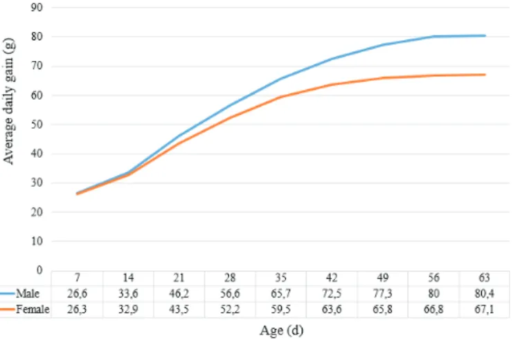
According to some reports, the disease occurs more frequently in males (Aziz & Barnes, 2007; Herdt
et al., 2009; Aitchison et al., 2014) and may affect
several strains (Wood et al., 2002; Gingerich, 2009).
The higher body weight normally observed in males (Figure 1) implies an increase in the weight needed to be supported by bones and joints and a greater possibility of trauma. The influence of high body weight has also been described in another condition that affects broilers named bacterial chondronecrosis with osteomyelitis (BCO) by Wideman & Prisby (2013).
Figure 1 – Average daily weight gain (g) of male and female broilers from seven to 63 days-old. Adapted from Cobb manual (2015).
The broilers affected by VO are usually older than 30 days-old, and outbreaks of the disease are reported
between three to 18 weeks-old (Herdt et al., 2009;
Armour et al., 2011; Robbins et al., 2012). However,
there is a report in a flock as young as 15 days-old (Gingerich, 2009).
Predisposing factors for VO are not well defined (Kense & Landman, 2011; Robbins et al., 2012); however, immunosuppression and environmental conditions have been identified as factors that contribute to the
occurrence of the disease (Stalker et al., 2010; Armour
et al., 2011). Any immunosuppressive condition can
naturally predispose to opportunistic infection by E.
cecorum, which is a normal intestinal commensal. However, a study demonstrated differences in the pathogenicity among commensal and clinical cases isolates, raising the question whether the emergence of clones is most likely the cause for the increased
occurrence of infections (Boerlin et al., 2012).
Some evidences suggest that the higher incidence of enterococci-associated diseases in poultry may be
due to horizontal spread of dominant E. cecorum
clones that exhibit high pathogenicity (Kense &
Landman, 2011; Boerlin et al., 2012). However, the
source of pathogenic E. cecorum remains unknown
(Borst et al., 2017). Moreover, bacterial strains with
genotypes similar to those isolated from VO cases were rarely recovered from the ceca of birds with VO and the presence of these isolates was not statistically
associated with a higher risk of disease (Borst et al.,
2012). These findings suggest that the long-term cecal carrier state of pathogenic clones may not be required
for the pathogenesis of VO caused by E. cecorum.
However, as the disease has a chronic character, taking weeks from the time of infection until the onset of clinical signs, pathogenic strains may be transient in the gastrointestinal tract and therefore not recoverable in
the moment of clinical presentation (Borst et al., 2012).
Field observations showed that the disease occurred
in successive flocks, suggesting the persistence of E.
cecorum on the farm (Herdt et al., 2009; Kense &
Landman, 2011). According to Borst et al. (2017),
persistence of E. cecorum in the environment between
flocks is unproven, although the repeated outbreaks on affected farms may be associated to environmental
contamination by pathogenic E. cecorum.
Despite their worldwide distribution, how
pathogenic clones of E. cecorum spread remains
undetermined. Epidemiological studies on different VO outbreaks suggest that mechanical spreading by biological vectors or inadequate biosafety may contribute to disease transmission, but horizontal transmission between geographically distant locations is considered unlikely (Borst et al., 2012).
Kense & Landman (2011) demonstrated that vertical transmission does not occur. Recently, Borst
et al. (2014) showed lower survival rate of SPF and
non-SPF chicken embryos inoculated with E. cecorum
isolated from vertebral lesions compared with those
inoculated with E. cecorum isolated from the intestines
of healthy birds. The embryos infected with pathogenic strains presented lesions of septicemia, such as hemorrhage and edema. In embryos inoculated with non-pathogenic strains, these lesions were found only 48 hours later.
Etiology
Enterococcus cecorum
Since 2002, E. cecorum has been found to be the
non-vertebral and non-vertebral osteomyelitis and arthritis in broiler and broiler breeders (Aziz & Barnes, 2007;
Herdt et al., 2009; Aziz & Barnes, 2009; Gingerich,
2009; Stalker et al., 2010; Martin et al., 2011; Boerlin
et al., 2012; Aitchison et al., 2014).
Enterococcus spp. are gram-positive and spherical bacteria, which occur alone, in pairs or short chains. They are non-motile, non-spore-forming, facultative anaerobic with diverse biochemical properties (Wages, 1998). However, the relationship between the biochemical characteristics and pathogenicity of
this genus remains unknown (Thayer et al., 2008).
Enterococcus spp. are ubiquitous in nature, with worldwide distribution in avian species. They are considered part of the normal intestinal microbiota of chickens and commonly found in poultry environments.
The frequency with different species of Enterococcus
spp. are isolated from the intestinal tract of healthy birds may vary according to age, but only a limited number of species are commonly isolated. The species
E. faecium, E. cecorum, E. faecalis, E. hirae and E. durans were regularly isolated in at least one of three different age groups (1 day-old, 3 to 4 weeks-old, and
older than 12 weeks-old) examined by Devriese et al.
(1991). Enterococci are also important as nosocomial pathogens that cause bacteremia, endocarditis and other infections. Some strains are resistant to many antibiotics and possess virulence factors, such as
adhesins, invasins, hemolysin, and pili (Franz et al.,
2011).
E. cecorum occurs more frequently in the intestines
of chickens older than 12 weeks of age (Devriese et
al., 1991) and has rarely been associated with clinical
disease in these birds (Devriese et al., 2002; Wood et
al., 2002; Chadfield et al., 2004; Thayer et al., 2008). This could explain the limited number of publications on its role in disease and its pathogenicity (Makrai
et al., 2011). Two main hypotheses were proposed
to explain the recent increase in the incidence of E.
cecorum infections: 1) changes in the host or in environmental factors; and 2) emergence of individual
clones with increased pathogenicity (Boerlin et al.,
2012). In order to test the second hypothesis, those
authors analyzed E. cecorum isolates recovered from
the ceca of healthy chickens and those with VO by
pulsed-field gel electrophoresis (PFGE). The E. cecorum
genotypes isolated from the vertebral lesions were significantly more similar to each other than those isolated from the ceca of healthy birds and of birds with VO, regardless of the affected broiler flock.
E. cecorum was also isolated from a broiler flock with pericarditis, hepatitis, femoral head necrosis and/ or VO and the authors concluded that bacteremia and generalized infection seem to be important steps in the pathogenesis of the infection caused by this bacterium in broilers (Jung & Rautenschlein, 2014). This was also
suggested by Borst et al. (2017), who observed that E.
cecorum isolates recovered from the intestine, spleen, and free thoracic vertebra (FTV) of broilers in natural cases of VO had matching genotypes, confirming that intestinal colonization with pathogenic strains precedes bacteremia and infection of the FTV.
Other etiologic agents
Infections by E. hirae are relatively frequent in
broilers, but its importance is not well understood. The incidence of diseases caused by this bacterium have increased in some countries, such as Norway,
where E. hirae was isolated from cases of osteomyelitis
in the proximal femur and endocarditis in broilers (Kolbjørnsen et al., 2011).
Braga et al. (2016b) recently reported E. hirae, E.
faecalis, Escherichia coli, and Staphylococcus aureus
in single or mixed culture from VO cases in broilers.
E. coli, which has also been isolated from VO cases
in poultry (Dinev, 2013; Braga et al., 2016a; Braga et
al., 2016b), is part of normal intestinal microbiota of
humans and many animal species. E. coli is a
gram-negative non-spore-forming bacillus, measuring 2-3 x 0.6 µm, and most strains are motile with peritrichous flagella (Barnes et al., 2008).
Several E. coli strains are able to express virulence
factors and cause intestinal or extra-intestinal diseases
(Ambrozic et al., 1998). E. coli is currently considered
the most important gram-negative bacterium in poultry due to its different pathogenicity mechanisms
and described diseases (Nakazato et al., 2009). In
avian species, E. coli pathogenic strains are named
Avian Pathogenic Escherichia coli (APEC), which are
responsible for extra-intestinal diseases generically
known as colibacillosis (Ewers et al., 2004). The presence
of E. coli in bone and synovial tissues is a common consequence of colisepticemia and the affected birds probably were not able to have completely eliminated
the bacterial infection (Barnes et al., 2008).
Staphylococcus pyogenes was isolated from VO cases in seven- to 16-weeks-old chickens (Carnaghan,
1966). Nairn (1973) reported the isolation of S. aureus
from vertebral lesions in turkeys naturally affected with locomotor disorder, and that the experimental inoculation of turkeys resulted in osteomyelitis in the vertebral body and long bones. Van Veen (1999)
detected the involvement of Aspergillus fumigatus in
VO outbreaks in two flocks of 17- to 19-weeks-old broilers.
Pathogenesis
The pathogenesis of VO in birds remains largely unknown, although important progress has been made
(Kense & Landman, 2011; Robbins et al., 2012; Jung
& Rautenschlein, 2014; Borst et al., 2017). There are
few publications on the pathogenicity of E. cecorum
(Makrai et al., 2011) and the knowledge of the genetic
basis for the recently acquired pathogenicity of certain
E. cecorum strains and the pathogenesis of vertebral lesions characteristic of the disease remains limited (Borst et al., 2012; Borst et al., 2015).
Recent molecular epidemiologic studies found that
pathogenic E. cecorum strains were genetically clonal and
suggested acquisition of specific virulence determinants
by pathogenic E. cecorum. This study also showed that
pathogenic isolates had smaller genomes with a higher guanidine-cytosine (GC) content and large regions of
synteny compared with commensal E. cecorum isolates.
Molecular phylogenetic analysis demonstrated that, at a threshold of 98% identity, 414 predicted proteins were
highly conserved in pathogenic E. cecorum but not in
commensal strains (Borst et al., 2015).
A recent study compared pathogenic and
non-pathogenic E. cecorum strains from different animal
species. Pathogenic and commensal E. cecorum
strains were not clearly separated from each other in a phylogenetic tree based on partial sequences of the 16S-rRNA-gene and their fatty acid profile. Also,
it was noted that all pathogenic E. cecorum strains
was not able to utilize mannitol, while 31.0% of the
commensal strains were mannitol positive (Jung et
al., 2017). This characteristic was also observed in
other studies regarding the phenotypic and genotypic
characterization of E. cecorum associated with infections
in poultry (Dolka et al., 2016). Jung et al. (2017) did
not find any significant difference in virulence factors between pathogenic and commensal isolates. On the
other hand, mean embryo lethality of pathogenic E.
cecorum isolates (39.7%) was significantly higher than that of commensal strains (18.9%). Those authors also reported that two different antisera were produced,
although none of the serotypes were predominantly found either in pathogenic or commensal isolates.
Molecular studies on conserved genes to speciate
enterococci suggested that pathogenic E. cecorum
might be considered a subspecies in view of the
divergence between commensal and pathogenic E.
cecorum genomes (Borst et al., 2015). In addition, pathogenic strains showed conserved potential mediators of virulence, including genes encoding predicted collagen-binding proteins, a capsular locus, and orthologs to the enterococcal polysaccharide antigen of E. faecalis and E. faecium (Borst et al., 2015).
The disease was experimentally reproduced by
Martin et al. (2011) by inoculating broilers with
E. cecorum by oral and intravenous routes. Gross lesions were found five weeks after the experimental infection in 6.1% and 2.9% of the broilers orally and intravenously inoculated, respectively. However, histologic lesions were found in 30.3% of the orally-inoculated broilers, and the authors suggest that macroscopic evidence of disease would be greater if the broilers were older.
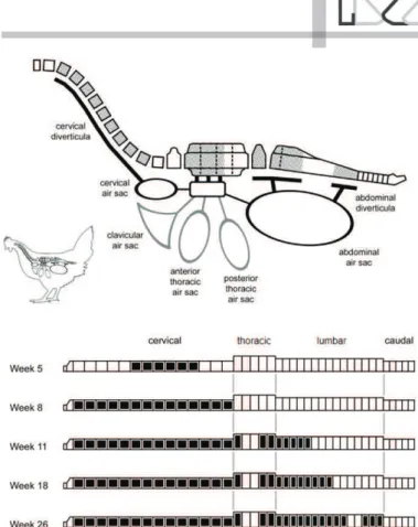
The FTV (T4) body is singly affected in VO and the reasons for this predilection are not fully understood. The FTV articulation is located between the immediately anterior fused thoracic vertebrae and the posterior synsacrum (Figure 2), enabling body position adjust-ments and flexibility during walk and flight, and is subject to greater biomechanical stress and microtraumas than any other vertebra. Excessive stress may lead to changes in blood flow, with the development of micro-thrombi, sequestrum and multiplication of bacteria
present in the blood (Aziz & Barnes, 2007; Stalker et
al., 2010; Wideman & Prisby, 2013; Aitchison et al.,
2014). This corroborates with a recent study reporting that broilers with osteochondrosis dissecans (OCD) were more susceptible to VO development than birds with no FTV lesions, and that suggested that intestinal colonization, bacteremia, and OCD of the FTV in early life are crucial to the pathogenesis of the disease (Borst
et al., 2017).
According to Stashak & Mayhew (1984), VO is usually secondary to the hematogenous dissemination of a microorganism. However, other theories have been proposed to explain how the bacteria reach the FTV. One of these theories suggests that the agent has access to the bones via bloodstream due to disruption
of the intestinal mucosal barrier (Stalker et al., 2010;
Martin et al., 2011), as occurs in coccidiosis or bacterial
enteritis (Gingerich, 2009). According to Martin et
al. (2011), any factor that negatively interferes with
balance may predispose broilers to the systemic
dissemination of E. cecorum. On the other hand, Borst
et al. (2017) observed that clinical intestinal disease
was not required to cause E. cecorum bacteremia.
A possible link of the VO with air sacs and pneumatic vertebra could exist (Aziz & Barnes, 2007). However, the pneumatization of the vertebra where the disease occurs (T4) begins only after eight weeks of age (Figure
2). The experimental inoculation of E. cecorum in
two-weeks-old broilers by air sac route did not result in VO
(Martin et al., 2011), suggesting that older broilers
should be inoculated for experimental studies. In a
study conducted by Tankson et al. (2002), E. faecalis,
E. durans, and E. coli were isolated from the heart and lungs in 15% of healthy chicks. However, there are no
studies that provide this information for E. cecorum.
Some aspects of the pathogenesis of BCO may aid the understanding of VO pathogenesis. This disease commonly affects the femur and the tibiotarsus, but it may affect the FTV. BCO begins with the degeneration and necrosis of the cartilage, followed by bacterial
invasion, and it is mainly associated with S. aureus, E.
coli and E. cecorum, often in mixed infections, as well
as with other bacteria (Wideman & Prisby, 2013). Also,
a recent study (Borst et al., 2017) demonstrated that
pathogenic E. cecorum was observed within OCD
lesions in the FTV in 1- to 3-weeks-old broilers with VO, and suggested that intestinal colonization, bacteremia, and OCD of the FTV in early life seem to be crucial for the pathogenesis of VO.
It is believed that the BCO begins with mechanical damage to the poorly-mineralized columns of chondrocytes present mainly in the proximal growth plate of fast-growing bones, such as the femur and tibia, and is followed by the colonization of the chondronecrotic clefts by opportunistic hematogenous bacteria (Carnaghan, 1966; McNamee & Smyth, 2000;
Dinev, 2009; Wideman Jr et al., 2012), which was
recently described in VO cases by Borst et al. (2017).
Terminal BCO presents itself as degeneration, necrosis and bacterial infection at the proximal ends (epiphyseal and metaphyseal growth plates) of the femur and tibiotarsus. A similar process may occur in the growth plates of other bones that are subject to severe torque and shear stresses, as occur in the FTV, which functions as a flexible pivot between the cranially-fused vertebrae of the notarium and the caudally-fused vertebrae of the synsacrum (Carnaghan, 1966; McNamee & Smyth,
2000; Dinev, 2009; Wideman Jr et al., 2012).
According to Barnes et al. (2008), the involvement of
E. coli in infectious processes of the bone and synovial tissues is a common consequence of colisepticemia. Osteomyelitis caused by the hematogenous spread of E. coli after infection by the hemorrhagic enteritis virus was experimentally reproduced in turkeys (Droual
et al., 1996). Some authors report that, although
the intravenous inoculation of E. coli promoted its
hematogenous spread to the bones and joints and reproduction of lesions, bird mortality caused by initial
sepsis is usually high (Bayyari et al., 1997). According
to Bayyari et al. (1997), that bacterium colonizes the
vascular branches that invade the growth plate of growing bones, causing an inflammatory response that results in osteomyelitis. The transphyseal vessels in birds may possibly serve as conduits for the spread of bacteria to the joint and surrounding soft tissues.
Clinicopathological changes
Clinical signs
The clinical signs are similar in all VO cases reported (Gingerich, 2009), although the age of onset of clinical presentation may vary. In cases of osteomyelitis and
arthritis caused by E. cecorum, Herdt et al. (2009)
reported that the clinical signs started in the first and second weeks of age, with a mortality rate of 7%. In
the osteomyelitis cases studied by Makrai et al. (2011),
the clinical signs started between 5-9 weeks up to
10-13 weeks of age, with a mortality rate ranging from 8% to 30%, which was higher than previously
reported (Wood et al., 2002; Herdt et al., 2009).
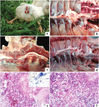
The major clinical sign observed is the limited mobility, with birds presenting mild to severe lameness. The affected birds frequently acquire the posture described as “sitting on their hocks”, characterized by cranially extended legs and support given by the tibiotarsus-metatarsus joints (Figure 3a) (Gingerich,
2009; Braga et al., 2016b). This is considered the classic
clinical presentation of the disease, which is similar to
that observed in birds with spondylolisthesis (Wood et
al., 2002; Gingerich, 2009).
Severely affected broilers may remain in lateral recumbency (Gingerich, 2009). They occasionally use their wings to help locomotion, which may result in
wing laceration and hematomas (Makrai et al., 2011).
One of the consequences of impaired locomotion is the difficulty to access water and feed, resulting in reduced growth rate and ultimately in death due to
dehydration or starvation (Barnes et al., 2008; Borst et
al., 2017).
Gross changes
The macroscopic examination of the thoracolumbar region of vertebral column of the affected broilers reveals gross changes in the FTV (T4), characterized by a palpable whitish to yellowish enlargement (Figure 3b). The sagittal section of this lesion shows caseonecrotic material inside the vertebral body characterized by yellow to gray, granular and friable exudate, which is surrounded by a thick whitish capsule of fibrous connective tissue (Figure 3b, inset) (Gingerich, 2009; Martin et al., 2011; Robbins et al., 2012, Braga et al., 2016b). The most pronounced lesion is characterized by the increased size of the vertebral body as a result of infection, which results in narrowing of the overlying spinal canal and compression of the spinal
cord (Makrai et al., 2011; Aitchison et al., 2014, Braga
et al., 2016b). In the early stages of the disease, there is no obvious increase of the vertebral body and spinal compression of the sagittal section is absent or mild. The body condition of the affected birds varies from
good to cachectic (Braga et al., 2016b).
Histopathology
The microscopic changes of VO observed with
the experimental reproduction of the disease by E.
cecorum infection were detailed by Martin et al.(2011) and were similar to those reported in natural cases
of the disease (Stalker et al., 2010; Robbins et al.,
2012; Aitchison et al., 2014, Braga et al., 2016b). On
the histopathologic examination, the FTV body and occasionally the adjacent vertebrae of notarium and synsacrum, presented necrotic tissue and exudate composed of fibrin, hemorrhage, and heterophils (Figure 3e and 3f). The bone tissue that forms the basis of the spinal canal is replaced by fibrous connective tissue and exudate, leading to spinal canal stenosis and spinal cord compression. In addition, there is fibrous connective tissue proliferation and bone remodeling in the areas surrounding the lesion. There are also areas of bone and cartilage tissue sequestrum within the exudate. When bacterial colonies are present (Figure 3e, inset), they are numerous and associated with the
sequestrated areas (Aitchison et al., 2014; Braga et
al., 2016b). In addition to these changes, Aitchison et
al. (2014) and Braga et al. (2016b) described reactive
osteoid formation and cartilaginous metaplasia in the areas where there was severe thickening of the vertebral body, resulting in areas of spinal cord compression. In these areas, there was axonal loss and degeneration, and the neuropil was disorganized and vacuolated, indicating a compressive effect.
Martin et al. (2011) reported histologic changes in broilers in the absence of macroscopic lesions, including mild histologic lesions in the subchondral vertebral areas, with no extension to the articular cartilage or adjacent vertebrae. Moderate to severe lymphocyte infiltration and diffuse fibroplasia in the affected vertebra with intralesional bacteria was confirmed in
half (4/8) of the cases. Braga et al. (2016b) detected
also lesions in adjacent vertebrae, characterized by degeneration and necrosis of the articular cartilage (T4/ T5), and occasional presence of clefts associated or not with hemorrhages and bacterial colonies. In the study
of Martin et al. (2011), osteochondrosis was present in
all birds, some of which presenting different degrees of subluxation FTV. A recent study on VO pathogenesis also described histologic lesions consistent with OCD, characterized by variably large cartilaginous clefts, which were often partially to completely filled with thrombocytes, fibrin, and erythrocytes. In some cases,
there were cocci consistent with E. cecorum colonizing
some of these clefts (Borst et al., 2017).
Diagnosis
VO may be suspected in birds sitting on their hocks (McNamee & Smyth, 2000). It should be emphasized that high-quality veterinary support and thorough pathological and bacteriological examinations are indispensable for the early diagnosis of VO in broiler flocks. This is essential for the success of antibiotic therapy in the early stages of the disease (Jung & Rautenschlein, 2014).
Broilers presenting locomotor disorders should be placed in orthostatic (standing) position and encouraged to move to assess gait and posture changes. These birds should be then euthanized and necropsied to examine possible gross changes in the locomotor system (axial and appendage skeleton), and the vertebral column sectioned along the longitudinal midline to examine the vertebral body and assess the degree of spinal cord compression (Gingerich, 2009;
Braga et al., 2016b). At necropsy, samples for bacterial
isolation and identification, polymerase chain reaction (PCR) tests, and histopathology should be collected.
For bacterial culture and isolation, swabs from caseonecrotic material of vertebral lesions should be aseptically collected and inoculated onto blood agar (BA) and MacConkey agar. It is recommended to incubate one BA plate under microaerophilic conditions (37 °C, 24 to 72 h) and the other plates under aerobic conditions at the same temperature and time to enable
the isolation of different etiologic agents (Braga et al.,
2016b). After initial bacterial growth, the colonies
must be Gram stained and submitted to catalase and oxidase tests. Bacterial isolates may be identified by biochemical tests, rapid ID 32 STREP (BioMerieux), VITEK 2 system (bioMérieux), and MALDI-TOF (AXIMA Assurance).
For DNA detection of the etiologic agent, vertebral lesion samples should be collected in sterile microtubes and frozen at -20 °C for DNA extraction and subsequent PCR testing. A multiplex PCR assay was described to
enable the detection of different species of Enterococcus
sp. (Jackson et al., 2004). Recently, Jung et al. (2017)
reported the use of a newly-developed quantitative TaqMan real-time PCR (qPCR) assay based on the 16S–
rRNA-gene for the detection of E. cecorum. According
to those authors, this new qPCR is highly specific, more sensitive than classical cultivation, and was able to determine colonization differences between a broiler
flock presenting a late outbreak of E. cecorum disease
compared to a healthy flock.
After sample collection for bacteriology and PCR, vertebral columns with gross lesions must be preserved in 10% neutral buffered formalin (48 to 72 h) for histopathological analysis. Vertebral columns should be decalcified in 24% formic acid before routine histological processing. The slides should be stained with hematoxylin-eosin and Goodpasture stains (Luna, 1968) to search for VO characteristic lesions (previously described in the histopathology subsection) and intralesional bacteria under a light microscope (Braga
et al., 2016b).
The differential diagnosis of VO includes other pathologies that may cause spinal cord compression or changes in nerves causing impaired mobility. One of these conditions is spondylolisthesis (“kinky back”),
characterized by subluxation of the FTV (Armour et
al., 2011; Robbins et al., 2012). Grossly, these cases
show varying extents of ventral dislocation of the FTV,
whose posterior end raises the 5th thoracic vertebra.
The dislocation may produce kyphotic angulation of the spinal canal and varying degrees of spinal cord compression (Figure 3c). Necrotic and inflammatory lesions of the vertebral body in broilers with spondylolisthesis may not be present (Dinev, 2012). Scoliosis characterized by lateral deviation of the spine (Figure 3d) should also be considered for differential
diagnosis of the aforementioned conditions (Droual et
al., 1991). Proper monitoring of the flock may aid the
in differential diagnosis. In Marek’s disease, no changes in the vertebral column are observed, but there is enlargement of peripheral nerves, which become greyish or yellowish with loss of striations, acquiring an edematous appearance in some cases (Schat & Nair, 2008).
Prevention, treatment and control
Information on prevention, treatment and control of the disease is limited, as the etiopathogenesis of
VO remains unclear (Aitchison et al., 2014), and most
studies are related only to infections caused by E.
cecorum. For the prevention of VO, recommendations on management practices to reduce the risk of developing the disease have been made, such as: 1) avoiding excessive feed restriction; 2) following the suggested weight gain standards and nutritional
recommendations (Martin et al., 2011); 3) promoting
adequate control of coccidiosis (Stalker et al., 2010); 4)
avoiding high housing density; 5) ensuring adequate access to feeders; and 6) preventing respiratory
diseases (Aitchison et al., 2014). All practices to
prevent bacterial infections that may cause bacteremia would probably also help to avoid bone and articular inflammation.
Antibiotics have been used to treat the bacterial infection in VO. Although several antibiotics have shown efficacy against the commonly described bacteria, it is difficult to achieve adequate concentrations of the antibiotics in the vertebral column. In the reported outbreaks of the disease, antibiotics were ineffective in reducing mortality possibly due to the inability of the antibiotic to effectively penetrate the anatomical areas where the bacterium is located or antimicrobial resistance of E. cecorum (Kense & Landman, 2011).
The antimicrobial susceptibility profiles of E. cecorum
isolated from outbreaks in different countries were
similar (Herdt et al., 2009; Aitchison et al., 2014).
Aitchison et al. (2014) reported that, after isolation and
identification of E. cecorum, it was difficult to perform
the antibiotic susceptibility test due to the growing
conditions of the bacterium. Makrai et al. (2011)
reported that, after the onset of the outbreak, broilers presenting clinical signs were separated from those clinically normal and the clinically normal were treated with different antibiotics (amoxycillin, amoxycillin with clavulanic acid, lincomycin or doxycycline), resulting in no new clinical case of the disease in the flock.
After the occurrence of the disease, the prevention of new cases requires repeated cycles of disinfection and usually occur after a single cleaning and disinfection procedure. Increased efforts in subsequent flocks
are required to eliminate the disease. Some practices that may reduce the risk of VO in subsequent flocks include: 1) emptying and completely disinfecting the broiler house; 2) changing or composting the litter; 3) adequate cleaning of water lines; and 4) continuously
sanitizing the water (Gingerich, 2009; Stalker et al.,
2010; Armour et al., 2011; Martin et al., 2011).
Antimicrobial resistance and public health
As previously mentioned, enterococci are normal bacteria of the gastrointestinal tract of animals and humans and are often considered as beneficial commensal organisms (Tannock, 1995). However, they may also be opportunistic pathogens, responsible for serious systemic infections and for the spread of antimicrobial resistance and virulence determinants (Wisplinghoff et al., 2004; Heuer et al., 2006). In recent years, enterococci have emerged as a major cause of
nosocomial infections, particularly E. faecalis (Kola et
al., 2010), causing extraintestinal infections in humans
(Creti et al., 2004). These bacteria have intrinsic
resistance to many antibiotics and have acquired new resistance genotypes, out of which vancomycin-resistant enterococci (VRE) are of special concern
(Cetinkaya et al., 2000; Willems & Bonten, 2007).
VRE have become a major problem in nosocomial infections. A retrospective study of ten human patients with osteomyelitis showed that eight cases were due
to infection by E. faecalis resistant to vancomycin, with
one death reported due to bacteremia (Holtom et al.,
2002).
The contamination of animals and their by-products by resistant bacteria and their possible foodborne transmission to humans are an animal and public
health concern (Foulquié-Moreno et al., 2006).
Enterococci may not only contaminate raw meat, but may also be present in processed meat products, such
as cured raw sausages or cooked products (Martin et
al., 2005; Barbosa et al., 2009; Ruiz-Moyano et al.,
2009). Although no food poisoning associated with
E. faecalis has been described in humans, a recent study performed in Brazil showed the presence of this bacterium in 42% of the chicken carcasses tested
(Campos et al., 2013). All strains were resistant to at
least one of the antibiotics tested, with the detection
of the antimicrobial resistance genes erm(B), vanC-1,
et al., 2013). Hayes et al. (2003) analyzed 981 raw meat samples of various species (chicken, turkey, swine and bovine) obtained in grocery stores and
isolated 1,357 Enterococcus spp. strains, including
E. faecalis (29%) and E. hirae (5.7%). Those authors also detected gentamicin resistance in 4% of the strains, most of which isolated from chicken meat.
Braga et al. (2017), analyzing E. faecalis isolates from
VO in broilers, demonstrated that the highest level of antibiotic resistance was against aminoglycosides, particularly gentamicin (40%).
CONCLUSION
VO is an emerging disease in poultry worldwide that still needs to be further elucidated. Many aspects of the etiopathogenesis of the disease remain unclear, limiting its prevention and control. Most reports
associate the disease to the infection by E. cecorum,
probably related to emerging clones with higher pathogenicity. However, other bacteria have been isolated from VO in broilers, raising questions about the etiology and pathogenesis of the disease. This highlights the need for isolation and identification of the etiologic agent in cases of VO, as well as studies for molecular characterization and antimicrobial resistance of the involved bacterium.
ACKNOWLEDGMENTS
J.F.V. Braga was a fellow of the Programa de Pós-graduação em Ciência Animal/Universidade Federal de Minas Gerais supported by Coordenação de Aperfeiçoamento de Pessoal de Nível Superior (CAPES). Financial support was provided by Conselho Nacional de Desenvolvimento Científico e Tecnológico (CNPq) under Grant number 14/2010. We also are thankful to Pró-Reitoria de Pesquisa da Universidade Federal de Minas Gerais.
REFERENCES
Aitchison H, Poolman P, Coetzer M, Griffiths C, Jacobs J, Meyer M., Bisschop S. Enterococcal-related vertebral osteoarthritis in South African broiler breeders: A case report. Journal of the South African Veterinary Association 2014;85(1):1-5.
Ambrozic J, Ostroversnik A, Starcic M, Kuhar I, Grabnar M, Zgur-Bertok D.
Escherichia coli Co IV plasmid pRK100: genetic organization, stability and conjugal transfer. Microbiology 1998;144(2):343–352.
Armour NK, Collet SR,Williams SM. Enterococcus cecorum- related arthritis and osteomyelitis in broilers and broiler breeders. The Poultry Informed Professional 2011;117:1–7.
Aziz T, Barnes HJ. Is spondylitis an emerging disease of broilers? World Poultry 2007;23:44–45.
Aziz T, Barnes HJ. Spondylitis is emerging in broilers.World Poultry 2009;25:14.
Barbosa J, Ferreira V, Teixeira P. Antibiotic susceptibility of Enterococci isolated from traditional fermented meat products. Food Microbiology 2009;26(5):527–532.
Barnes HJ, Vaillancourt JP, Gross WB. Colibacillosis. In: Saif YM, Fadly AM, Glisson JR, McDougald LR, Nolan LK, Swayne DE, editors. Diseases of poultry. 12th ed. Ames: Blackwell Publishing; 2008. p.631-656.
Bayyari GR, Huff WE, Rath NC, Balog JM, Newberry LA, Villines JD, et al. Immune and physiological responses of turkeys with green-liver osteomyelitis complex. Poultry Science1997;76(2):280-288.
Boerlin P, Nicholson V, Brash M, Slavic D, Boyen F, Sanei B, et al. Diversity of Enterococcus cecorum from chickens. Veterinary Microbiology 2012;157(3-4):405–411.
Borst LB, Suyemoto MM, Keelara S, Dunningan SE, Guy JS, Barnes HJ. A chicken embryo lethality assay for pathogenic Enterococcus cecorum. Avian Diseases 2014;58(2):244-248.
Borst LB, Suyemoto MM, Robbins KM, Lyman RL, Martin MP, Barnes HJ. Molecular epidemiology of Enterococcus cecorum isolates recovered from enterococcal spondylitis outbreaks in the southeastern United States. Avian Pathology 2012;41(5):479-485.
Borst LB, Suyemoto MM, Sarsour AH, Harris MC, Martin MP, Strickland JD, et al. Pathogenesis of enterococcal spondylitis caused by Enterococcus cecorumin broiler chickens. Veterinary Pathology 2017;54(1):61-73. Borst LB, Suyemoto MM, Scholl EH, Fuller FJ, Barnes HJ. Comparative
genomic analysis identifies divergent genomic features of pathogenic
Enterococcus cecorum including a type IC CRISPR-Cas system, a capsule locus, an epa-like locus, and putative host tissue binding proteins. PLoSOne 2015;10(4):e0121294.
Braga JFV, Chanteloup NK, Trotereau A, Baucheron S, Guabiraba R, Ecco R, et al. Diversity of Escherichia coli strains involved in vertebral osteomyelitis and arthritis in broilers in Brazil. BMC Veterinary Research 2016a;12(1):1-12.
Braga JFV, Leal CAG, Silva CC, Fernandes AA, Martins NRS, Ecco R. Genetic diversity and antimicrobial resistance profile of Enterococcus faecalis
isolated from broilers with vertebral osteomyelitis in Southeast Brazil. Avian Pathology 2017;24:1-27.
Braga JFV, Silva CC, Teixeira MPF, Martins NRS, Ecco R. Vertebral osteomyelitis associated with single and mixed bacterial infection in broilers. Avian Pathology 2016b;45(6):640-648.
Campos ACFB, Souza NR, Silva PHC, Santana AP. Resistência antimicrobiana em Enterococcus faecalis e Enterococcus faecium isolados de carcaças de frango. Pesquisa Veterinária Brasileira 2013;33(5):575-580. Carnaghan RBA. Spinal cord compression in fowls due to spondylitis
caused by Staphylococcus pyogenes. Journal of Comparative Pathology 1966;76(1):9-14.
Caudry SD, Stanisich VA. Incidence of antibiotic-resistant Escherichia coli associated with frozen chicken carcasses and characterization of conjugative R plasmids derived from such strains. Antimicrobial Agents and Chemotherapy 1979;16(6):701-709.
Cetinkaya Y, Falk P, Mayhall CG. Vancomycin-resistant enterococci. Clinical Microbiology Reviews 2000;13(4):686-707.
Cobb. Cobb500: broiler performance & nutrition supplement. São José do Rio Preto; 2015 [cited 2016 Jan 11]. Available from: http://www.cobb-vantress.com/docs/default-source/cobb-500-guides/Cobb500_Broiler_ Performance_And_Nutrition_Supplement.pdf .
Creti R, Imperi M, Bertuccini L, Fabretti F, Orefici G, Di Rosa R, Baldassarri L. Survey for virulence determinants among Enterococcus faecalis
isolated from different sources. Journal of Medical Microbiology 2004;53(1):13–20.
Devriese LA, Cauwerts K, Hermans K, Wood AM. Enterococcus cecorum
septicemia as a cause of bone and joint lesions resulting in lameness in broiler chickens. Vlaams Diergeneeskundig Tijdschrift 2002;71(3):219– 221.
Devriese LA, Hommez J, Wijfels R, Haesebrouck F. Composition of the enterococcal and streptococcal intestinal flora of poultry. Journal of Applied Bacteriology 1991;71(1):46–50.
Dho-Moulin M, Fairbrother JM. Avian pathogenic Escherichia coli (APEC). Veterinary Research 1999;30(2-3):299-316.
Dinev I. Clinical and morphological investigations on the prevalence of lameness associated with femoral head necrosis in broilers. British Poultry Science 2009;50(3):284–290.
Dinev I. Pathomorphological investigations on the incidence of axial skeleton pathology associated with posterior paralysis in commercial broiler chickens. Poultry Science 2013;50(4):283-289.
Dinev I. Pathomorphological investigations on the incidence of clinical spondylolisthesis (kinky back) in different commercial broiler strains. Revue de MédecineVétérinaire 2012;163(11):511-515.
Dolka B, Chrobak-Chmiel D, Makrai L, Szeleszczuk P. Phenotypic and genotypic characterization of Enterococcus cecorum strains associated with infections in poultry. BMC Veterinary Research 2016;12:129. Droual R, Bickford AA, Farver TB. Scoliosis and tibiotarsal deformities in
broiler chickens. Avian Diseases 1991;35(1): 23-30.
Droual R, Chin RP, Rezvani M. Synovitis, osteomyelitis, and green liver in turkeys associated with Escherichia coli. Avian Diseases 1996;40(2):417-424.
Ewers C, Janssen T, Kiessling S, Philipp HC,Wieler LH. Molecular epidemiology of avian pathogenic Escherichia coli (APEC) isolated from colisepticemia in poultry. Veterinary Microbiology 2004;104(1-2):91– 101.
Foulquié-Moreno MR, Sarantinopoulos P, Tsakalidou E, De Vuyst L. The role and application of enterococci in food and health. International Journal of Food Microbiology 2006;106(1):1-24.
Franz CMAP, Huch M, Abriouel H, Holzapfel W,Gálvez A. Enterococci as probiotics and their implications in food safety. International Journal of Food Microbiology 2011;151(2):125–140.
Gingerich EN, Barnes JH, Owen RL, Rankin SC. Spinal abscesses due to
Enterococcus cecorum in broiler chickens: an emerging disease? Proceedings of the American Association of Avian Pathologists; 2009; Seattle. USA: American Association of Avian Pathologists; 2009. Hayes JR, English LL, Carter PJ, Proescholdt T, Lee KY, Wagner DD, http://
www.ncbi.nlm.nih.gov/pubmed/?term=White%20DG%5BAuthor %5D&cauthor=true&cauthor_uid=14660361 et al. Prevalence and antimicrobial resistance of enterococcus species isolated from retail meats. Applied Environmental Microbiology 2003;69(12):7153-7160. Herdt P, Defoort P, Van Steelant J, Swam H, Tanghe L, Van Goethem S, et
al. Enterococcus cecorum osteomyelitis and arthritis in broiler chickens. Vlaams Diergeneeskundig Tijdschrift 2009;78(1):44–48.
Heuer OE, Hammerum AM, Collignon P, Wegener HC (2006). Human health hazard from antimicrobial-resistant enterococci in animals and food. Clinical Infectious Diseases 2006;43(7):911-916.
Holtom PD, Zamorano D, Patzakis MJ. Osteomyelitis attributable to vancomycin-resistant enterococci. Clinical Orthopaedics and Related Research 2002;(403):38-44.
Jackson CR, Fedorka-Cray PJ, Barrett JB. Use of a Genus- and Species-Specific Multiplex PCR for Identification of enterococci. Journal of Clinical Microbiology 2004;42(8):3558–3565.
Jung A, Martin M, Ryl lM. Comparison of pathogenic and non-pathogenic
Enterococcus cecorum strains from different animal species. BMC Microbiology 2017;17(1):33.
Jung A, Rautenschlein S. Comprehensive report of an Enterococcus cecorum infection in a broiler flock in Northern Germany. BMC Veterinary Research 2014;10:311.
Kense MJ, Landman WJM. Enterococcus cecorum infections in broiler breeders and their offspring: molecular epidemiology. Avian Pathology 2011;40(6):603–612.
Kola A, Schwab F, Barwolff S, Eckmanns T, Weist K, Dinger E, Klare I, et al. Is there an association between nosocomial infection rates and bacterial cross transmissions? Critical Care Medicine 2010;38(1):46–50. Kolbjørnsen Ø, David B, Gilhuus M. Bacterial osteomyelitis in a
3-week-old broiler chicken associated with Enterococcus hirae. Veterinary Pathology 2011;48(6):1134-1137.
Luna LG. Manual of histologic staining methods of the Armed Forces Institute of Pathology. 3rd ed. New York: McGraw-Hill; 1968.
Makrai L, Nemes C, Simon A, Ivanics E, Dudás Z, Fodor L, et al. Association of Enterococcus cecorum with vertebral osteomyelitis and spondylolisthesis in broiler parent chicks. Acta Veterinaria Hungarica 2011;59(1):11–21.
Martin B, Garriga M, Hugas M, Aymerich T. Genetic diversity and safety aspects of enterococci from slightly fermented sausages. Journal of Applied Microbiology 2005;98(5):1177–1190.
Martin LT, Martin MP, Barnes HJ. Experimental reproduction of Enterococcal spondylitis in male broiler breeder chickens. Avian Diseases 2011;55(2):273–278.
McGaw L. Use of plant-derived extracts and essential oils against multidrug resistant bacteria affecting animal health and production. In: Rai MK, Kon KV, editors. Fighting multidrug resistance with herbal extracts, essential oils and their components. London: Academic Press; 2013. p.191–203.
McNamee PT, Smyth JA. Bacterial chondronecrosis with osteomyelitis (“femoral head necrosis”) of broiler chickens: a review. Avian Pathology 2000;29(4):253–270.
Nairn ME. Bacterial osteomyelitis and synovitis of the turkeys. Avian Diseases 1973;17(3):504-517.
Nakazato G, Campos TA, Stehling EG, Brocchi M, da Silveira WD. Virulence factors of avian pathogenic Escherichia coli (APEC). Pesquisa Veterinária Brasileira 2009;29(7):479-486.
Robbins KM, Suyemoto MM, Lyman RL, Martin MP, Barnes HJ, Borst LB. An outbreak and source investigation of enterococcal spondylitis in broilers caused by Enterococcus cecorum. Avian Diseases 2012;56(4):768-773. Ruiz-Moyano S, Martin A, Benito MJ, Aranda E, Casquette R, Cordoba G.
Schat KA, Nair V. Marek´s disease. In: Saif YM, Fadly AM, Glisson JR, McDougald LR, Nolan LK, Swayne DE, editors. Diseases of poultry. 12th
ed. Ames: Blackwell Publishing; 2008. p. 452-514.
Stalker MJ, Brash ML, Weisz A, Ouckama RM, Slavic D. Arthritis and osteomyelitis associated with Enterococcus cecoruminfection in broiler and broiler breeder chickens in Ontario, Canada. Journal of Veterinary Diagnostic Investigation 2010;22(4):643–645.
Stashak TS, Mayhew IG. The nervous system. In :Jennings PB, Saunders WB, editors. The practice of large animal surgery. Philadelphia: WB Saunders; 1984. p.1013-1016.
Talebi A, Taifebagherlu J, Sharifi A, Delkhosh-Kasmaie F. Spondylitis in broiler breeder farms in West-Azerbaijan province, Iran: Clinical Report. Veterinary Research Forum 2016;7(4):353-355.
Tankson JD, Thaxton JP,Vizzier-Thaxton Y. Bacteria in heart and lungs of young chicks. Journal of Applied Microbiology 2002;92(3):443-450. Tannock GW. Normal microflora: an introduction to microbes inhabiting the
human body. London: Chapman and Hall;1995.
Thayer SG, Waltman WD, Wages DP.Streptococcus and Enterococcus. In:Saif YM, Fadly AM, Glisson JR, McDougald LR, Nolan LK, Swayne DE, editors. Diseases of poultry. 12th ed. Ames: Blackwell Publishing;
2008. p.900–908.
Van Veen L, Dwars RM, Fabri THF. Mycotic spondylitis in broilers caused by Aspergillus fumigatus resulting in partial anterior and posterior paralysis. Avian Pathology 1999;28(5):487-490.
Wages DP. Streptococcosis. In: Swayne DE, Glisson JR, Jackwood MW, Person JE, Reed WM, editors. Isolation and identification of avian pathogens. 4th ed. Kennett Square: American Association of Avian
Pathologists; 1998. p. 58-60.
Wedel MJ. Postcranial pneumaticity in dinosaurs and the origin of the avian lung [thesis]. Berkeley (USA): University of California; 2007.
Wideman Jr RF, Hamal KR, Stark JM, Blankenship J, Lester H, Mitchell KN, et al. A wire-flooring model for inducing lameness in broilers: Evaluation of probiotics as a prophylactic treatment. Poultry Science 2012;91(4):870–883.
Wideman RF, Prisby RD. Bone circulatory disturbances in the development of spontaneous bacterial chondronecrosis with osteomyelitis: a translational model for the pathogenesis of femoral head necrosis. Frontiers in Endocrinology 2013;3:1-14.
Willems RJL, Bonten MJM. Glycopeptide-resistant enterococci: deciphering virulence, resistance and epidemicity. Current Opinion in Infectious Diseases 2007;20(4):384-390.
Wisplinghoff H, Bischoff T, Tallent SM, Seifert H, Wenzel RP, Edmond MB. Nosocomial bloodstream infections in US hospitals: analysis of 24,179 cases from a prospective nationwide surveillance study. Clinical Infectious Diseases 2004;39(3):309-317.