J of Evolution of Med and Dent Sci/ eISSN- 2278-4802, pISSN- 2278-4748/ Vol. 4/ Issue 42/ May 25, 2015 Page 7396
STUDY OF THE OUTCOME OF VARIOUS SURGICAL PROCEDURES FOR
SIMPLE CONGENITAL BLEPHAROPTOSIS
Ruchi Kabra1, Parth Khatri2
HOW TO CITE THIS ARTICLE:
Ruchi Kabra, Parth Khatri. “Study of the Outcome of Various Surgical Procedures for Simple Congenital Blepharoptosis . Journal of Evolution of Medical and Dental Sciences 2015; Vol. 4, Issue 42, May 25;
Page: 7396-7401, DOI: 10.14260/jemds/2015/1072
ABSTRACT: AIM & OBJECTIVE: The aim of the study is to evaluate the functional and cosmetic outcome and compare the complications of various ptosis surgeries done for simple congenital ptosis at a tertiary care hospital. METHODS: 46 patients with 52 eyelids having undergone ptosis surgery for congenital simple blepharoptosis were included in our study. Patients of all age groups and either sex which post-operatively completed a follow–up period at 1, 3, 6 months were included in the study and grouped under four groups-levator resection, silicon sling, Fascia Lata sling, Fasanella Servat surgery. The postoperative outcome and complications were noted in all cases. RESULTS: Out of 52 eyelids operated for blepharoptosis, 45 cases (86.54%) had good, 5 (9.62%) had fair and 2 (4.35%) had poor post-operative outcome. Though on applying the chi square test there was no significant difference observed in the cosmetic and functional outcome between all the groups but the maximum number of complications was noted in the silicon sling group. CONCLUSION: With the selection of the right surgery depending on amount of ptosis and levator muscle action we could achieve good cosmetic correction and minimal complications in most of our cases.
KEYWORDS: Ptosis, blepharoptosis, levator resection, fascia lata sling, silicon rod sling, Fasanella Servat surgery.
INTRODUCTION: Ptosis, an abbreviation for the term blepharoptosis, refers to vertical narrowing of the palpebral fissure secondary to drooping of the upper eyelid to a lower than normal position. Ptosis is considered congenital if present at birth or if it is diagnosed within the first year of life.[1] Congenital ptosis is generally unilateral (70%), but may be bilateral, and can be isolated or associated with disease of one or more of the extraocular muscles and/or other systemic conditions.[2,3] More severe forms may involve hypoplasia of the levator palpebrae superioris muscle or tendon with a minimal or absent eyelid crease.[4]
Treatments of blepharoptosis have been under development for more than 100 years and are still being refined. Management is still challenging for the oculoplastic surgeon as there are different circumstances and guidelines relevant to the repair of upper eyelid ptosis.[1,5-9]
The original surgical technique for the correction of ptosis utilized resection of upper lid skin to more effectively allow the frontalis muscle to elevate the eyelid. Failure of skin excision methods led to the modern, muscle-based surgical techniques.[10-13] Maintenance of correct eyelid position is an important consideration when selecting a technique to correct congenital ptosis. Other considerations which have shaped the evolution of these surgical techniques include the need for cosmetically acceptable results, preservation of the normal eyelid crease, maintenance of the normal tear film, and prevention of exposure keratopathy by prevention of over correction.[1]
J of Evolution of Med and Dent Sci/ eISSN- 2278-4802, pISSN- 2278-4748/ Vol. 4/ Issue 42/ May 25, 2015 Page 7397 MATERIALS AND METHODS: This was a prospective nonrandomised interventional single center study. The study was conducted at M & J Western Regional Institute of Ophthalmology, Civil Hospital, Ahmedabad. An appropriate consent from the patients were taken prior to surgery. Ethical guidelines were followed and a no objection certificate was sought from the institution for publication of this data.
In this study 46 patients of all age and sex who were operated for simple congenital blepharoptosis by the same consultant and followed up to a postoperative period of minimal 6 months were included in the study to analyse the postoperative complications of various surgical techniques. The exclusion criterion included patients operated for acquired ptosis, or complicated ptosis as in monoelevation defect syndrome or ptosis associated with Marcus Gunn Jaw Winking Phenomenon. Patients who were operated for congenital simple ptosis but did not follow up a period of 6 months were also excluded from the study.
A routine ophthalmological examination including a visual acuity measurement, a detailed slit lamp examination and a fundus examination was done preoperatively for all the patients. A detailed ptosis analysis regarding its amount, type and severity was assessed and measured preoperatively. All preoperative data regarding palpebral fissure heights, marginal reflex distance (MRD1) , absence or presence of lid crease, levator function, Bells phenomenon, were entered in the case records prior to any surgical intervention. The method of surgical procedures to be done in congenital simple ptosis depended on the amount of ptosis and the amount of levator action.
Ptosis was graded as:
1. Mild ptosis-drooping of lid was 2mm or less from the normal position of rest in primary gaze. 2. Moderate ptosis -drooping of lid between 2mm to 4mm from the normal position of rest in
primary gaze.
3. Severe ptosis -drooping of lid equal to more than 4mm from the normal position of rest in primary gaze.
Post-operatively the data regarding amount of ptosis correction achieved, presence of any complication of visual significance and cosmetic appearance were evaluated to define success of the surgery. The post-operative correction in amount of blepharoptosis in our study was defined as:
1. Good: If postoperative correction is equal to the amount of preoperative ptosis or overcorrection or under correction of 1 mm with normal lid crease and no to less than 2mmlagophthalmos.
2. Fair: Under correction or overcorrection up to 2 mm with cosmetically acceptable lid crease and 2 to 3 mm lagophthalmos.
3. Poor: Under correction or overcorrection by more than 2 mm with cosmetically unacceptable lid crease and gross lagophthalmos.
All complications of blepharoptosis were noted in form of cosmetic appearance, under
correction, lid fold, and bell’s phenomenon, synkinetic movement, lid lag, lagophthalmos, lid notching,
entropion, prolapsed of fornix, over correction, exposure keratitis, fat prolapse, sling exposure, granuloma and infection.
J of Evolution of Med and Dent Sci/ eISSN- 2278-4802, pISSN- 2278-4748/ Vol. 4/ Issue 42/ May 25, 2015 Page 7398 RESULTS: A total of 46 patients with 52 eyelids were enrolled for the study. Age of the patients ranged from 5-55 years with a mean of 15 years and median of 12 years. Maximum number of patients was in the 5-20 years age group. 40(86.96%) patients had unilateral presentation and 6(13.04%) patients had bilateral presentation. There were 26 males and 20 females in the study. 5(9.61%) patients had mild ptosis (Less than or equal to 2mm), 12(23.07%) had moderate ptosis (Between 2 -4 mm) and 35(67.30%) had severe ptosis (Equal to or more than 4mm).
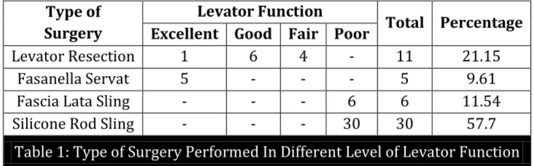
Table 1: Shows the various types of surgeries performed in different level of levator function.
Type of Surgery
Levator Function
Total Percentage Excellent Good Fair Poor
Levator Resection 1 6 4 - 11 21.15
Fasanella Servat 5 - - - 5 9.61
Fascia Lata Sling - - - 6 6 11.54 Silicone Rod Sling - - - 30 30 57.7
Table 1: Type of Surgery Performed In Different Level of Levator Function
In our study we found that in the levator resection group 9 cases out of 11(81.82%) patients had a good postoperative outcome, and 2(18.18%) cases had a fair postoperative outcome. All 5 cases (100%) that underwent a Fasanella Servat Surgery had a good correction of ptosis surgery. Amongst the patients operated for sling surgeries, all patients who underwent fascia lata sling surgery had good correction, where as in patients where silicon rod sling was performed 25(83.33%) cases had a good correction whereas 3(10%) cases had fair correction, 2(6.67%) cases had a poor correction. A chi-square test was applied to find if there was any significant difference in the postoperative outcomes in all the four groups. The p value was 0.72 and was not significant. From the chi-square test it can be concluded that the outcome difference between these four surgeries is not significant statistically.
Type of Surgery
Post-Operative Outcome
Good Fair Poor
Levator Resection 9(81.82%) 2(18.18%) - Fasanella Servat 5(100%) - - Fascia Lata Sling 6(100%) - - Silicone Rod Sling 25(83.33%) 3(10%) 2(6.67%)
Table 2: Post-Operative Outcome of Lid Elevation Achieved At 6 Months
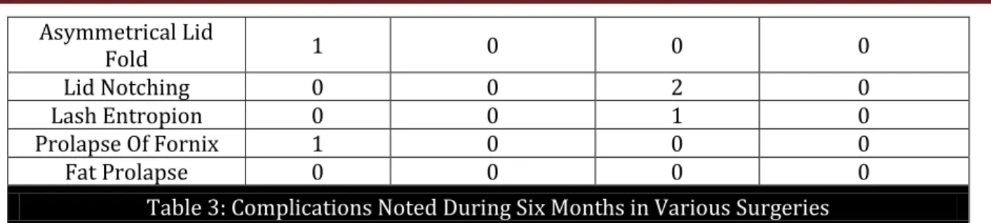
The various other complications noted in the postoperative period after surgeries were noted and tabulated in Table 3.
Complications Levator
Resection
Fasanella Servat Surgery
Silicone Sling Surgery
Fascia Lata Sling Surgery
Lagophthalmos 0 0 5 1
Suture Granuloma 0 0 3 0
Silicone Sleeve
J of Evolution of Med and Dent Sci/ eISSN- 2278-4802, pISSN- 2278-4748/ Vol. 4/ Issue 42/ May 25, 2015 Page 7399
Asymmetrical Lid
Fold 1 0 0 0
Lid Notching 0 0 2 0
Lash Entropion 0 0 1 0
Prolapse Of Fornix 1 0 0 0
Fat Prolapse 0 0 0 0
Table 3: Complications Noted During Six Months in Various Surgeries
DISCUSSION: Cosmesis and visual improvement were the predominant indications for the surgery in our study. Depending on the levator function and amount of ptosis, surgery was performed. In case of excellent levator function with mild ptosis Fasanella Servat surgery was done on 5(10.87%) eyelids. In case of good-fair levator function with mild-moderate ptosis we performed levator resection surgery on 11(23.91%) eyelids. In case of poor levator function with severe ptosis fascia lata sling surgery was done on 6(13.04%) eyelids, silicone rod sling surgery was performed on 30(65.23%) eyelids.
In a similar kind of study, Geoff M Whitehouse performed retrospective study of visual and surgical outcome following the surgical correction of isolated congenital ptosis, at The Children’s Hospital, Camperdown, Australia. In their study 30(37.5%) eyes underwent levator resection procedure, 40(50%) eyes underwent a brow suspension using donor stored fascia lata, and in 10(12.5%) eyes a brow suspension was performed using mersilene mesh.[14]
In our study 52 eyelids of 46 patients were included. In Geoff study total 80 eyelids of 65 patients were included.[14]
In our study the age of patients was 5-55 years with mean age 15.85 years. In Geoff study the mean age was 4.5 years.[14] This late age of presentation points to a possible delay in seeking healthcare facilities in western part of our country. It also points to the lack of awareness about the possible visual and facial cosmetic abnormality associated with ptosis. We also noted especially in the poor socio-economic class of our patients, as youngsters approach a marriageable age group only then generally for cosmetic concern medical help is sought for.
Amongst all the four groups, a good postoperative correction was achieved in 100% of the subjects in Fasanella Servat. In another study by Pang et al. The authors retrospectively reviewed 169 charts of 2 surgeons from patients who had undergone a Fasanella-Servat procedure for mild to moderate ptosis. Surgical success was defined as lid symmetry within 0.5 mm or correction of eyelid contour abnormality from previous surgery or trauma. With a mean follow-up of 7 months, success was achieved in 89.5% of cases (137/153). Postoperative problems included dry eye symptoms (6/144 patients), contour abnormalities in 12 lids, and dermatochalasis in 10 lids.[15]
J of Evolution of Med and Dent Sci/ eISSN- 2278-4802, pISSN- 2278-4748/ Vol. 4/ Issue 42/ May 25, 2015 Page 7400
period of 22 months, with only 4 eyes requiring revision. Silicon band extrusion, however, was reported in 3 (5%) eyes.[17]
In our study out of 30 silicone sling surgeries, 5(09.617%) patient had lagophthalmos, 3(10%) patient had suture granuloma and 1(3.34%) patient had silicone sleeve slippage. Suture granuloma excision performed in total 3 patients. 3 patients underwent re-surgery, 1 for silicone
sleeve slippage, and 2 for significant under correction. In Geoff’s study, in fascia lata sling surgery 4%
patients had suture granuloma and 2% patients had infection. In mersilene mesh sling surgery, 35% patients had suture granuloma. In levator resection 3.3% patients had wound dehiscence and 6.6% patients had excess skin.[14]
Hence, fromour observations during study the few conclusions we could summarise are as given below:
Good bell’s phenomenon is an essential prerequisite to prevent postoperative corneal exposure
and associated complication if full correction of ptosis is aimed at.
In case of excellent levator function (>12mm) Fasanella Servat surgery gives good result.
In case of good to fair levator function (4-11mm) levator muscle resection surgery gives good result.
In case of poor levator function (<4mm) fascia lata sling surgery or silicon rod sling surgery gives good result.
No significant difference on applying the chi square test was observed in the cosmetic and functional outcome between all the groups.
For severe congenital ptosis repair silicon sling material instead of fascia lata can be also be safely used. We also recommend that sling material should be placed in deeper tissue plane to decrease the incidence of infection, extrusion and granuloma formation.
CONCLUSION: Ptosis surgery has to be customised according to the amount of ptosis, levator action, and the clinical and surgical experience. Familiarity with the advantages and disadvantages of each of these techniques, as well as meticulous patient selection is the key to achieve a successful outcome with a low rate of complications.
ACKNOWLEDGEMENT: We acknowledge the immense help received from the scholars whose articles are cited and included in the references of this manuscript. We are also very grateful to all the authors/editors/publishers of all the articles, journals and books from where the literature for this article has been reviewed and discussed.
REFERENCES:
1. Allard FD, Durairaj VD. Current Techniques in Surgical Correction of Congenital Ptosis. Middle East African Journal of Ophthalmology.2010; 17 (2): 129-133.
2. Sakol PJ, Mannor G, Massaro BM. Congenital and acquired blepharoptosis. Curr Opin Ophthalmol.1999; 10: 335–9.
3. Smith B, McCord CD, Baylis H. Surgical treatment of blepharoptosis. Am J Ophthalmol.1969; 68: 92–9.
4. Gureico JR, Martyn LJ. Congenital Malformation of eye and orbit. Otolaryngol Clinic North Am.2007;40:113-40.
J of Evolution of Med and Dent Sci/ eISSN- 2278-4802, pISSN- 2278-4748/ Vol. 4/ Issue 42/ May 25, 2015 Page 7401
6. Finsterer J. Ptosis: causes, presentation, and management. Aesthetic Plast Surg. 2003; 27: 193– 204.[PubMed]
7. Fasanella RM, Servat J. Levator resection for minimal ptosis. Another simplified operation. Arch Ophthalmol. 1961; 65: 493–496.
8. Song R, Song Y. Treatment of blepharoptosis. Direct transplantation of the frontalis muscle to the upper eyelid. Clin Plast Surg.1982; 9: 45–48.
9. Zou LY, Chang TS. Frontalismyo fascial flap from eyebrow region for the correction of ptosis of the upper eyelid. Eur J Plast Surg. 1988; 11: 73–78.
10.Park DH, Choi WS, Yoon SH, et al. Comparison of levator resection and frontalis muscle transfer in the treatment of severe blepharoptosis. Ann Plast Surg. 2007; 59: 388–392.
11.Park DH, Lee SJ, Song CH. Recurrence of blepharoptosis after a superiorly based muscle flap: treatment by frontalis muscle advancement. Plast Reconstr Surg. 2005; 116: 1954–1959. 12.Park DH, Ahn KY, Han DG, et al. Blepharoptosis repair by selective use of superiorly based
muscle flap. Plast Reconstr Surg. 1998; 101: 592–603.
13.Park DH, Choi SS. Correction of recurrent blepharoptosis using an orbicularis oculi muscle flap and a frontalismusculofascial flap. Ann Plast Surg. 2002; 49: 604–611.
14.Whitehouse, G. M., Grigg, J. R. and Martin, F. J. (1995), congenital ptosis: results of surgical management. Australian and New Zealand Journal of Ophthalmology.1995; 23: 309–314. 15.Pang NK1, Newsom RW, Oestreicher JH, Chung HT, Harvey JT. Fasanella-Servat procedure:
indications, efficacy, and complications. 2008; 43 (1): 84-8.
16.M. E. Wilson and R. W. Johnson. Congenital ptosis: long-term results of treatment using lyophilized fascia lata for frontalis suspensions. Ophthalmology.1991. 98 (8): 1234–1237. 17.N. Wasserman, M. D. Sprunger, and E. M. Helveston. Comparision of materials used in frontalis
suspension. Archives of Ophthalmology.2001; 119: 687–691.
18.S. Crawford, Frontalis slingoperation, Journal of Pediatric Ophthalmology and Strabismus.1982.19 (5); 253–255.
19.S. Wagner, J. A. Mauriello, L. B. Nelson, et al. Treatment of congenital ptosis with frontalis suspension: a comparision of suspensory materials. Ophthalmology. 1984; 91 (2): 245–248.
AUTHORS:
1. Ruchi Kabra 2. Parth Khatri
PARTICULARS OF CONTRIBUTORS:
1. Assistant Professor, Oculoplastic Unit, M & J Western Regional Institute of
Ophthalmology, Civil Hospital, Ahmedabad. 2. Resident Doctor, Oculoplastic Unit, M & J
Western Regional Institute of
Ophthalmology, Civil Hospital, Ahmedabad.
FINANCIAL OR OTHER
COMPETING INTERESTS: None
NAME ADDRESS EMAIL ID OF THE CORRESPONDING AUTHOR:
Dr. Ruchi Kabra,
# 603, Madhuram Tower, Circuit House Road,
Shahibaug-380004, Ahmedabad. E-mail: [email protected]