INGRID NEY KRAMER DE MELLO
AVALIAÇÃO DE DOIS ISOLADOS DO FUNGO NEMATÓFAGO Duddingtonia flagrans NO CONTROLE DE LARVAS INFECTANTES DE Ancylostoma spp. DE CÃES
VIÇOSA
MINAS GERAIS – BRASIL 2013
Ficha catalográfica preparada pela Seção de Catalogação e Classificação da Biblioteca Central da UFV
T
Mello, Ingrid Ney Kramer de, 1985-
M527a Avaliação de dois isolados do fungo nematófago
2013 Duddingtonia flagrans no controle de larvas infectantes de Ancylostoma spp. de cães / Ingrid Ney Kramer de Mello. –
Viçosa, MG, 2013.
viii, 44f. : il. ; 29cm.
Orientador: Jackson Victor de Araújo
Dissertação (mestrado) - Universidade Federal de Viçosa. Inclui bibliografia.
1. Cão - Doenças. 2. Duddingtonia flagrans. 3. Ancylostoma. I. Universidade Federal de Viçosa.
Departamento de Veterinária. Programa de Pós-Graduação em Medicina Veterinária. II. Título.
AGRADECIMENTOS
A Deus, toda honra, glória e louvor. Senhor, obrigada pelo fim de mais essa etapa.
Ao meu amado esposo Alexson, pelo carinho, dedicação, paciência e incentivo.
À Universidade Federal de Viçosa, em particular ao Departamento de Veterinária e ao Laboratório Bionema pela estrutura disponibilizada.
À CAPES pela concessão da bolsa.
À minha querida mãe, pela vida e por ser tão dedicada e batalhadora. Abriu mão de muitas coisas para me proporcionar estudo de qualidade.
Ao meu querido pai, por todo o carinho e amor. Meu eterno agradecimento pelos momentos em que esteve ao meu lado, me apoiando e me fazendo acreditar que nada é impossível. Se estivesse aqui, com certeza estaria muito orgulhoso.
Aos meus irmãos, Felipe e Lorena, cunhados, cunhadas e sobrinhos pelo apoio e torcida.
Ao Professor Jackson Victor de Araújo, pela oportunidade, orientação, amizade e por acreditar em meu potencial.
Aos amigos do laboratório de Parasitologia Veterinária: Juliana Milani Araujo, Alexandre Tavela, Fernanda Mara Fernandes, Manoel, Anderson Silva Dias, Rosane, Alessandra, Lorendane, e Wendeo Ferreira da Silveira, obrigado aos que, em algum momento, contribuíram na realização deste trabalho e outros que durante nosso convívio tornaram-no um momento de descontração e alegria. Em especial agradeço ao Fabio Ribeiro Braga pela orientação segura e sugestões na execução dos experimentos e escrita dos artigos.
Aos funcionários do laboratório de Parasitologia do Departamento de Veterinária, José Geraldo e Ademir, pela colaboração.
Ao Professor Leandro Grassi de Freitas por ter aberto as portas de seu laboratório para a realização do trabalho, além da orientação, confiança, incentivo, conselhos e amizade.
As secretárias da Pós-Graduação do Departamento de Veterinária Rosi e Beth, pelo carinho e gentileza.
Aos amigos Caio Lins e Rafaela Schaedler Lins pela amizade, apoio e por tornar a vida em Viçosa mais alegre.
BIOGRAFIA
INGRID NEY KRAMER DE MELLO, Filha de Nilza Ney Kramer e Sizomar Kramer, nasceu em Guarapari- Espirito Santo, em 8 de Maio de 1985.
Em Julho de 2009 graduou-se em Medicina Veterinária pela Universidade Federal do Espirito Santo.
SUMÁRIO
RESUMO...vii
ABSTRACT...viii
INTRODUÇÃO GERAL... 1
Capítulo 1 - Different structures of the nematophagous fungus Duddingtonia flagrans in the control of infective larvae of Ancylostoma spp... 5
Abstract... 6
Introduction... 6
Material and Methods... 7
Results... 10
Discussion... 11
References... 13
Capítulo 2- Biological Control of infective larvae of Ancylostoma spp. in beach sand... 17
Abstract... 18
Introduction... 18
Methods... 20
Results... 22
Discussion... 24
References... 26
Capítulo 3 - Effect of the fungus Duddingtonia flagrans on infective larvae Ancylostoma spp. of dogs under semi-natural conditions... 28
Abstract... 29
Introduction... 29
Materials and Methods... 30
Results... 32
Discussion... 33
References... 35
CONCLUSÕES... 38
REFERÊNCIAS BIBLIOGRÁFICAS... 39
RESUMO
MELLO, Ingrid Ney Kramer de, M.Sc., Universidade Federal de Viçosa, fevereiro de
2013. Avaliação de dois isolados do fungo nematófago Duddingtonia flagrans no
controle de larvas infectantes de Ancylostoma spp. de cães. Orientador: Jackson Victor de Araújo. Coorientadores: Leandro Grassi de Freitas e Fabio Ribeiro Braga.
Os nematóides do gênero Ancylostoma são endoparasitas de cães e também
geohelmintos zoonóticos que podem infectar o ser humano. O controle destes nematóides em estágio adulto é baseado na utilização de anti-helmínticos. No entanto, o uso de agentes biocontroladores pode ser uma medida complementar para reduzir a população em estágios pré-parasitários em desenvolvimento no ambiente. Este estudo
objetivou avaliar o fungo predador Duddingtonia flagrans no controle da forma larval
infectante (L3) de Ancylostoma spp., em areia de praia. Foi avaliada a infectividade in
vitro de dois isolados do fungo nematófago D. flagrans (AC001 e CG768) sobre larvas
infectantes (L3) de Ancylostoma spp. de cães. Utilizou-se como inóculos fúngicos
estruturas vegetativas (micélio), reprodutivas (conídios) e de sobrevivência (clamidósporos). A interação foi avaliada ao final de 10 dias de incubação em placas de Petri contendo meio ágar-água 2% em temperatura de 25 °C. O antagonismo em condições semi-naturais foi avaliado por meio da utilização de uma produção massal de inóculo fúngico em grãos de milho moído. O fungo foi incorporado à areia, em grãos colonizados de milho moído na concentração de 15.000 clamidósporos/grama de areia.
Essa concentração se mostrou a mais efetiva em ensaio in vitro preliminar (redução de
59,2%). Os resultados mostraram a eficiência do fungo D. flagrans no controle de
larvas infectantes de Ancylostoma spp. em areia de praia. Isso sugere que isolados desse
fungo podem ser utilizados como parte de um programa de controle de Ancylostoma
spp. no ambiente.
ABSTRACT
MELLO, Ingrid Ney Kramer de, M.Sc., Universidade Federal de Viçosa, February of
2013. Evaluation of two isolates of the nematophagous fungus Duddingtonia
flagrans in the control of infective larvae of Ancylostoma spp. dogs. Adviser: Jackson Victor de Araújo. Co-advisers: Leandro Grassi de Freitas and Fabio Ribeiro Braga
The nematodes of the genus Ancylostoma are endoparasites of dogs and also zoonotic
geohelminths that can infect humans. The control these nematodes in adult stage is based on the use of anthelmintics. However, the use of biocontrol agents may be an additional action to reduce the population in pre-parasitic stages developing in the
environment. This study aimed to evaluate the Duddingtonia flagrans predator fungus
for the control of the larval form (L3) of Ancylostoma spp., in beach sand. We evaluated
the in vitro infectivity of two isolates of the nematophagous fungus D. flagrans (AC001
and CG768) on infective larvae (L3) of Ancylostoma spp. dogs. Vegetative structures
(mycelium), reproductive (conidia) and survival (chlamydospores) were used as fungal inoculum. The interaction was evaluated at the end of 10 days of incubation in Petri dishes containing agar-water 2% medium in a temperature of 25 ° C. The antagonism in semi-natural conditions was assessed by use of a mass production of fungal inoculum in grains of milled maize. The fungus was introduced into the sand in colonized milled maize at the concentration of 15,000 chlamydospores / gram of sand. This concentration
was the most effective in the preliminary in vitro assay (reduction of 59.2%). The
results showed the efficiency of the fungus D. flagrans in the control of infective larvae
of Ancylostoma spp. in beach sand. This suggests that isolates of this fungus may be
INTRODUÇÃO GERAL
Os cães são hospedeiros de inúmeros parasitas e estão envolvidos na transmissão involuntária de mais de 60 infecções zoonóticas (Macpherson et al., 2005). Dentre elas, as parasitoses gastrintestinais causadas por geohelmintos, estão entre as mais prevalentes e importantes infecções parasitárias destes animais (Oliveira-Sequeira, 2002).
Dentre os nematóides parasitas intestinais que utilizam o cão como hospedeiro
definitivo e são zoonóticos, os do gênero Ancylostoma têm requerido importante
atenção médica e veterinária (Katagiri & Oliveira-Sequeira, 2008; Mulvenna et al. 2009). Este gênero está classificado zoologicamente no filo Nematoda, classe Secementea, ordem Strongylida, superfamilia Ancylostomatoidea, família Ancylostomatidae e subfamilia Ancylostomatinae (Maggenti, 1981). Os membros desta subfamília utilizam apenas carnívoros como hospedeiros definitivos (Lichtenfels, 1980).
As espécies A. braziliense, A. caninum e A. ceylanicum são as mais prevalentes nestes
animais (Baker et al. 1989; Traub et al. 2005). Segundo Urquhart et al. (1998) os ancilostomídeos são responsáveis por ampla morbidade e mortalidade em cães sendo
que a espécie A. caninum é a mais patogênica devido à maior espoliação sanguínea.
Além de sua importância em saúde animal este parasito tem se destacado como agente causador da Síndrome da larva migrans cutânea. Esta síndrome ocorre por meio
do contato direto da pele humana com a L3 ativa de Ancylostoma spp. presente em solos
contaminados por fezes de cães (Robertson e Thompson, 2002). Após a penetração na epiderme, a larva migra no tecido subcutâneo ocasionando reações inflamatórias caracterizadas por intenso prurido e erupções serpiginosas (Mattone-Volpe, 1998).
Além disso, adultos imaturos de A. caninum podem ser ocasionalmente encontrados no
intestino causando enterite eosinofílica (EE), caracterizada por dor abdominal aguda associada a eosinofilia periférica, anorexia, náusea e diarreia (Prociv & Croese, 1996;
Croese et al, 1994;. Landmann & Prociv, 2003). O A. caninum também foi reportado
como agente da neuroretinite subaguda unilateral difusa que é uma doença causada pela presença de uma larva no espaço subretiniano (Casella et al. 2001; Venkatesh et al., 2005; Vedantham et al., 2006).
Santarém et al., 2004) uma vez que maior prevalência de parasitismo é observada nestes animais em comparação aos domiciliados, em virtude de não receberem tratamento
antiparasitário e da facilidade com que circulam por áreas públicas (Palmer et al.,2008).
Portanto, o solo de áreas públicas representa um foco potencial de transmissão para a população humana e animal pelo fato de estar sendo freqüentemente infestado por ovos veiculados nas fezes de cães que comumente estão infectados (Robertson et al., 2000; Blazius et al., 2005; Macpherson, 2005). Surtos de larva migrans cutânea (LMC) são relatados e relacionados à atividade em caixas de areia contaminadas com fezes de cães. (Lima et al., 1984; Nunes et al., 2000; Araújo et al., 2000; Santarém et al., 2004). Ainda, diversos autores têm relatado elevada prevalência de ovos e ou larvas de Ancylostoma spp. em areias de praças públicas e em canteiros de praias em diferentes
regiões do Brasil (Araújo et al., 1999; Scaini et al., 2003; González et al., 2004; Castro
et al., 2005; Santos et al., 2006; Silva et al., 2009).
O aparecimento de relatos de resistência às drogas anti-helmínticas levou pesquisadores de todo o mundo a buscar medidas alternativas para o controle de endoparasitoses de animais domésticos, visando à diminuição do emprego de quimioterápicos (Mota et al., 2003). Dentre as propostas que tem sido trabalhadas com o intuito de melhorar esse controle, sugere-se o controle biológico, como uma alternativa viável e promissora que reduz as infecções causadas por helmintos parasitos gastrintestinais, e cuja ação se dá por meio de organismos vivos como os fungos nematófagos que atuam como antagonistas naturais no ambiente (Araújo et al., 2004). Pesquisas a campo ou sob condições experimentais mostram que espécies de fungos nematófagos são bons agentes de controle biológico de nematóides de animais (Larsen, 1999).
2006). Segundo Gray (1987) este grupo diferencia suas hifas vegetativas em seis estruturas de captura (armadilhas): hifas adesivas não diferenciadas; ramificações de hifas que sofrem anastomose, formando redes adesivas tridimensionais; ramificações adesivas, onde em algumas vezes podem se unir formando redes adesivas simples bidimensionais; nódulos adesivos; anéis constritores e anéis não constritores. Entretanto o tipo de armadilha mais encontrado em fungos predadores são as redes adesivas (Mota et al., 2003).
O segundo grupo, denominados fungos endoparasitos, é capaz de infectar os nematóides através de esporos, que uma vez ingeridos desenvolvem hifas responsáveis pela absorção do conteúdo interno do nematóide. Estes fungos não produzem hifas vegetativas fora do corpo do hospedeiro, mas somente hifas férteis ou conidióforos contendo esporos. O terceiro grupo de fungos é denominado oportunistas, parasitos de ovos (Araújo et al., 1995). As hifas penetram a casca do ovo, através dos pequenos poros existentes na camada vitelínica, causando alteração na permeabilidade da casca e expandindo seu volume. A hifa aumenta de tamanho ao passar pela camada vitelínica e atravessa a camada adjacente quitínica e lipídica. Como conseqüência do processo, a camada vitelínica se divide, a camada de quitina se torna vacuolizada e a camada de lipídios se torna dispersa. Estes tipos de fungos colonizam o conteúdo do ovo, ou ainda a larva em desenvolvimento no seu interior (Morgan-Jones & Rodríguez-Kábana, 1988).
Os fungos nematófagos podem apresentar esporos bastante diversificados no tamanho, coloração, forma e resistência no ambiente. A maioria dos fungos nematófagos apresenta esporos secos, emergindo de estruturas de frutificação, denominadas conidióforos, essenciais na dispersão aérea dos conídios. Os conidióforos crescem verticalmente, em direção perpendicular ao substrato o qual o isolado foi cultivado. Algumas espécies produzem conidióforos contendo apenas um conídio em sua extremidade, outras espécies apresentam cachos de conídios em toda a estrutura do conidióforo. Estruturas denominadas clamidósporos também podem ser produzidas. Estes são esporos de parede espessa, diferenciada a partir das hifas, aparecem em condições de estresse extremo e podem dar origem a hifas, conidióforos e conídios (Barron, 1977).
nematóides que parasitam animais domésticos, reduzindo de forma efetiva a sua população tanto em condições laboratoriais quanto em condições a campo, além disso, possuem a vantagem de apresentar maior potencial de industrialização (Larsen, 1999).
No grupo de fungos predadores, a espécie Duddingtonia flagrans tem sido a
mais estudada, havendo características que propiciam o seu uso como agente de controle biológico de parasitos de animais, destacando-se a produção de grande número de clamidósporos que resistem as condições adversas (Sanyal et al. 2008). O potencial desta espécie como biocontrolador está consolidado e tem sido relatado por vários
autores em experimentos in vitro (Araújo et al., 2004; Araújo et al., 2006;) e em
condições in vivo (Mendoza-de-Gives et al., 1998; Dias et al., 2007).
Devido à capacidade dos clamidósporos de D. flagrans suportarem condições
adversas, eles têm sido administrados na alimentação de bovinos, ovinos. caprinos, eqüinos e suínos por resistirem à passagem através do trato gastrintestinal destes animais e posteriormente germinarem nas fezes, formando armadilhas (Campos et al., 2009; Silva et al., 2010; Braga et al., 2010; Ferreira et al., 2011).
Em cães, a administração oral de fungos biocontroladores não é uma opção prática devido ao elevado número de cães errantes os quais tem grande importância na disseminação de helmintos no ambiente devido à maior freqüência de parasitismo em conseqüência do abandono (Labruna et al., 2006). Isto justifica a utilização de uma abordagem em que o fungo seja incorporado à areia, o que deve ser feito juntamente com um substrato de crescimento fúngico para favorecer seu crescimento e estabelecimento.
O controle biológico de parasitas de cães no ambiente por fungos nematófagos é promissor. Entretanto ele deve ser visto como parte de um programa integrado em complementação ao controle químico de endoparasitas em cães domiciliados e ao controle da população de cães errantes, uma vez que reduz satisfatoriamente as formas pré-parasitarias no ambiente (Waler & Larsen, 1993).
Capítulo 1
Different structures of the nematophagous fungus Duddingtonia flagrans in the control
of infective larvae of Ancylostoma spp.
Abstract
The infectivity of two isolates of the nematophagous fungus Duddingtonia flagrans
(AC001 and CG768) on infective larvae (L3) of Ancylostoma spp. was evaluated in vitro
using mycelium, conidia and chlamydospores as fungal inocula. Evaluation of the
isolates was performed at the end of 10 days of interaction between the fungus and nematodes in Petri dishes containing water-agar 2% at 25°C. The control without
fungus contained only 1 x 10³ L3/Petri dish, while the other treatments contained the
sameconcentration of L3 in mycelial discs of 5 mm in diameter, and 1 x 10³ conidia or
1 x 10³ chlamydospores/Petri dish. Both isolates had a significant effect (p < 0.05) on
reducing the average of L3 recovered. The treatment with chlamydospores of the isolate
CG768 showed the highest predatory activity by reducing the number of L3 of
Ancylostoma spp. recovered by 80.7% when compared to the control treatment, without fungus. Moreover, the predatory activity of the isolate AC001 exceeded that of CG768 when conidia were used as an inoculum source, reducing the recovery by 73.58% and
52.93%, respectively. The D. flagrans fungus may be used as an alternative for
biologicalcontrol of L3 form Ancylostoma spp.
Keywords: Nematophagous fungi, Duddingtonia flagrans, Ancylostoma spp., larva migrans, biological control, dogs.
Introduction
The syndrome of cutaneous larva migrans is an important public health problem in
most countries around the world. This syndrome is caused by infective larvae (L3) in the
third stage of some geohelminths, mainly Ancylostoma caninum and Ancylostoma
braziliensis species, where the dog is a definitive host and human beings are eventually infected (Acha and Szyfres, 2003). The risk of infection is higher in children because of their frequent contact with soil (Capuano and Rocha, 2006).
This syndrome occurs through direct contact of human skin with L3 of
The emergence of reports on resistance to anti-helminthic drugs has led to the search of alternative sustainable strategies for control of animal-parasitic nematodes, highlighting the use of nematophagous fungi (Waller and Larsen, 1993). Field studies have shown that nematophagous fungi species are good biological control agents of animal-parasite nematodes (Larsen, 1999). These fungi control the parasites by destroying their pre-parasitic stages free living in the environment, which limits exposure of hosts to the agent of the disease and prevents new infections. In this
context, predatory nematophagous fungi such as the genera Duddingtonia, Arthrobotrys
and Monacrosporium have been considered promising (Mota et al., 2003).
Potential use of the species Duddingtonia flagrans as a biocontrol agent of
animal-parasitic helminthes is established and has been reported by several authors in in
vitro experiments (Araújo et al., 2004; Araújo et al., 2006; Braga et al., 2010) and in vivo conditions and in the field (Mendoza-de-Gives et al., 1998; Dias et al., 2007). In
these in vitro studies, conidia have been commonly used as the inoculum source.
Although other inoculum such as chlamydospores have been previously used in in vivo
studies for the control of Haemonchus contortus in sheep (Campos et al., 2009) and
mycelia in the control of Ancylostoma spp. in dogs (Carvalho et al., 2009), it is not
known if the type of inoculum influences predatory ability.
This study aimed to compare the in vitro infectivity of two isolates of the
predatory nematophagous fungus Duddingtonia flagrans (AC001 and CG768) on L3 of
Ancylostoma spp. and to evaluate which inoculum type (chlamydospores, mycelia or conidia) provides a higher nematophagous activity of this fungus.
Material and Methods Fungus
Two isolates of the nematophagous fungus D. flagrans (CG768 and AC001)
obtained from samples of Brazilian soils and animal feces were used in this experiment. They were isolated using the soil-sprinkling method of Duddington (1955), modified by Santos et al. (1991). The isolate AC001 was stored in the dark at 4ºC in test tubes containing 2% corn-meal-agar (2% CMA). The isolate CG768 was stored in same
conditions described above, but on integral rice grains in 5 mL BD Vacutainer® glass
Mycelia production
Culture disks of the isolate AC001 (5 mm in diameter) and one rice grain colonized by isolate CG768 were transferred to 50 mm × 10 mm Petri dishes containing 2% water-agar (2% WA) culture medium. These plates were incubated for 7 days at 26 °C in a dark room. After mycelial growth, a 5 mm diameter culture disk of one isolate was transferred to a 90 mm x 15 mm Petri dish containing a 2% CMA. Next a 5 mm diameter culture disk of this new culture was transferred to 50 mm × 10 mm polypropylene Petri dishes containing 2% WA and incubated for 10 days as previously described.
Conidia and chlamydospore production
Culture disks of the isolate AC001 (5 mm in diameter) and one rice grain colonized by the isolate CG768 were transferred to 50 mm × 10 mm Petri dishes containing 2% WA culture medium. These plates were incubated for 7 days at 26 ºC in a dark room. After mycelial growth, a 5 mm diameter culture disk was transferred to a 90 mm x 15 mm Petri dish containing a 2% potato dextrose agar. These plates were incubated for 10 and 20 days at 26 ºC in a dark room, to obtain conidia and chlamydospores, respectively.
At the end of this period, the surface of the fungal colony was gently scraped with a fine sterile brush and washed with 10 mL sterile ultrapure water. The fungal suspension was filtered through gauze (two layers) to eliminate mycelial fragments,
according to Maciel et al. (2009). Next two aliquots of the conidia suspension were
counted in a Neubauer chamber and the suspension was adjusted to the desired concentration in the experiment. The same procedure described above was used to obtain the chlamydospore concentration from fungal colonies with 20 days of incubation.
Ancylostoma spp. L3
on positive thermo/hydrotropism of the larvae and gravity sedimentation. After 12 hours
the sediment containing L3 was transferred to centrifuge tubes and washed in sterile
ultrapure water 5 times followed by centrifugation at 1000 rpm for 5 min. The L3 was
filtered, decontaminated for 10 minutes with 10 mL of 0.5% (v/v) sodium hypochlorite solution, as described by Barçante et al. (2003), and then rewashed five times in sterile
ultrapure water. The L3 suspension was homogenized and 3 aliquots of 10 μL were
placed on a microscope glass slide marked with longitudinal lines to facilitate counting.
Each aliquot was covered with a glass coverslip after adding 10 μL of lugol's solution to
kill L3. They were then counted using a light microscope (40x magnification) and the
concentration of the suspension was adjusted to 1,000 L3/10 μL. The viability of L3 was
verified by microscopic examination before inoculating the dishes.
Experimental assay
The interaction experiment between D. flagrans isolates and Ancylostoma spp. L3
consisted of seven treatments as follows:
(1) Colony of the isolate CG768 with 10 days of grown and 1 x 10³ L3;
(2) Colony of the isolate AC001 with 10 days of grown and 1 x 10³ L3;
(3) 1 x 10³ conidia of the isolate CG768 and 1 x 10³ L3;
(4) 1 x 10³ conidia of the isolate AC001 and 1 x 10³ L3;
(5) 1 x 10³ chlamydospores of the isolate CG768 and 1 x 10³ L3;
(6) 1 x 10³ chlamydospores of the isolate AC001 and 1 x 10³ L3;
(7) Control without fungus and 1 x 10³ L3.
The assay was performed in 50 mm x 10 mm polypropylene Petri dishes containing 2% WA. Plates were sealed with polyvinyl chloride (PVC) transparent film and incubated for 10 days at 26 °C in a dark room. At the end of the interaction period,
the non-predated L3 were harvested from the 2% WA medium by the Baermann funnel
technique, as described before. After 12 hours of larval concentration under gravity in 5 mL vacutainer-like glass tubes connected to a funnel, 3 mL of water without larvae were discarded and a drop of Lugol`s solution was added to the remaining volume to
kill Ancylostoma spp. L3. Then, the larvae were counted in a Peter’s counting slide
Statistical method
The experiment was arranged in a completely randomized design with 10 replications per treatment, where each experimental plot consisted of a Petri dish. After the data were analyzed by ANOVA, the Tukey’s test was used for comparison among the averages of the treatments and the Dunnett’s test for comparison among these and the fungus-free control, both at 5% significance level.
Results
The two isolates (CG768 and AC001) of D. flagrans fungus produced
significantly reduced (p<0.05) in the reduction of the average number of L3 of Ancylostoma spp., but they presented different efficiency in the predation of in vitro
tests (Table 1). The reduction in the average number of L3 ranged from 30.5% to
80.7%, according to the type of inoculum (conidia, chlamydospores or mycelium) and isolate fungal used.
Table 1
Average values, standard deviation (±) and reduction percentage (%) of Ancylostoma
spp. dog infective larvae recovered from 2% water-agar culture medium by the Baerman method after 10 days of interaction in Petri dishes containing fungal isolates Duddingtonia flagrans (isolates CG768 and AC001) at three fungal inocula structures (conidia, chlamydospores and mycelium) in comparasion to fungus-free control treatment.
Fungal isolates Treatments
Conidia Chlamydospores Mycelium
CG768 a195,2B ± 73,79 (52,93) b79,8C ± 11,37 (80,76) a183,9C ± 28,89 (55,7)
AC001 c109,6C ± 41,37 (73,58) b201,5B ± 62,43 (51,5) a288,4B ± 38,63 (30,5)
Control 414,7A ± 66,74
Colonies of the CG768 isolate, developed from the chlamydospore inoculum,
showed the most predatory activity and resulted in a reduction of L3 of Ancylostoma
spp. of 80.7%. On the other hand, the predation activity of AC001 isolated surpassed that of the CG768 isolated, when conidia was used as the inoculum.
The lowest percentages of reduction in the average number of L3 of Ancylostoma spp. were observed when the fungi had mycelium as the inoculum. This
lower efficiency was observed for both isolated of D. flagrans.
Discussion
Results showed the potential use of two isolates of D. flagrans for the control of
L3 of Ancylostoma spp. These results are consistent with those encountered by Maciel et
al. (2006) and Carvalho et al. (2009), in which the species D. flagrans showed high
predatory activity against Ancylostoma spp. when comparing this fungus species with
two others (Arthrobotrys robusta and Monacrosporium thaumasium).
The predatory activity of isolates of D. flagrans on Ancylostoma spp. larvae in
vitro is related to the formation of traps by the fungus. Previous studies report the presence of traps after 24 hours of exposure of larvae to fungal isolates (Maciel et al., 2006; Carvalho et al., 2009). This strategy of predatory nematophagous fungi was also
observed by Araújo et al. (2006), who used these microorganisms to control Cooperia
and Oesophagostomum in cattle. Traps are formed in response to the presence of nematodes and may occur as a result of limited nutritional conditions and/or lack of water (Scholler and Rubbner, 1994; Gronvold et al., 1996). The mobility of nematode acts as a stimulus to the formation of these structures (Nansen et al., 1988). Thus, in this
study, the presence of L3 of Ancylostoma spp. in dishes containing a poor nutritional
environment (2% WA) was essential for the formation of traps, as analyzed by Maciel et al. (2006).
In this study, although the isolate CG768 showed to be effective in reducing the
average of L3 larvae of Ancylostoma spp., an average of 50% were recovered. This
percentage was lower than that reported in similar studies conducted by Maciel et al.
(2006), in which this isolate demonstrated high efficiency in the capture of Ancylostoma
conducted by Carvalho et al. (2009), in which the same isolate demonstrated high
efficiency in the predation of L3 of Ancylostoma spp. (reduction percentage of 87.02%)
when treated with conidia.
Campos et al. (2009) evaluated the dynamics conidia transition, chlamydospores
and mycelium of the fungus D. flagrans (isolated CG768) in the digestive duct in goats
and the effect of different structures on larval development of Haemonchus contortus in
coprocultures, and found a higher reduction percentage of larval development in the group treated with chlamydospores (61.23%).
The use of chlamydospores, in this experiment in vitro, indicated a better
performance of the isolate CG768 in the reduction of L3, while the activity of the isolate
AC001 was more favored when using conidia. Works have shown that intra-specific variation in the predatory activity of fungi are common (Araújo et al., 1993, 1994; Mendoza-de-Gives et al., 1994; Larsen, 2000), regardless of the species of fungus or nematode used in this experiment. These variations may be related to different factors, such as experimental design, loss of viability during storage in the laboratory (Gronvold et al., 1996) and antigenic variation present in different isolates of the same fungal species (Mendoza-de-Gives et al., 1999). Furthermore, while some isolates need only a few conidia to achieve the desired effect, others require large amounts of inoculum to achieve the same effect (Maciel et al., 2009).
In in vitro studies, fungi are not exposed to adverse factors of the environment (competition with other organisms and pH and temperature changes), which may help to
explain the higher reduction percentage of L3 when using conidia inoculum of isolate
AC 001, compared to the use of chlamydospores. It is possible that the greater strength of the chlamydospore structure exceeds that of conidia and mycelia in field conditions.
Moreover, studies subjected to in vitro conditions are essential to select fungal isolates
with greatest potential for application in biological control programs. In this sense, the results of the present study show that the isolate CG768 had its better activity when using chlamydospores as a source of inoculum. However, the isolated AC001 showed to
be more efficient for predation of L3 of Ancylostoma spp. when conidia served as the
source of inoculum. D. flagrans fungi can be used as an alternative for biological
References
Acha, P. N., Szyfres, B. 2003. Zoonoses and communicable diseases common to man and animals, 3rd Edn. Pan-american Health Organization, Washington.
Araújo, J. V., Santos, M. A., Ferraz, S., Maia, A.S., 1993. Antagonistic Effect of
Predacious Fungi Arthrobotrys on Infective Haemonchus placei Larvae. Journal of
Helminthology 67, 136-138.
Araújo, J. V., Santos, M. A., Ferraz, S., Maia, A.S., 1994. Biological control in vitro of
infective Haemonchus Placei larvae by predacious fungi Arthrobotrys Musiformis.
Arquivo Brasileiro de Medicina Veterinária e Zootecnia 46, 197-204.
Araujo, J. V., Assis, R. C. L., Campos, A.K., Mota, M., 2004. Atividade in vitro dos
fungos nematófagos dos gêneros Arthrobotrys, Duddingtonia e Monacrosporium sobre
nematóides trichostrongilídeos (Nematoda: Trichostrongyloidea) parasitos gastrintestinais de bovinos. Revista Brasileira de Parasitologia Veterinária 13, 65-71.
Araújo, J. V., Freitas, B. W., Vieira, T. C., Campos, A. K., 2006. Avaliação do fungo
predador de nematóides Duddingtonia flagrans sobre larvas infectantes de Haemonchus
contortus e Strongyloides papillosus de caprinos. Revista Brasileira de Parasitologia Veterinária 15, 76-79.
Barçante, J. P. M., Barçante, T. M., Dias, S. R. C., Vieira, L. Q., Lima, W. S., Negrão-
Corrêa, D., 2003. A method to obtain axenic Angiostrongylus vasorum first-stage larvae
from dog feces. Parasitology Research 89, 89-93.
Braga, F. R., Araújo, J. V., Silva, A. R., Carvalho, R. O., Araujo, J. M., Ferreira, S. R.,
Benjamin, L. A., 2010. Predatory activity of the nematophagous fungus Duddingtonia
flagrans on horse cyathostomin infective larvae. Tropical Animal Health and Production 42, 1161-1165.
Campos, A. K., Araújo, J. V., Guimarães, M. P., Dias, A. S. 2009. Resistance of
different fungal structures of Duddingtonia flagrans to the digestive process and
predatory ability on larvae of Haemonchus contortus and Strongyloides papillosus in
Capuano, D. M., Rocha, G. M. 2006. Ocorrência de parasitas com potencial zoonótico em fezes de cães coletadas em áreas públicas do município de Ribeirão Preto, SP, Brasil. Revista Brasileira de Epidemiologia 9, 81-86.
Carvalho, R. O., Araújo, J. V., Braga, F. R., Araújo, J. M., Silva, A. R., Tavela, A. O.
2009. Predatory activity of nemathophagous fungi on infective larvae of Ancylostoma
spp.: evaluation in vitro and after passing through the gastrointestinal tract of dogs. Journal of Helminthology 83, 231-236.
Dias, A. S., Araujo, J. V., Campos, A. K., Braga, F. R. 2007. Application of a
Formulation of the Nematophagous Fungus Duddingtonia flagrans in the Control of
Cattle Gastrointestinal Nematodiosis. World Journal of Microbiology and Biotechnology 23, 1245-1252.
Duddington, C. L. 1955. Notes on the tecnique of handling predaceous fungi. Transactions of Brithish Mycology Society 38, 97- 103.
Gronvold, J., Henriksen, S. A., Larsen, M., Nansen, P., Wolstrup, J. 1996. Aspects of biological control - With special reference to arthropods, protozoans and helminths of domesticated animals. Veterinary Parasitology 64, 47- 64.
Larsen, M. 1999. Biological Control of Helminths. International Journal for Parasitology 29, 139-146.
Larsen, M. 2000. Prospects for controlling animal parasitic nematodes by predacious microfungi. Parasitology 120, 121-131.
Maciel, A. S., Araújo, J. V., Cecon, P. R. 2006. Atividade predatória in vitro dos fungos
Arthrobotrys robusta, Duddingtonia flagrans e Monacrosporium thaumasium sobre
larvas infectantes de Ancylostoma spp. de cães. Revista Brasileira de Parasitologia
Veterinária 15, 71-75.
Mattone- Volpe, F. 1998. Cutaneous larva migrans infection in the pediatric foot. A
review and two cases reports. Journal of the American Pediatric Medical Association
88, 228- 231.
Mendoza-de-Gives, P., Zavaleta-Mejia, E., Herrera-Rodrigues, D., Quiroz-Romero, H.
1994. In vitro trapping capability of Arthrobotrys spp on infective larvae of
Haemonchus contortus and Nacobus aberrans. Journal of Helminthology 68, 223-229.
Mendoza-de-Gives, P., Flores-Crespo, J., Herrera-Rodrigues, D., Vasquez-Prats, V., Liebano-Hernandez, E., Ontiverosfernadez, G. E. 1998. Biological control of Haemonchus contortus infective larvae in ovine faeces by administering oral suspension of Duddingtonia flagrans chlamydospores to sheep. Journal of Helminthology 72, 343- 347.
Mendoza-de-Gives, P., Davies, K. G., Clark, S. J., Behnke, J. M. 1999. Predatory
behavior of trapping fungi against srf mutants of Caenorhabditis elegans and different
plant and animal parasitic nematodes. Parasitology 119, 95-104.
Mota, M. A., Campos, A. K., Araújo, J. V. 2003. Controle biológico de helmintos parasitos de animais: estágio atual e perspectivas futuras. Pesquisa Veterinária Brasileira 23, 93-100.
Nansen, P., Gronvold, J., Henriksen, S. A., Wolstrup, J. 1988. Interactions between the
predacious fungus Arthrobotrys oligospora and third-stage larvae of a series of animal
parasitic nematodes. Veterinary Parasitology 26, 329-337.
Peters, B. G. 1952. Toxicity tests with vinegar eelworm. Counting and culturing. Journal of Helminthology 26, 97-110.
Robertson, I. D., Thompson, R. C. 2002. Enteric parasitic zoonoses of domesticated dogs and cats. Microbes and Infection 4, 867- 873.
Scholler, M., Rubner, A. 1994. Predacious activity of the nematode destroying fungus Arthrobotrys oligospora in dependence of the médium composition. Microbiological Research 149, 145-149.
Capítulo 2
Biological Control of infective larvae of Ancylostoma spp. in beach sand
Abstract
Background: Geohelminths are parasites that stand out for their prevalence and wide
distribution, depending on the soil for their transmission. Aims: The aim of this study
was to evaluate the predatory capacity of the fungal isolate of the genus Duddingtonia
(CG768) on third stage larvae (L3) of Ancylostoma spp. in beach sand under laboratory
conditions. Methods: In the first assay, five treatments were formed with the fungus and
one treatment was the control without the fungus. The treatments contained 5000, 10000, 15000, 20000 or 25000 chlamydospores of the fungal isolate and 1000 Ancylostoma spp. L3 in pots containing 30 g of sand, and the control treatment
contained only 1000 Ancylostoma spp. L3 and distilled water in pots with 30 g of sand.
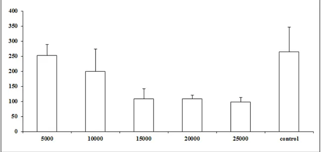
Results: Evidence of predatory activity was observed at the end of 15 days and the percentages of reduction for the five treatments were, respectively, 4.5 %, 24.5%,
59.2%, 58.8% and 63% the number of L3. Statistical difference was observed (p <0.01)
only at concentrations 15000, 20000 and 25000 in relation to control treatment. The second assay consisted of two treatments, in Petri dishes of 9 cm in diameter containing agar water 2% medium. In the first treatment, each Petri dish contained 500 Ancylostoma spp. L3 and 5 g of sand containing the isolate CG 768 at a concentration of 25,000 chlamydospores /g of sand, and the second treatment the control without fungus,
contained only 500 L3. After 7 days non-predated L3 were recovered from the Petri
dishes by the method of Baermann. The use of the fungus reduced (p <0.01) the average
number of Ancylostoma spp., L3 in 84%. Conclusions: The results of this study confirm
earlier work on the efficiency of the Duddingtonia genus in control of Ancylostoma spp.
infective larvae.
Keywords: Nematophagous fungi, Duddingtonia flagrans, Ancylostoma spp.,sand.
Introduction
Geohelminths are parasites that stand out for their prevalence and wide
distribution, and depend on the soil for their transmission. According to Silva et al.1
In this context, the hookworm have a cosmopolitan distribution and are more prevalent in tropical and subtropical regions where the soil presents temperature and moisture conditions suitable for the development of pre-parasitic forms (egg and/or
larvae) 2,3. On the other hand, the literature has reported that larvae and eggs of these
gastrointestinal parasites are commonly found in samples of sand. Moreover, this environment serves not only as a source of leisure but also presents risks to animal and
human health 4,5. Also in relation to this fact, Guimarães et al.6 reported that in several
cities of the country a considerable canine population circulates through the streets and public squares, where often their habits of defecation contaminate the soil with various types and potentially zoonotic parasitic forms. These authors observed the occurrence of Toxocara spp., and eggs or larvae of Ancylostoma spp. in 69.6% of soil samples collected in public squares. In this context these same authors showed that infected samples of sand from schools or kindergartens were positive only with larvae of Ancylostoma spp. In recent work Silva et al.7 demonstrated contamination of the sand
on the beaches of southeastern of Pernambuco state by larvae of Ancylostoma spp.
Regarding the control of these parasites, the same is based on the use of antihelminthic drugs, however, there are problems with parasitic resistance already installed in production animals, and that in the future may represent a problem for the
combat of parasitic nematodes of dogs and cats. High resistance of Ancylostoma
caninum to anthelmintic-therapy with pyrantel was demonstrated by Kopp et al.8. Thus, much has been researched about the use of biological control in environmental
decontamination of eggs and larvae of the genera Ancylostoma and Toxocara spp.9
However, it should be emphasized that biological control by nematophagous fungi is of the type "classic", ie, an environmental flood of organisms antagonists present in the environment and whose action is concentrated in the faecal environment by reducing the
amount of pre-parasitic forms of helminths10,11. Thus, stands out the fungus
Duddingtonia flagrans extensively studied as biocontroller organism under laboratory
and natural conditions12,13. However, there are no reports regarding the biological
control using nematophagous fungi on beach sand, representing a new approach.
The aim of this study was to evaluate the effect of different concentrations of
chlamydospores of the fungus D. flagrans (isolate CG 768) on the destruction of
Methods
Organism
The predator nematophagous fungus Duddingtonia flagrans (CG768) obtained
from samples of Brazilian soil and of animal feces was used in this experiment. The
isolate was stored on integral rice grains inside 5 mL BD Vacutainer® glass tubes
(Becton Dickinson, Brazil) containing blue silica gel, based on the conservation
technique described by Smith and Onions14. The species was chosen for this study due
to the success in the destruction of Ancylostoma spp. third stage larvae observed in
previous studies and its great capacity for production of chlamydospores.
Production of chlamydospores
The chlamydospore production was performed in 2.5 L plastic bags with biological filter for aeration. Milled maize was used as substrate. Bags containing 300 g of substrate and 110 ml of distilled water were closed and sterilized for 30 minutes at
121 ◦ C. The sterilized substrate in each bag was inoculated with six mycelium discs of
about 5 mm diameter taken from the edges of culture D. flagrans after growth on plates
containing agar-water medium 2% during 7 days at 25 °C. The bags were then closed to
allow growth of D. flagrans on the substrate for 15 days in the dark at 25 ◦ C. The
substrate was agitated every five days until the end of the incubation period to ensure homogeneous mycelial growth. After the incubation period, six samples of 1 g of colonized substrate were transferred of each bag to an Erlenmeyer flask containing 10 mL of distilled water and 0.2% (v / v) dispersing polysorbate (Tween ® 80) and shaken
by two minutes for dispersion of chlamydospores. Then two aliquots of 10μL were
placed in a Neubauer chamber to estimate the number of chlamydospores per gram of substrate.
Ancylostoma spp. L3
Ancylostoma spp. L3 was obtained from fresh feces of naturally infected stray dogs, by vermiculite coproculture kept for 10 days at 26 ºC in a BOD. After this period,
active larvae were harvested from culture using the Baermann funnel technique The L3
lugol's solution to kill L3. Then, they were counted using a light microscope (40x
magnification) and the concentration of the suspension was adjusted for 1,000 L3/10 µL.
Collection and preparation of sand samples
Throughout the test sand samples were collected in the resort city of Guarapari, Espírito Santo state and submitted to the particle size and chemical by the methodology
of Ruiz15 (coarse sand: 76%; fine sand: 22%; silt 0%; clay: 2%; sodium: 342 mg/dm3;
phosphorus: 34,15 mg/dm3; potassium: 20 mg/dm3; calcium: 0,46 cmol/ dm3;
magnesium: 0,32 cmol/ dm3; aluminum: 0 cmol/ dm3; ph: 8,2). The beaches of this
resort are much visited by tourists throughout the year, and in this context we chose to perform this work. Samples were collected at a depth of 0 to 20 cm and stored in plastic bags. The samples were autoclaved for 1 hour in order to eliminate possible pre-parasitic forms.
Experimental assay
Assay A
Five treatment groups and one control group were formed in accordance with the
following description: Group 1 (5000 chlamydospores of the fungus D. flagrans per
gram of sand and 1000 Ancylostoma spp., L3); Group 2 (10000 chlamydospores of the
fungus D. flagrans per gram of sand and 1000 Ancylostoma spp., L3); Group 3 (15000
chlamydospores of the fungus D. flagrans per gram of sand and 1000 Ancylostoma spp.
L3); Group 4 (20000 chlamydospores of the fungus D. flagrans per gram of sand and
1000 Ancylostoma spp. L3); Group 5 (25000 chlamydospores of the fungus D. flagrans
per gram of sand and 1000 Ancylostoma spp. L3) e group 6 (control) containing only
distilled water and 1000 L3.
The present assay consisted of 30 g of sand inside of transparent polypropylene
pots (PP size). The sand in each jar was artificially infected with 1000 Ancylostoma spp.
L3. Then the pots were closed with their own plastic lids and incubated for 15 days in
Assay B
Two groups were formed into Petri dishes of 9cm in diameter containing 2% agar-water medium, with 6 replicates for each group. In the treated group, each Petri dish
contained 500 Ancylostoma spp. L3 and 5 g of sand containing the isolated CG 768 at a
concentration of 25 000 chlamydospores/g of sand, and the control group (without fungus)
contained only 500 L3 in the plates with WA 2%. The plates were maintained for 7 days in
BOD in the dark at 25 °C. At the end of this period, were recovered the non-predated L3
from the content of Petri dishes by the method of Baermann.
Statistical analysis
The data obtained in the assays A and B were statistically interpreted by
analysis of variance in levels of significance of 1% probability16. The predation
efficiency of L3 compared to control was evaluated by Tukey's test at 1% probability.
Subsequently the percent reduction from the average of L3 was calculated according to
the following formula:
% Reduction = (Average of L3 recovered from control – Average of L3 recovered from treatment) x 100
Average of L3 recovered from control
Results
The tested fungal isolate D. flagrans (CG768) was able to prey on the Ancylostoma
spp. L3 in both in vitro experimental assays. In the assay A, the proof of predatory
activity was observed at the end of the experiment (fifteen days), where we observed the
following percentages of reduction of L3 for each concentration of chlamydospores used
Figure 1. Average number of non-preyed infective larvae of Ancylostoma spp. recovered of sand by Baermann method on the fifteenth day of treatment after
interaction with the fungus Duddingtonia flagrans (CG768) and control (without
fungus). Bars represent the standard deviation. Asterisk denotes difference (p <0.01) of the treated groups compared to control group.
In the assay B, a significant difference (p <0.01) was observed between the treatment with the fungus and the control treatment, reducing in 84% the average
number of Ancylostoma spp. L3. This fact proves that the fungus was able to establish
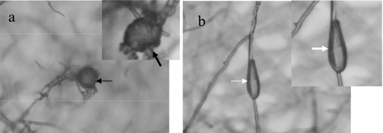
Figure 2 (a-b). Formation of trap (black arrow) by the tested fungal isolate of Duddingtonia flagrans (CG768) and third stage larvae of Ancylostoma spp. preyed (white arrow).
Discussion
Sand of public areas represent a potential source of transmission to human and animal population because it is frequently infested by zoonotic parasite eggs such as Ancylostoma spp. in the faeces of dogs that are commonly infected17,18. The predation and population reduction of this nematode in the beach sand, as observed in this work, justify this approach as an important tool to decontaminate the environment.
The are some reports about the of success use of nematophagous fungi in the control of potential zoonotic geohelminths, especially in an experimental model with
dogs9,19. On the other hand, most studies are carried out under laboratorial conditions
and even partially natural conditions, since it is observed the viability of tested
nematophagous fungi after passage through the gastrointestinal tract. Carvalho et al.9
demonstrated that the fungus D. flagrans (AC001) was able to withstand the passage
through the gastrointestinal tract of dogs and was viable in predation on Ancylostoma
spp L3 under laboratory conditions. In that work, using another approach, those authors
have observed that the administration of 0.5 g/10 kg of mycelial mass containing the
fungus D. flagrans (isolate AC001) was effective in reducing egg counts per gram of
faeces and in the recovery of A. caninum larvae in treated animals compared to the
one isolate of nematophagous fungus was able to pass through the gastrointestinal tract
of dogs and show its predation on Toxocara canis eggs an in vitro assay.
Nematophagous fungi should be applied in the environment (soil or sand) together with a fungal growth substrate, in order to promote their growth and therefore promote their establishment. However, until now there are no studies about the biological control of hookworm in beach sand, serving as a constant source of contamination. In the constitution of the sand used in this work were shown only minerals, without evidence of organic matter that could help in the proliferation of the fungus. Therefore, the main nutrients for the fungus were the substrate (corn meal) and the nematode.
Moreover, there is a lack of studies on the use of different concentrations of
chlamydospores.In recent work, Maciel et al.20 tried to determine the best dose of the
fungus D. flagrans comparing different concentrations of the fungus, but no differences
were observed between the concentrations of 10,000 to 25,000 chlamydospores per gram of soil in microcosm setups, but the control of the nematode ranged from 72,0% to 79,4% at the concentration of 10,000 chlamydospores/g of soil.
In the present work, differences could not be observed (p <0.01) in the concentrations from 15000 to 25000 of chlamydospores/g of soil, the highest nematode
L3 predation. However, these differences are interesting from the biological point of
view and even when dealing with sand and not soil as described above. Furthermore, the authors of the present study suggest that possibly the environment of "soil" may be rich in organic matter and can directly influence the development of fungal isolates. Thus, further studies using the material "beach sand" should be conducted in order to
observe the predatory activity of the fungus D. flagrans.
References
1. Silva JP, Marzochi MCA, Santos ECL. Avaliação da Contaminação Experimental
de Areias de Praias por Enteroparasitas. Pesquisa de Ovos de Helmintos. Cad Saude
Publica 1991;7:90-99.
2. Soulsby EJL. Helminths, arthropods and protozoa of domesticated animals.
London: Baillière Tindall; 1982.
3. Gomes JF, Hoshino-Shimizu S, Dias LC, Araujo AJ, Castilho VL, Neves FA.
Evaluation of a novel kit (TF-test) for the diagnosis of intestinal parasitic infections. J Clin Lab Anal 2004;18:132-138.
4. Santos NM, Silva VMG, The TS, Santos AB, Souza TP. Contaminação das praias
por parasitos caninos de importância zoonótica na orla da parte alta da cidade de
Salvador-Ba. R Ci Med Biol 2006;5:40-47.
5. Maikai BV, Umoh JU, Ajanusi OJ, Ajogi I. Public health implications of soil
contaminated with helminth eggs in the metropolis of Kaduna, Nigeria. J
Helminthol 2008;82:113-118.
6. Guimarães AM, Alves EGLA, Rezende GF, Rodrigues MC. Ovos de Toxocara sp. e
larvas de Ancylostoma sp. em praça pública de Lavras, MG. Rev Saude Publica
2005;39:293-295.
7. Silva PF, Cavalcanti IMD, Irmão JI, Rocha FJS. Common beach sand contamination
due to enteroparasites on the southern coast of Pernambuco State, Brazil. Rev Inst
Med Trop S Paulo 2009; 51:217- 218.
8. Kopp SR, Kotze AC, McCarthy JS, Coleman GT. High-level pyrantel resistance in
the hookworm Ancylostoma caninum. Vet Parasitol 2007;143:299-304.
9. Carvalho RO, Araújo JV, Braga FR, Araujo JM, Silva AR, Tavela AO. Predatory
activity of nematophagous fungi on infective larvae of Ancylostoma sp.: evaluation
in vitro and after passing through the gastrointestinal tract of dogs. J Helminthol
2009;83:231–236.
10. Larsen M. Biological control of helminths. Int J Parasitol 1999;29:139– 146.
11. Araújo JV, Assis RCL, Campos AK, Mota MA. Atividade in vitro dos fungos
nematófagos dos gêneros Arthrobotrys, Duddingtonia e Monacrosporium sobre nematoides trichostrongilídeos (Nematoda: Trichostrongyloidea) parasitos
gastrintestinais de bovinos. Rev Bras Parasitol Vet 2004;13:65-71.
Proteolitic action of the crude extract Duddingtonia flagrans on Cyathostomin
(Nematoda:Cyathostominae) in coprocultures. Rev Bras Parasitol Vet 2012;21:1-4.
13. Tavela AO, Araújo JV, Braga FR, Araujo JM, Queiroz LM, Silveira WF, Borges, LA. In vitro association of nematophagous fungi Duddingtonia flagrans (AC001), Monacrosporium thaumasium (NF34) and Pochonia chlamydosporia (VC1) to
control horse cyathostomin (Nematoda: Strongylidae). Biocontrol Sci Technol
2012;22:607-610.
14. Smith D, Onions AHS. The Preservation and Maintenance of Living Fungi. Surrey:
Commonwealth Mycological Institute; 1983.
15. Ruiz HA. Incremento da exatidão da análise granulométrica do solo por meio da
coleta da suspensão (silte + argila). R Bras Ci Solo 2005;29:297-300.
16. Ayres M, Ayres JRM, Ayres DL, Santos AS. Aplicações estatísticas nas áreas de
ciências Biológicas. Belém: Sociedade Civil mamirauá; 2003.
17. Santarém VA, Giuffrida R, Zanin GA. Larva migrans cutânea: ocorrência de casos
humanos e identificação de larvas de Ancylostoma spp. em parque público do
município de Tacibá, São Paulo. Rev Soc Bras Med Trop 2004;37:179-181.
18. Blazius RD, Emerick S, Prophiro JS, Roosevelt P, Romão T, Silva OS. Ocorrência de protozoários e helmintos em amostras de fezes de cães errantes da cidade de
Itapema, Santa Catarina. Rev Soc Bras Med Trop 2005;38:73-74.
19. Araujo JM, Araújo JV, Braga FR, Araújo DM, Ferreira SR, Soares FEF, et al.
Survival of Pochonia chlamydosporia in the gastrointestinal tract of experimentally
treated dogs. Res Vet Sci 2012;12:803-806.
20. Maciel AS, Freitas LG, Campos AK, Lopes EA, Araújo JV. The biological control of Ancylostoma spp. dog infective larvae by Duddingtonia flagrans in a soil
Capítulo 3
Effect of the fungus Duddingtonia flagrans on infective larvae Ancylostoma spp. of
Abstract
Nematophagous fungi have performed their role in the capture and destruction of geohelminths. The objective of this study was to evaluate the effect of nematophagous
fungus Duddingtonia flagrans on infective larvae of Ancylostoma spp. of dogs in
semi-natural conditions. The experiment consisted of two treatments, one with fungus at 15,000 chlamydospores/g sand and other without the fungus, both in 100 g of sand,
infested with 2,000 L3 of Ancylostoma spp. Sand samples from each treatment were
collected right after the incorporation of chlamydospores into the sand and at the end of
assay for determination of fungus population. For isolation of D. flagrans was applied
the technique of serial dilution and plating on the semi-selective culture medium. The
fungal isolate (CG768) showed predatory activity on the Ancylotoma spp. L3 and
reduced in of 47.9% the number of nematodes recovered when compared to the control group (p <0.05). The biological control with nematophagous fungi may become a tool in combating geohelminths of dogs, working directly in the free life stages of Ancylostoma spp.
Keywords: Biological control, Nematophagous fungi, Duddingtonia flagrans, Ancylostoma spp., sand.
Introduction
The nematophagous fungi are widely distributed geographically around the world, inhabiting all types of soil, especially those rich in organic matter (NORDBRING-HERTZ et al., 2006; SANYAL et al., 2008). These organisms are nutritionally versatile and their good saprophytic capacity allows them to survive in the soil for long periods in the absence of nematodes (GRAY, 1987). Most species of nematophagous fungi is predatory (LARSEN, 1999) and is characterized by developing an extensive system of vegetative hyphae, along which morphological differentiation occurs in structures called "traps" that capture and retain living nematodes to nourish themselves of nematode internal contents (BARRON, 1977). This change from a saprophytic phase to a parasitic phase with morphological differentiation in traps and use of nematodes as food, influenced by biotic and abiotic factors, provides a nutritional advantage for these microorganisms in soil (NORDBRING-HERTZ et al., 2006).
conduct part of their life cycle in the soil. In this context, the Duddingtonia flagrans
species has been tested on various conditions (natural and laboratory) (PAZ-SILVA et al., 2011), however, this is the first report of the study on sand.
The survival of these parasites is influenced not only by physical and chemical factors such as temperature, pH, moisture and aeration (CARVALHO et al., 2009; BRAGA et al., 2011; ARAUJO et al., 2012). In another context, the survival of a nematode in the external environment may be influenced by the presence or absence of known agents as biocontrollers and in this sense, stand out particularly: bacteria, fungi, nematodes, insects, mites and protozoa (GRONVOLD et al., 1996; STROMBERG, 1997; KERRY, 1987). However, the main biocontrollers of nematodes are the nematophagous fungi (NORDBRING-HERTZ et al., 1988).
Thus, considering this fact, parasites of the genus Ancylostoma have part of their
life cycle in the soil (SOULSBY, 1982) and these fungi could become part of an effective and complementary alternative in its control (CARVALHO et al., 2010; LARSEN, 2000).
The objective of this study was to evaluate the effect of nematophagous fungus Duddingtonia flagrans on infective larvae of Ancylostoma spp. of dogs in semi-natural conditions.
Materials and Methods Fungal inoculum
In present assay, it was used the isolate CG768 of the nematophagous fungus Duddingtonia flagrans obtained from samples of Brazilian soil and of animal feces were used in this experiment. The isolate was stored on integral rice grains inside 5 mL
BD Vacutainer® glass tubes (Becton Dickinson, Brazil) containing blue silica gel, based
on the conservation technique described by Smith and Onions (1983). Production of chlamydospores
The chlamydospore production was performed in 2.5 L plastic bags with biological filter. Milled maize was used as substrate. Bags containing 300 g of substrate and 110 ml of distilled water were closed and sterilized for 30 minutes at 121 °C. The sterilized substrate in each bag was inoculated with six mycelium discs of about 5 mm
containing agar-water medium at 2% during 7 days at 25 °C. The bags were then closed
to allow growth of D. flagrans on the substrate for 15 days in the dark at 25 ° C. The
substrate was agitated every five days until the end of the incubation period to ensure homogeneous mycelial growth. After the incubation period, six samples of 1 g of colonized substrate of each bag were transferred to an Erlenmeyer flask containing 10 mL of distilled water and 0.2% (v/v) dispersing polysorbate (Tween ® 80) and shaken
by two minutes for dispersion of chlamydospores. Then two aliquots of 10μL were
placed in a Neubauer chamber to estimate the number of chlamydospores per gram of substrate.
Ancylostoma spp. L3
The Ancylostoma spp. L3 was obtained from fresh feces of naturally infected stray dogs, by vermiculite coproculture kept for 10 days at 26 ºC in a BOD. After this period, active larvae were harvested from culture using the Baermann funnel technique
The L3 suspension was homogenized and 3 aliquots of 10 µL were placed on a
microscope glass slide. Each aliquot was covered with a glass coverslip after adding 10
µL of lugol's solution to kill L3. Thus, they were counted and identified under a light
microscope with the magnification of 40X, allowing the estimation of the total number
of larvae in the suspension. The motility of Ancylostoma spp. was checked by
microscopical examination, before they were used in the experiment. Experimental procedure
Sand samples were used (coarse sand: 76%; fine sand: 22%; silt 0%; clay: 2%;
sodium: 342 mg/dm3; phosphorus: 34,15 mg/dm3; potassium: 20 mg/dm3; calcium: 0,46
cmol/ dm3; magnesium: 0,32 cmol/ dm3; aluminum: 0 cmol/ dm3; ph: 8,2). These
samples were derived from the resort city of Guarapari, state of Espírito Santo and were submitted regarding the particle size analysis and chemical analysis by methodology of Ruiz (2005). Samples were collected at a depth of 0 to 20 cm and stored in plastic bags. The minimum and maximum temperatures were measured daily by a mercury column thermometer.
group was formed, containing infested sand with Ancylostoma spp., without the fungus.
Next, the opened pots were placed in black polypropylene pots of 350 cm ³ and kept
inside a greenhouse protected from direct sunlight for a period of 15 days.The sand of
the pots was daily irrigated to prevent dehydration of the larvae and to allow germination of chlamydospores. At the end of the incubation period the larvae were recovered from the sand by the Baermann’s funnel technique (Baermann, 1917) and counting was performed as described above.
Sand samples from each experimental group were collected right after the incorporation of chlamydospores to sand and the end of assay for determination of
population of the fungus. For isolation of D. flagrans, 1 g samples of sand were put into
test tubes containing 9 ml of sterile water and shaken for 3 minutes at stirrer.
Subsequently, the serial dilution technique was employed to 1x10⁻², and an aliquot of 1
mL was spread on Petri dishes containing semi-selective culture medium, with the aid of a Drigalsky strap as Gaspard et al. (1990), with three replicates per treatment. The plates were stored at 25 ºC until the growth of colony forming units (CFU).
The colonies were then pricked out to medium plates containing potato dextrose agar (PDA) and incubated at 25 ºC. After seven days, the fungal colonies were identified according to morphological characteristics of the fungus.
Statistical analysis
The data obtained in the test were interpreted by analysis of variance at a significance level of 1% probability (AYRES et al., 2003). The destruction efficiency of
L3 compared to control was assessed by Tukey's test at 1% probability. Thereafter, the
percent reduction from the average of L3 was calculated according to the following
formula:
% Reduction = (Average L3 recovered from control – Average L3 recovered from treatment) x 100
Average L3 recovered from control
Results
In the presente work, the fungal isolate used (CG768) showed predatory activity
on the Ancylotoma spp. L3 and it was observed the percentage reduction of 47.9% in the
The isolate evaluated has established itself in the sand throughout the trial period (Table 1). This fact was confirmed by plating sand in semi-selective culture medium
and by observation of colony forming units (CFU) of D. flagrans. Even after 7 days of
plating were observed vegetative and resistance structures, proving that the fungus was able to establish itself in the sand from the substrate used (Figure 1).
Table 1. Population of the fungus Duddingtonia flagrans after sand infestation with 15.000 chlamydospores/g of sand and after 15 days. Averages of three replications.
Log CFU/g of sand Experimental group
Test setup After 15 days
Treated 5,38 3,95
Control 0 0
Figure 1. Chlamydospores and conidia of the fungus Duddingtonia flagrans observed after 7 days after plating in semi-selective medium. (a) Chlamydospore in germination phase (black arrow) and (b) conidia (white arrow).
Discussion
The results of this study revealed that the fungus D. flagrans was able to survive
in the sand even in non-favorable conditions (absence of organic matter). The
incorporation of the fungus in the sand along with milled maize was crucial for its establishment and nematophagous activity. This is due to the fact of nematophagous fungi are saprophytic and because they feed on other sources of nutrients in the absence of nematodes in order to ensure their survival in the environment (NORDBRING-HERTZ, 1988)
Environmental temperature directly affects the growth and production of traps by the fungus. According Gronvold et al. (1996) and Morgan et al. (1997), the optimal
growth temperature for D. flagrans is between 25-33 ºC. In present assay, it was
observed that the average temperature was 31ºC. These results are in agreement with previous studies mentioned above.
Regarding the results in respect of the fungus population tested (CG768) at the end of the test, it was observed that after 15 days it was still viable. Also according to Dallemole-Giaretta (2008), the population of nematophagous fungi in soil by means of CFU counts of isolates tested, in this specific case CG768, strongly suggests the establishment of the fungus on soil. Thus, in the present work, even using a poor soil in organic matter, certain types of nematophagous fungi can come to settle. In this sense, the authors call attention to the abundant production of chlamydospores (resistant structures) that are peculiar to their nature (ARAÚJO et al., 2004).
On the other hand, the authors agree that the use of certain sources of N: C is
important for the proper establishment of the fungus D. flagrans in poor soil. In this
context, Braga et al. (2011) reported that the species D. flagrans might require a lot of
sources of N: C, which on the one hand, suggests that this fungus has not a nutritional demanding character, being this an interesting premise from an economic point of view.
Regarding the use of Ancylostoma spp. L3, the authors suggest that more and
more bioassays involving potentially zoonotic nematodes may become an important tool of control in the future, since the nematophagous fungi are natural organisms and antagonists of these parasites in the soil (ARAUJO et al., 2012; CIARMELA et al., 2008). On the other hand, no one knows the real interaction of predatory
nematophagous fungi in a soil poor in organic matter on zoonotic nematode L3, and this
is the first report.
Accordingly Soulsby (1982) and Bowman (2009) the survival of Ancylostoma