Sporozoite Conversion to Liver Stages in the Malaria
Parasite
Katja Mu¨ller, Kai Matuschewski, Olivier Silvie*¤
Max Planck Institute for Infection Biology, Parasitology Unit, Berlin, Germany
Abstract
Malaria is a vector-borne infectious disease caused by unicellular, obligate intracellular parasites of the genusPlasmodium. During host switch the malaria parasite employs specialized latent stages that colonize the new host environment. Previous work has established that gametocytes, sexually differentiated stages that are taken up by the mosquito vector, control expression of genes required for mosquito colonization by translational repression. Sexual parasite development is controlled by a DEAD-box RNA helicase of the DDX6 family, termed DOZI. Latency of sporozoites, the transmission stage injected during an infectious blood meal, is controlled by the eIF2alpha kinase IK2, a general inhibitor of protein synthesis. Whether RNA-binding proteins participate in translational regulation in sporozoites remains to be studied. Here, we investigated the roles of two RNA-binding proteins of the Puf-family,PlasmodiumPuf1 and Puf2, during sporozoite stage conversion. Our data reveal that, in the rodent malaria parasiteP. berghei,Puf2participates in the regulation of IK2and inhibits premature sporozoite transformation. Inside mosquito salivary glandspuf2(-)sporozoites transform over time to round forms resembling early intra-hepatic stages. As a result, mutant parasites display strong defects in initiating a malaria infection. In contrast, Puf1is dispensablein vivo throughout the entirePlasmodium life cycle. Our findings support the notion of a central role forPuf2in parasite latency during switch between the insect and mammalian hosts.
Citation:Mu¨ller K, Matuschewski K, Silvie O (2011) The Puf-Family RNA-Binding Protein Puf2 Controls Sporozoite Conversion to Liver Stages in the Malaria Parasite. PLoS ONE 6(5): e19860. doi:10.1371/journal.pone.0019860
Editor:Anne Charlotte Gruner, Agency for Science, Technology and Research (A*STAR), Singapore ReceivedFebruary 3, 2011;AcceptedApril 6, 2011;PublishedMay 18, 2011
Copyright:ß2011 Mu¨ller et al. This is an open-access article distributed under the terms of the Creative Commons Attribution License, which permits unrestricted use, distribution, and reproduction in any medium, provided the original author and source are credited.
Funding:This work was supported by the Max Planck Society, and in part by grants from the European Commission (EviMalaR,#34), the Joachim Siebeneicher Foundation and the Chica and Heinz Schaller Foundation. OS was a recipient of a Marie Curie Intra-European fellowship and an EMBO Long-Term Fellowship. The funders had no role in study design, data collection and analysis, decision to publish, or preparation of the manuscript.
Competing Interests:The authors have declared that no competing interests exist.
* E-mail: olivier.silvie@inserm.fr
¤ Current address: INSERM UMR_S 945 "Immunity and infection", Centre Hospitalier Universitaire Pitie´-Salpeˆtrie`re, Faculte´ de Me´decine et Universite´ Pierre et Marie Curie, Paris, France
Introduction
Plasmodiumparasites, the causative agents of malaria, are tran-smitted by femaleAnophelesmosquitoes. During the probing phase prior to the blood meal, sporozoites are injected into the skin of the mammalian host [1]. The motile sporozoites actively migrate in the skin, enter the peripheral blood circulation, and then rapidly reach the liver. Sporozoites invade hepatocytes by forming a parasitophorous vacuole (PV) [2], where they transform into replicative exo-erythrocytic forms (EEFs). After intense multipli-cation during 2–6 days, depending on the Plasmodium species, mature EEFs release thousands of merozoites, which invade erythrocytes and initiate the pathogenic blood stage cycle [3].
Plasmodiumsporozoites are formed inside oocysts in the mosquito midgut, but become fully infective only after colonization of the insect salivary glands. This maturation process is associated with the up-regulation of a specific subset of genes, referred to as Up-regulated in Infective Sporozoites (UIS) genes [4]. Regulation of gene expression inPlasmodiumremains poorly understood. Genome sequencing data initially revealed a paucity of specific transcription factors in Plasmodium [5]. Recently however, a family of genes related to the plant Apetala-2 (AP2) transcription factors has been identified inPlasmodiumand related apicomplexan parasites [6,7],
and proposed to play a central role during life cycle progression. Molecular genetic studies have demonstrated vital roles of two stage-specific AP2 factors inPlasmodium berghei, a rodent malaria parasite widely used as a model [8,9]. One of these factors, the AP2-Sp transcription factor, is required during sporozoite differentiation and binds to a specific DNA sequence found in the promoter region of many genes expressed in sporozoites, including, but not restricted to,UISgenes [8]. Intriguingly, genes containing AP2-Sp binding sites are associated with a wide range of biological processes, such as sporozoite formation, host cell invasion or liver stage development. This observation strongly suggests that additional mechanisms participate in the fine-tuning of gene expression during sporozoite development and stage conversion. Another factor, called SLARP or SAP1, controls the expression of a subset of genes in sporozoites, and plays a critical role during intrahepatic development of the parasite [10,11]. It is still unclear whether SLARP/SAP1 acts on a transcriptional or a post-transcriptional level. The cellular localization of SLARP/ SAP1 remains controversial [10,11], and the absence of any domain known to bind nucleic acids suggests an indirect role.
phosphorylates the translation initiation factor eIF2alpha and down-regulates protein synthesis [12,13].P. bergheilackingUIS1/IK2display a partial loss of infectivity associated with premature transformation of sporozoites in the mosquito salivary glands [12]. The contribution of RNA-binding prxoteins in translational regulation has not been studied in sporozoites yet, but has been well characterized in Plasmodium sexual stages. In female gametocytes, many transcripts encoding ookinete proteins are translationally repressed by a DEAD-box RNA helicase called DOZI, which binds to the 39untranslated region (UTR) of target mRNAs such as P28 and blocks their translation until occurrence of gamete fertilization and differentiation into a zygote and ookinete [14,15]. Whether DOZI plays a role in sporozoites is not known, but other RNA-binding proteins may participate in translational regulation in sporozoites, including members of the Puf-family.
Puf proteins are evolutionary conserved in eukaryotes and are characterized by the presence of a RNA-binding Puf domain, named after the Drosophila melanogaster protein Pumilio and the Caenorhabditis elegans protein fem-3 binding factor (FBF), and consisting of eight imperfect repeats of 36 amino acids (PFAM: PF00806) [16,17]. Puf proteins typically bind to the 39 UTR of target mRNAs and repress their translation or induce their degradation (reviewed in [18] and [19]).Plasmodiumparasites possess two genes encoding proteins with Puf domains,Puf1andPuf2[20]. In P. falciparum, both Puf1(PFE0935c) and Puf2 (PFD0825c) are differentially expressed in gametocytes [20,21]. Targeted gene disruption in P. falciparum recently revealed a role of PfPuf2 in repressing gametocytogenesis and male gametocyte differentiation in the human malaria parasite [22]. Whether the Puf2 protein plays additional, perhaps vital, roles in subsequent life cycle stages remains to be shown. Interestingly, microarray data indicate thatPuf2 is most highly expressed in P. falciparumsporozoites [23], and inP. berghei, expression of both Puf1 (PBANKA_123350) and Puf2 (PBANKA_071920) has been reported in sporozoites, wherePuf1 was initially identified asUIS9[4,24]. In this study, we used a reverse genetic approach to investigate the roles of Puf1 and Puf2 inP. berghei, with the aim to identify potential mRNA binding proteins that play critical roles in sporozoite stage conversion.
Results
Targeted gene deletion ofP. berghei Puf1andPuf2
We first assessed the expression ofPuf1andPuf2duringP. berghei development in the insect vector, in comparison to DOZI and UIS1/IK2, using quantitative RT-PCR (Figure 1). Similarly to UIS1/IK2[12], we found thatPuf1andPuf2are upregulated inP. bergheisalivary gland sporozoites (Figure 1). This was expected for Puf1, which was initially described asUIS9 [4,24]. Furthermore, Puf1was also upregulated in gametocytes and ookinetes, similarly toIK2andDOZI. In good agreement with published microarray data [24], only low levels of DOZImRNA were detected in P. bergheisporozoites (Figure 1). In contrast toPuf1andPuf2,DOZI steady state mRNA levels were down-regulated in infectious salivary gland-associated sporozoites resulting in,100 fold lower levels in the latent transmission stage. Together, the expression profiling indicated that both Puf members could play a role in sporozoite stage conversion, as has been described previously for the eIF2alpha kinaseUIS1/IK2[12].
In order to investigate the functional importance ofPuf1/UIS9 and Puf2 in P. berghei, we generated loss-of-function mutants (Figure 2). We used a replacement strategy to disrupt the endogenousPuf1(Figure 2A) orPuf2(Figure 2B) gene copy by double crossover homologous recombination [25]. Targeting constructs containing 59 and 39 fragments of either Puf1orPuf2
flanking a pyrimethamine-resistance cassette were used to transfect P. bergheiparasites that constitutively express GFP (ANKA cl507) [26]. Recombinant parasites were selected with pyrimethamine in the mouse drinking water, and cloned by limiting dilutions. For both genes we were successful in generating clonal knockout parasite populations, as demonstrated by PCR and Southern blot analysis of genomic DNA (Figures 2C–F). For Puf2 we also generated a second independent knockout clone, which was phenotypically identical to the first puf2(-) clonal parasite line (unpublished data). This indicates thatPuf1andPuf2do not play any vital role during P. berghei erythrocytic stages, in good agreement with successful generation ofPfpuf2(-)parasites [22].
puf1(-) andpuf2(-) parasites produce gametocytes that develop to sporozoites in mosquitoes
puf1(-) and puf2(-) parasites were indistinguishable from WT parasites in development and growth of asexual blood stages and produced gametocytes. BecausePfPuf2 has been shown to control gametocytogenesis inP. falciparum[22], we analyzed in more detail the sexual development ofP. berghei puf2(-) parasites. After injection of 107 infected erythrocytes intravenously into groups of five C57BL/6 mice, parasitemia at day 4 were similar in mice infected with WT or puf2(-) (Figure 3A). However, the proportion of gametocytes among all parasite stages was significantly higher in puf2(-)than in WT parasites (Figure 3B). We then examined the ability of mature male gametocytes to exflagellate in puf2(-) parasites. The number of exflagellation centers in mouse blood was significantly higher forpuf2(-) parasites than for WT parasites (Figure 3C), suggesting that male gametocytes contribute to the increased gametocytogenesis inPbpuf2(-) parasites, in full support of the data reported forP. falciparum Puf2-deficient parasites [22]. After transmission toAnopheles stephensimosquitoes, bothpuf1(-) and puf2(-) parasite lines produced oocysts and high numbers of
Figure 1.Puf1andPuf2 are upregulated inP. berghei sporo-zoites.Shown is an expression profiling of selected transcripts of RNA regulatory proteins, the DDX6-family DEAD-box helicaseDOZI[15], the Puf proteinsPuf1andPuf2[20], and of the eIF2alpha kinaseUIS1/IK2
that controls sporozoite latency [12].P. bergheipurified gametocytes, ookinetes, oocysts and salivary gland sporozoites were analyzed by RT-qPCR using primers specific forDOZI,UIS1/IK2,Puf1andPuf2. Expression data from two independent experiments are shown and were normalized to the level ofGFPtranscripts, which are expressed under the control of theEF1alphapromoter [26].
doi:10.1371/journal.pone.0019860.g001
sporozoites (Table 1). The number of puf2(-) oocysts was significantly higher than for WT, consistent with the higher gametocyte rates. Intriguingly, we found lower numbers of oocysts and salivary gland sporozoites in puf1(-)-infected mosquitoes, as compared to WT parasites (Table 1). Although the differences were not statistically significant, we cannot exclude an effect ofpuf1 depletion on oocyst development and sporogony.
Liver infection is impaired inPuf2-deficient parasites We then analysed the infectivity ofpuf1(-) andpuf2(-) sporozoites to susceptible mice. C57BL/6 mice were injected intravenously with 1,000 WT,puf1(-) orpuf2(-)P. bergheisporozoites, or exposed to the bites of 10 infected mosquitoes, the natural transmission route (Table 1). Emergence of erythrocytic stages, resulting from complete liver stage development, was monitored by microscopic examination of daily blood smears. With both inoculation routes, all mice injected withpuf1(-) sporozoites developed a parasitemia, with no delay as compared to WT parasites (Table 1). In contrast, only a fraction of the mice injected withpuf2(-) sporozoites developed a parasitemia, with a two-day delay as compared to WT, indicative of at least 100-fold reduction of infectivity (Table 1). Moreover,puf2(-) sporozoites isolated late after mosquito infection (at day 25) were not capable of inducing a blood stage infection in mice.
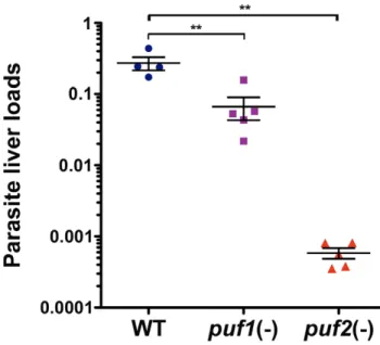
We next injected C57BL/6 mice intravenously with WT,puf1(-) orpuf2(-) sporozoites isolated on day 18 from mosquito salivary glands. Forty-two hours after infection, livers were removed and the parasite loads were quantified by RT-qPCR. As shown in Figure 4, thepuf2(-) liver loads were extremely reduced (,500 fold) as compared to WT, confirming that infectivity of puf2(-) sporozoites to C57BL/6 mice is severely impaired. The reduction of parasite liver loads as measured by RT-qPCR is consistent with the delay or absence of parasitemia in mice injected withpuf2(-) sporozoites (Table 1), therefore we assume that the absence of Puf2did not interfere with 18S rRNA quantification. Interestingly, we also observed a significant, although less pronounced (,4 fold), reduction ofpuf1(-)parasite liver loads (Figure 4). Our findings demonstrate thatPbPuf2plays an importantin vivorole only in the pre-erythrocytic phase of the Plasmodium life cycle. In contrast, Puf1/UIS9 appears to be dispensable for parasite life cycle progression, at least under the conditions tested.
We also determined theIn vitroinfectivity ofpuf1(-)andpuf2(-) sporozoites isolated on day 22 from mosquito salivary glands, in cultured HepG2 hepatoma cells (Figure 5). Both puf1(-) and puf2(-)sporozoites entered hepatoma cells as efficiently as WT, as evidenced by similar numbers of infected cells at early time points (4–6 hours) (Figure 5A). While the number of EEFs at later time
Figure 2. Targeted gene deletion ofPuf1/UIS9andPuf2inP. berghei.(A–B) Replacement strategy to generate thepuf1(-)andpuf2(-)parasites.
P. berghei PUF1gene (A) consists of five exons encoding an 1183 amino-acid protein (PBANKA_123350), whereasPUF2(B) consists of four exons encoding a 477 amino-acid protein (PBANKA_071920). The PUF domains are shown in blue. For each gene, the wild-type (WT) genomic locus was targeted with a replacement plasmid containing 59and 39regions ofPUF1orPUF2and a positive selectable marker,Toxoplasma gondii dhfr/tsor humanDHFR, respectively. Upon a double crossover event, thePUF1orPUF2gene is replaced by the selectable marker. Replacement- and wild type-specific test primer combinations and expected PCR fragments (WT, 59integration and 39integration) are indicated by arrows and lines, respectively. Restriction sites, Southern probes and expected restriction fragments are also shown.S, SpeI;X, XhoI;A, AfeI;E, EcoRV. (C)Puf1replacement-specific PCR analysis. Confirmation of the predicted gene targeting is achieved by specific primer combinations (59and 39integration), which can only amplify a signal from the recombinant locus. A wild type-specific PCR reaction confirms the absence of residual wild-type parasites in the clonalpuf1(-)
population. (D) Southern blot analysis of genomic DNA isolated from WT,puf1(-)andpuf2(-)parasites, using digoxigenin-labelled probes specific for
Puf1. After digest withSpeI andXhoI, thePuf1probe hybridizes to a 8.3 or a 6.9 kb fragment in WT andpuf1(-)parasites, respectively. (E)Puf2
replacement-specific PCR analysis. Confirmation of the predicted gene targeting is achieved by specific primer combinations (59and 39integration), which can only amplify a signal from the recombinant locus. A wild type-specific PCR reaction confirms the absence of residual wild-type parasites in the clonalpuf2(-)population. (F) Southern blot analysis of genomic DNA isolated from WT,puf1(-)andpuf2(-)parasites, using digoxigenin-labelled probes specific forPuf2. After digest withAfeI andEcoRV, thePuf2probe hybridizes to a 8.4 kb fragment in WT and a 4.0 kb fragment inpuf2(-)
parasites.
doi:10.1371/journal.pone.0019860.g002
Table 1.Loss of infectivity ofpuf2(-)sporozoites in C57BL/6 mice.
Parasites
Number of oocysts/ mosquitoa (mean±SD)
Number of salivary gland sporozoites/mosquitoa
(mean±SD) Route of injectionb
Number of infected/ Number of injected
Prepatency period (days)c
WT 182 (6164) 31,600 (618,600) bites (d 21) 3/3 3
i.v. (d 18) 2/2 3
i.v. (d 21) 6/6 3.5
i.v. (d 25) 4/4 3
puf1(-) 137 (6100) 11,400 (64,700) bites (d 17) 3/3 3
i.v. (d 21) 6/6 3.5
puf2(-) 320 (6234)d 25,000 (
618,800) bites (d 21) 3/4 (5)
i.v. (d 18) 2/4 (5)
i.v. (d 25) 0/4 NA
aThe number of midgut oocysts and salivary gland sporozoites was determined at d10–14 and d18–25, respectively, after the infectious blood meal, from at least three independent feeding experiments.
bC57BL/6 mice were exposed to the bites of 10 infected mosquitoes or injected intravenously (i.v.) with 1,000 sporozoites, 18–25 days after mosquito infection. cThe prepatent period is defined as the number of days after sporozoite inoculation until detection of infected erythrocytes by microscopic blood smear examination.
Brackets indicate that not all animals became infected. NA, not applicable. dp
,0.05 in comparison to WT, as determined by Kruskal-Wallis followed by Dunn’s test. doi:10.1371/journal.pone.0019860.t001
points (24–48 hours) was similar in WT- and puf1(-)-infected cultures (Figure 5A), it was reduced in the case ofpuf2(-)parasites (Figure 5B). Whereas early after infection a vast majority (81%
63%; n = 122) of intracellular WT sporozoites expressed UIS4, a transmembrane protein that localizes to the membrane of the PV [27], only half of puf2(-) parasites were stained with UIS4 antibodies (53%69%; n = 127). This indicates that a substantial fraction ofpuf2(-)sporozoites failed to form and/or remodel the PV in vitro, which probably explains the reduced EEF numbers quantified at later time points. In addition, we cannot exclude a moderate impairment during liver stage development in puf2(-) parasites, as suggested by the reduction of EEF numbers observed between 24 and 48 hours post-infectionin vitro. Nevertheless, most puf2(-) sporozoites that formed a PV and expressed UIS4 were capable of developing into EEFs like WT and puf1(-) parasites (Figure 5C). Taken together, our data indicate thatPuf2plays a critical role during transmission of P. berghei sporozoites to the mammalian host, but is not required for liver stage development per se.
puf2(-)sporozoites transform prematurely in the mosquito In vivodata suggested that, over time,Puf2-knockout sporozoites rapidly loose infectivity in the mosquito (Table 1). To better characterize this phenomenon, we carefully analyzed puf2(-) sporozoite development in the mosquito (Figure 6). Strikingly, we observed that a major proportion ofpuf2(-)sporozoites showed signs of premature transformation, characterized by a bulb-like aspect or even complete rounding-up (Figure 6A). In WT parasites, transformation of sporozoites is typically observed at 37uC in culture medium, irrespective of the presence of host cells [28]. Inpuf2(-)-infected mosquitoes, however, the proportion of transformed sporozoites increased over time during the course of infection in the mosquitoes, which are kept at 20uC (Figure 6B). Quantification of partial and complete transformation in all three parasite populations revealed that at day 29 almost all puf2(-) Figure 3. Gametocytogenesis is increased inpuf2(-)parasites.
Groups of C57BL/6 mice (n = 5) were injected intravenously with 107 WT orpuf2(-)infected erythrocytes. Blood was collected from the mice 4 days later to determine the parasitemia (A), the proportion of gametocytes among parasites (B), and the number of exflagellation centers perml of blood (C). Results are expressed as mean+/2SEM.
**,p,0.01 (Mann-Whitney test). doi:10.1371/journal.pone.0019860.g003
Figure 4. Liver infection is severely impaired inpuf2(-)parasites. Parasite loads were determined by RT-qPCR analysis of mouse livers (n = 4 or 5 per group) harvested 42 hours after intravenous injection of 10,000 WT,puf1(-)orpuf2(-)sporozoites. Results are expressed as the relative expression ofPb18S normalized to mouse GAPDH. The means
sporozoites had transformed, whereas only a minor fraction of WT and puf1(-)sporozoites exhibited signs of premature transforma-tion (Figure 6B). Interestingly, we did not observe expression of the liver stage marker UIS4 or nuclear divisions, as seen in EEFs (Figure 5C), in the transformedpuf2(-)sporozoites (Figure 6A).
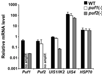
puf2(-)sporozoites have reduced levels ofPuf1andUIS1/ IK2mRNA
The phenotype ofpuf2(-)parasites is essentially identical to that of parasites that contain a targeted deletion of the kinaseUIS1/IK2[12]. Similarly topuf2(-)parasites,ik2(-) sporozoites transform prematurely in the mosquito salivary glands and have a decreased infectivityin vivo but notin vitro[12]. Therefore, we sought to test expression ofIK2in puf2(-) sporozoites, in comparison to WT and puf1(-) sporozoites, using RT-qPCR. As expected from gene deletion, noPuf1andPuf2 mRNA were detected inpuf1(-)andpuf2(-)sporozoites, respectively (Figure 7). Whereas expression of UIS1/IK2was not modified in
puf1(-)sporozoites, we observed a,14 fold reduction ofUIS1/IK2 mRNA inPuf2-deficient sporozoites as compared to WT (Figure 7). Additionally, we found a,17 fold reduction ofPuf1transcript levels in puf2(-) parasites. Conversely, Puf2 transcript levels were not affected in the absence ofPuf1(Figure 7). As controls,UIS4and HSP70mRNA levels were similar in the mutant and WT sporozoites. Altogether, these data indicate thatPuf2regulates a subset of genes in P. bergheisporozoites, includingPuf1and the kinaseUIS1/IK2. The latter probably explains, at least in part, why the phenotype ofpuf2(-) sporozoites recapitulates that ofIK2-knockout parasites.
Discussion
Plasmodiumsporozoites must persist and remain infectious within the salivary glands of the mosquito for many days until they are eventually transmitted to a mammalian host. Inside the warm-blooded host they need to quickly leave the site of deposition in
Figure 5.puf2(-)sporozoites are infectivein vitro.(A) HepG2 cells were infected with WT orpuf1(-)sporozoites and the numbers of infected cells were determined at 6, 24 and 48 h post-infection (p.i.). Results are expressed as the mean number of infected cells in triplicate wells+/2SD. (B) HepG2 cells were infected with WT orpuf2(-)sporozoites and the numbers of infected cells were determined at 4, 24 and 48 h post-infection (p.i.). Results are expressed as the mean number of infected cells in triplicate wells+/2SD. (C) Confocal microscopy analysis of HepG2 cells cultured for 5 and 24 hours post-infection (p.i.) with WT,puf1(-)orpuf2(-)sporozoites, using antibodies against UIS4 (red), CSP (5 h p.i., green) or HSP70 (24 h p.i., green). Nuclei were stained with DRAQ5 (blue). Bars, 10mm.
doi:10.1371/journal.pone.0019860.g005
order to travel to the liver, invade hepatocytes and differentiate into liver stages [29]. The transient developmental arrest of sporozoites inside mosquito salivary glands, termed latency [12], implies efficient control mechanisms to prevent premature
transformation before transmission and during transmigration before reaching a suitable host cell. In this study, we identified a factor controlling sporozoite latency inP. berghei,the RNA-binding protein Puf2. In the absence of Puf2, sporozoites transform
Figure 6. Premature transformation ofpuf2(-)sporozoites in the mosquito.(A) Fluorescence microscopy of WT andpuf2(-)sporozoites isolated from mosquito salivary glands 25 days after infection, and labelled with anti-UIS4 (red) and anti-CSP (green) antibodies. Nuclei were stained with DRAQ5 (blue). Bars, 10mm. (B) The proportion of non-transformed, partially transformed and fully transformed sporozoites was determined by
microscopic examination of sporozoites isolated from mosquito salivary glands 18, 21, 25 or 29 days after infection with WT,puf1(-)orpuf2(-)
parasites.
prematurely in the mosquito, resulting in a severe loss of infectivity.
Sporozoite conversion into liver stages requires initial remod-elling of the parasite pellicle, with disassembly of the inner membrane complex (IMC) and appearance of a bulb that progressively enlarges until the initially elongated sporozoite has transformed into a round form [28,30]. Previous work has shown that transformation of salivary gland sporozoites is induced at 37uC in culture medium, irrespective of the presence of host cells [28]. It should be noted that differentiation into EEFs involves additional events, including expression of liver stage specific proteins, onset of nuclear divisions and parasite growth. None of these events are observed in axenic culture conditions [28], where instead extracellular sporozoites die rapidly after transformation [31].
We show thatpuf2(-)sporozoites transform prematurely in the mosquito salivary glands, as evidenced by the characteristic bulb-like structures and rounding-up of the parasites. Premature transformation probably impairs the sporozoite functions that depend on IMC integrity, such as parasite motility, cell traversal and invasion, thus resulting in a loss of infectivity. In the absence ofPuf2, the proportion of transformed salivary gland sporozoites increases over time, which correlates with a progressive loss of infectivity to mice. Interestingly, although mostpuf2(-)sporozoites eventually transform into completely round forms, these forms do not progress to EEF differentiation, as shown by minimal expression of the liver stage marker UIS4 and absence of nuclear division or growth. In contrast, normal differentiation of puf2(-) parasites is observed once sporozoites invade cultured hepatoma cells. Collectively, these data strongly suggest that Puf2 plays a major role in preventing premature remodelling of the sporozoites prior to liver infection, but is not required for EEF differentiation. The defects observed inpuf2(-)parasites are reminiscent of those described in IK2-knockout parasites [12]. Both puf2(-) and ik2(-) sporozoites transform prematurely in the mosquito and display greatly reduced infectivity to mice. However, both loss-of-function mutants are able to invade and differentiate into EEFs in cultured cellsin vitro,indicating that they do not play any essential role after
host cell infection. These observations, combined with a major down-regulation ofIK2expression in puf2(-)sporozoites, suggest that the phenotype ofPuf2-deficient parasites can be explained, to a large extent, byIK2depletion.
How IK2 prevents sporozoite transformation has yet to be determined. Phosphorylation of the alpha subunit of eIF2 by distinct kinases, such asPlasmodiumIK2, is a central mechanism in stress-induced translational regulation [32], including in protozo-ans. For example, the eIF2alpha kinase IK1 regulates responses to starvation stress in P. falciparum blood stages [13], and in Toxoplasma gondii, phosphorylation of eIF2alpha promotes survival of extracellular tachyzoites [33]. Our data corroborate the findings of Zhanget al. [12], which together suggest that a similar stress response operates in sporozoites to maintain them in a quiescent stage.
The founding member of the Puf family,Drosophila melanogaster Pumilio (DmPUM), regulates, amongst other functions, abdominal development in the fly via translational repression of the maternally inherited hunchback (hb) mRNA [34]. The Puf domain of DmPUM binds to a nanos response element (NRE) sequence located in the 39UTR ofhbmRNA. Biochemical data, such asin vitrobinding assays using recombinant Puf domains expressed in bacteria and heterologous in vivo studies using the yeast three-hybrid system, have demonstrated intrinsic binding activity of the P. falciparum PfPuf1 and PfPuf2 to the NRE sequence [20,21]. Signature RNA sequences that are recognized by the Puf domain vary between species and members of the Puf family, but typically contain a UGUR motif [35,36,37,38]. A large number of Plasmodium genes contain UGUR motifs in their 39 UTR, but their functional significance remains uncertain, especially in the context of the exceptional AT-richness of thePlasmodiumgenome. Therefore, endogenous targets of Plasmodium Puf proteins still remain elusive. The P. falciparum[22] and P. berghei(this study) mutants now constitute potential tools to identify Puf2 target genes in Plasmodium. In sporozoites, Puf2 regulates at least two other genes in addition toIK2and Puf1. Indeed, using RT-qPCR, we found a 4-fold reduction ofSpectandSpect2mRNA levels inpuf2(-) sporozoites, whereas actin andAMA1were not affected (unpub-lished data). Reduced expression ofSpectandSpect2genes, which are both essential for sporozoite cell traversal and migration to the liver [29,39,40], may also contribute to the loss of infectivity of puf2(-) parasitesin vivo.
Whereas Puf proteins typically modulate target mRNA expression by either promoting mRNA turnover or translational repression, they can also activate gene expression or control mRNA subcellular localization (reviewed in [18] and [19]). Our results are not compatible with a role of Puf2 in repressingIK2, because puf2(-) and ik2(-)share a similar phenotype. Puf2 may instead participate in stabilization ofIK2transcripts. Alternatively, depletion ofIK2mRNA inpuf2(-)could be an indirect effect due to activation of an upstream factor that regulatesIK2.
Whereas Drosophila encodes only DmPUM, many organisms, includingC. elegans, contain two or more genes encoding Puf proteins, which can fulfil partly redundant functions [41]. Therefore, presence of twoPufgenes in thePlasmodiumgenome might be explained by overlapping or distinct roles. However, our molecular genetics data clearly exclude a vital role for Puf1 under normal conditions throughout theP. bergheilife cycle.Puf1may be critical under specific conditions, similarly to the role of IK1 in P. falciparum during starvation-induced stress [13]. Puf2 might compensate for the absence of Puf1 inpuf1(-)parasites, but notvice versa. While Puf1 in principle might be able to functionally complement forPuf2function, depletion of Puf1 at the mRNA level precludes a hypothetical functional overlapin vivo.In this regard, it should be noted thatP. Figure 7.puf2(-)sporozoites have reduced levels ofPuf1and
UIS1/IK2mRNA.Shown is an expression profiling by RT-qPCR analysis ofPuf1,Puf2,UIS1/IK2,UIS4andHSP70mRNA levels in WT,puf1(-)and
puf2(-) P. bergheisporozoites. Expression data from three independent experiments are shown and were normalized to the level of GFP
transcripts, which are expressed under the control of the EF1alpha promoter [26]. no amplif., no amplification.
doi:10.1371/journal.pone.0019860.g007
bergheiPuf1 and Puf2 proteins are very different in size (1183 versus 477 amino acids, respectively), and share only little homology (,27% identity) restricted to the Puf domains.
P. falciparumparasites that lackPuf2show increased gametocyte rates and a bias towards male gametocytes [22]. These observations fit with the proposed unifying, and perhaps ancestral, role of Pufs in promoting cell proliferation and repressing differentiation [18]. Our findings that Puf2 inhibits sporozoite transformation further support the notion of a central role in suppression of cellular differentiation. Because of the published data fromP.falciparum puf2(-)parasites we did not investigate sexual development and differentiation of Pbpuf2(-) parasites in great detail other than to confirm the previous findings, i.e. an increase in gametocytogenesis inpuf2(-) parasites, partly due to increased male gametocyte differentiation. In the previous study, life cycle progression of Pfpuf2(-) parasites beyond gametocytogenesis was not analyzed [22]. Based on our results in the rodent malaria model system, we predict thatP. falciparumsporozoites lackingPuf2 will present a similar phenotype, that is premature sporozoite transformation in the mosquito and decreased infectivity. Therefore, our findings might be of considerable interest in the context of development of genetically attenuated parasites for vaccination [42].
In conclusion, we show here that Puf2 plays a major role in controlling sporozoite latency during host switch, possibly through the regulation of IK2. Our results also highlight the functional importance of post-transcriptional regulation of gene expression during transmission of the malaria parasite between hosts.
Materials and Methods
Ethics statement
All animal work was conducted in accordance with the German ‘Tierschutzgesetz in der Fassung vom 18. Mai 2006 (BGBl. I S. 1207)’, which implements the directive 86/609/EEC from the European Union and the European Convention for the protection of vertebrate animals used for experimental and other scientific purposes. The protocol was approved by the ethics committee of MPI-IB and the Berlin state authorities (LAGeSo Reg#G0469/09).
Experimental animals, parasites and cell lines
Female NMRI and C57BL/6 mice were from Charles River Laboratories. We usedP. bergheiANKA clone 507 parasites, which constitutively express the green fluorescent protein (GFP) [26]. HepG2 cells (ATCC HB-8065) were cultured as described [43].
P. berghei Puf1andPuf2gene deletion
A targeting construct forPuf1gene knockout was generated by inserting a 503-bp 59fragment and a 575-bp 39fragment on either side of aT. gondii DHFR/TSexpression cassette. A construct for Puf2 gene knockout was generated by inserting a 1001-bp 59 fragment and a 945-bp 39 fragment on either side of a human DHFRexpression cassette. Oligonucleotide sequences are indicat-ed in Table S1.P. bergheiparasites were transfected with linearized plasmids, using the NucleofectorH device (Amaxa GmbH) as described [44], injected intravenously into naı¨ve NMRI mice, and selected by pyrimethamine treatment in the drinking water. Clonal parasite populations were obtained by limiting dilution series and intravenous injection of one parasite in 10 recipient NMRI mice. Onepuf1(-)and twopuf2(-)clonal parasite lines were established and phenotypically characterized. Genotyping of WT and recombinant parasites was performed by PCR and Southern blot analysis of genomic DNA. Standard Southern blot analysis was
performed using the PCR DIG Probe synthesis kit and the DIG Luminescent Detection kit (Roche), according to the manufactur-er’s instructions.
Real time quantitative RT-PCR
Parasite total RNA was extracted with the RNeasy kit (Qiagen) and reverse transcribed with the RETROScript kit (Ambion). Real time PCR was performed on cDNA preparations as described [11], using the StepOnePlusTM Real-Time PCR System and Power SYBRH Green PCR Master Mix (Applied Biosystems), according to the manufacturer’s instructions. Expression data were normalized using the constitutively expressedGFPgene.
Immunofluorescence
Parasites were fixed in 4% paraformaldehyde (PFA) and permeabilized with 1% Triton X-100. Immunofluorescence was then carried out using previously described monoclonal antibodies againstP. bergheiCSP [45] and HSP70 [46]. Polyclonal anti-UIS4 antibodies were raised in rabbits immunized with two synthetic peptides from P. berghei UIS4 (CLFTDEHKDEINDNIV and CNNVYNMENKSFGPYI) (Eurogentec). DRAQ5 (Biostatus) was used to stain nuclei. Confocal pictures were obtained with a Leica TCS-SP microscope equipped with appropriate filters, and processed with Photoshop software (Adobe Inc.).
Parasite growth and sexual development
C57BL/6 mice (n = 5) were injected intravenously with 107 infected erythrocytes. Four days later, the parasitemia was determined by microscopic examination of Giemsa-stained blood smears. To analyse exflagellation of male gametocytes, five microliters of tail blood were diluted 1:25 in RPMI 1640 containing 10% FCS and 50mM xanthurenic acid, and adjusted to pH 8.0. After 12 min incubation at room temperature, exflagellation centers were counted in a Neubauer chamber. Mean parasitemia and gametocyte rates were compared using the Mann-Whitney non-parametric test. After parasite transmission to Anopheles stephensimosquitoes, the numbers of midgut oocysts and salivary gland sporozoites were determined at day 10–14 and day 18–25, respectively, and compared using the Kruskal-Wallis followed by Dunn’s multiple comparison tests.
Analysis of sporozoitein vivoinfectivity
C57BL/6 mice were injected intravenously with 1,000 WT or mutant sporozoites isolated from the salivary glands of infected mosquitoes, or exposed to 10 infected mosquito bites, as indicated. Infection was then monitored daily by examination of Giemsa-stained blood smears. The delay of patency was defined as the time before detection of at least one erythrocytic stage in the smears. For quantification of parasite liver loads by real time RT-PCR, C57BL/6 mice were infected intravenously with 10,000 sporozo-ites. At 42 hours post-infection, livers were harvested, total RNA was extracted with the RNeasy kit (Qiagen) and cDNA synthesized with the RETROScript kit (Ambion). Real-time PCR was then performed with the StepOnePlusTMReal-Time PCR System and Power SYBRH Green PCR Master Mix (Applied Biosystems), using primers specific forP. berghei18S rRNA and mouse GAPDH, as described [47]. Liver parasite loads were compared using the Mann-Whitney non-parametric test.
Supporting Information
Acknowledgments
We thank Dr. Georgina Montagna for critically reading the manuscript, Marion Hliscs for providing cDNA from P. berghei gametocytes and ookinetes, Dr. Taco Kooij for providing plasmids, and Dr. Sylvie Briquet for technical assistance.
Author Contributions
Conceived and designed the experiments: K. Mu¨ller K. Matuschewski OS. Performed the experiments: K. Mu¨ller OS. Analyzed the data: K. Mu¨ller K. Matuschewski OS. Wrote the paper: K. Matuschewski OS.
References
1. Amino R, Thiberge S, Martin B, Celli S, Shorte S, et al. (2006) Quantitative imaging ofPlasmodiumtransmission from mosquito to mammal. Nat Med 12: 220–224.
2. Frevert U, Engelmann S, Zougbede S, Stange J, Ng B, et al. (2005) Intravital observation ofPlasmodium bergheisporozoite infection of the liver. PLoS Biol 3: e192.
3. Silvie O, Mota MM, Matuschewski K, Prudencio M (2008) Interactions of the malaria parasite and its mammalian host. Curr Opin Microbiol 11: 352–359. 4. Matuschewski K, Ross J, Brown SM, Kaiser K, Nussenzweig V, et al. (2002)
Infectivity-associated changes in the transcriptional repertoire of the malaria parasite sporozoite stage. J Biol Chem 277: 41948–41953.
5. Coleman BI, Duraisingh MT (2008) Transcriptional control and gene silencing inPlasmodium falciparum. Cell Microbiol 10: 1935–1946.
6. Balaji S, Babu MM, Iyer LM, Aravind L (2005) Discovery of the principal specific transcription factors ofApicomplexaand their implication for the evolution of the AP2-integrase DNA binding domains. Nucleic Acids Res 33: 3994–4006. 7. De Silva EK, Gehrke AR, Olszewski K, Leon I, Chahal JS, et al. (2008) Specific DNA-binding by apicomplexan AP2 transcription factors. Proc Natl Acad Sci U S A 105: 8393–8398.
8. Yuda M, Iwanaga S, Shigenobu S, Kato T, Kaneko I (2010) Transcription factor AP2-Sp and its target genes in malarial sporozoites. Mol Microbiol 75: 854–863.
9. Yuda M, Iwanaga S, Shigenobu S, Mair GR, Janse CJ, et al. (2009) Identification of a transcription factor in the mosquito-invasive stage of malaria parasites. Mol Microbiol 71: 1402–1414.
10. Aly AS, Mikolajczak SA, Rivera HS, Camargo N, Jacobs-Lorena V, et al. (2008) Targeted deletion of SAP1 abolishes the expression of infectivity factors necessary for successful malaria parasite liver infection. Mol Microbiol 69: 152–163.
11. Silvie O, Goetz K, Matuschewski K (2008) A sporozoite asparagine-rich protein controls initiation of Plasmodium liver stage development. PLoS Pathog 4: e1000086.
12. Zhang M, Fennell C, Ranford-Cartwright L, Sakthivel R, Gueirard P, et al. (2010) ThePlasmodiumeukaryotic initiation factor-2alpha kinase IK2 controls the latency of sporozoites in the mosquito salivary glands. J Exp Med 207: 1465–1474.
13. Fennell C, Babbitt S, Russo I, Wilkes J, Ranford-Cartwright L, et al. (2009) PfeIK1, a eukaryotic initiation factor 2alpha kinase of the human malaria parasitePlasmodium falciparum, regulates stress-response to amino-acid starvation. Malar J 8: 99.
14. Braks JA, Mair GR, Franke-Fayard B, Janse CJ, Waters AP (2008) A conserved U-rich RNA region implicated in regulation of translation inPlasmodiumfemale gametocytes. Nucleic Acids Res 36: 1176–1186.
15. Mair GR, Braks JA, Garver LS, Wiegant JC, Hall N, et al. (2006) Regulation of sexual development of Plasmodium by translational repression. Science 313: 667–669.
16. Zamore PD, Williamson JR, Lehmann R (1997) The Pumilio protein binds RNA through a conserved domain that defines a new class of RNA-binding proteins. Rna 3: 1421–1433.
17. Zhang B, Gallegos M, Puoti A, Durkin E, Fields S, et al. (1997) A conserved RNA-binding protein that regulates sexual fates in theC. eleganshermaphrodite germ line. Nature 390: 477–484.
18. Wickens M, Bernstein DS, Kimble J, Parker R (2002) A PUF family portrait: 3’UTR regulation as a way of life. Trends Genet 18: 150–157.
19. Quenault T, Lithgow T, Traven A (2010) PUF proteins: repression, activation and mRNA localization. Trends Cell Biol.
20. Cui L, Fan Q, Li J (2002) The malaria parasitePlasmodium falciparumencodes members of the Puf RNA-binding protein family with conserved RNA binding activity. Nucleic Acids Res 30: 4607–4617.
21. Fan Q, Li J, Kariuki M, Cui L (2004) Characterization of PfPuf2, member of the Puf family RNA-binding proteins from the malaria parasitePlasmodium falciparum. DNA Cell Biol 23: 753–760.
22. Miao J, Li J, Fan Q, Li X, Li X, et al. (2010) The Puf-family RNA-binding protein PfPuf2 regulates sexual development and sex differentiation in the malaria parasitePlasmodium falciparum. J Cell Sci 123: 1039–1049.
23. Le Roch KG, Zhou Y, Blair PL, Grainger M, Moch JK, et al. (2003) Discovery of gene function by expression profiling of the malaria parasite life cycle. Science 301: 1503–1508.
24. Hall N, Karras M, Raine JD, Carlton JM, Kooij TW, et al. (2005) A comprehensive survey of thePlasmodiumlife cycle by genomic, transcriptomic, and proteomic analyses. Science 307: 82–86.
25. Menard R, Janse C (1997) Gene targeting in malaria parasites. Methods 13: 148–157.
26. Janse CJ, Franke-Fayard B, Mair GR, Ramesar J, Thiel C, et al. (2006) High efficiency transfection ofPlasmodium bergheifacilitates novel selection procedures. Mol Biochem Parasitol 145: 60–70.
27. Mueller AK, Camargo N, Kaiser K, Andorfer C, Frevert U, et al. (2005) Plasmodium liver stage developmental arrest by depletion of a protein at the parasite-host interface. Proc Natl Acad Sci U S A 102: 3022–3027. 28. Kaiser K, Camargo N, Kappe SH (2003) Transformation of sporozoites into
early exoerythrocytic malaria parasites does not require host cells. J Exp Med 197: 1045–1050.
29. Amino R, Giovannini D, Thiberge S, Gueirard P, Boisson B, et al. (2008) Host cell traversal is important for progression of the malaria parasite through the dermis to the liver. Cell Host Microbe 3: 88–96.
30. Gantt SM, Myung JM, Briones MR, Li WD, Corey EJ, et al. (1998) Proteasome inhibitors block development ofPlasmodiumspp. Antimicrob Agents Chemother 42: 2731–2738.
31. Hegge S, Kudryashev M, Barniol L, Frischknecht F (2010) Key factors regulatingPlasmodium bergheisporozoite survival and transformation revealed by an automated visual assay. Faseb J 24: 5003–5012.
32. Wek RC, Jiang HY, Anthony TG (2006) Coping with stress: eIF2 kinases and translational control. Biochem Soc Trans 34: 7–11.
33. Joyce BR, Queener SF, Wek RC, Sullivan WJ, Jr. (2010) Phosphorylation of eukaryotic initiation factor-2{alpha} promotes the extracellular survival of obligate intracellular parasiteToxoplasma gondii. Proc Natl Acad Sci U S A 107: 17200–17205.
34. Murata Y, Wharton RP (1995) Binding of pumilio to maternal hunchback mRNA is required for posterior patterning in Drosophilaembryos. Cell 80: 747–756.
35. Wang Y, Opperman L, Wickens M, Hall TM (2009) Structural basis for specific recognition of multiple mRNA targets by a PUF regulatory protein. Proc Natl Acad Sci U S A 106: 20186–20191.
36. Wang X, McLachlan J, Zamore PD, Hall TM (2002) Modular recognition of RNA by a human pumilio-homology domain. Cell 110: 501–512.
37. Gerber AP, Luschnig S, Krasnow MA, Brown PO, Herschlag D (2006) Genome-wide identification of mRNAs associated with the translational regulator PUMILIO inDrosophila melanogaster. Proc Natl Acad Sci U S A 103: 4487–4492.
38. Koh YY, Opperman L, Stumpf C, Mandan A, Keles S, et al. (2009) A singleC. elegansPUF protein binds RNA in multiple modes. Rna 15: 1090–1099. 39. Ishino T, Chinzei Y, Yuda M (2005) APlasmodiumsporozoite protein with a
membrane attack complex domain is required for breaching the liver sinusoidal cell layer prior to hepatocyte infection. Cell Microbiol 7: 199–208.
40. Ishino T, Yano K, Chinzei Y, Yuda M (2004) Cell-passage activity is required for the malarial parasite to cross the liver sinusoidal cell layer. PLoS Biol 2: E4. 41. Lamont LB, Crittenden SL, Bernstein D, Wickens M, Kimble J (2004) FBF-1 and FBF-2 regulate the size of the mitotic region in theC. elegansgermline. Dev Cell 7: 697–707.
42. Hafalla JC, Silvie O, Matuschewski K (2011) Cell biology and immunology of malaria. Immunol Rev 240: 297–316.
43. Silvie O, Franetich JF, Boucheix C, Rubinstein E, Mazier D (2007) Alternative invasion pathways forPlasmodium bergheisporozoites. Int J Parasitol 37: 173–182. 44. Janse CJ, Ramesar J, Waters AP (2006) High-efficiency transfection and drug selection of genetically transformed blood stages of the rodent malaria parasite Plasmodium berghei. Nat Protoc 1: 346–356.
45. Potocnjak P, Yoshida N, Nussenzweig RS, Nussenzweig V (1980) Monovalent fragments (Fab) of monoclonal antibodies to a sporozoite surface antigen (Pb44) protect mice against malarial infection. J Exp Med 151: 1504–1513. 46. Tsuji M, Mattei D, Nussenzweig RS, Eichinger D, Zavala F (1994)
Demonstration of heat-shock protein 70 in the sporozoite stage of malaria parasites. Parasitol Res 80: 16–21.
47. Putrianti ED, Silvie O, Kordes M, Borrmann S, Matuschewski K (2009) Vaccine-like immunity against malaria by repeated causal-prophylactic treatment of liver-stagePlasmodiumparasites. J Infect Dis 199: 899–903.