Fascicle VI – Food Technology (2015), 39(1), 9-24
REVIEW PAPER
ANTHOCYANINS: NATURALLY OCCURING FRUIT PIGMENTS WITH FUNCTIONAL PROPERTIES
MIHAELA TURTURICĂ*, ANA MARIA OANCEA, GABRIELA RÂPEANU,
GABRIELA BAHRIM
”Dunărea de Jos” University of Galati, Faculty of Food Science and Engineering, Domnească Street, 111, RO -800201, Galati, Romania
*Corresponding author: [email protected]
Received on 18th September 2014 Revised on 2nd July 2015
Anthocyanin is a water-soluble pigment existing in plants, and has various health benefits to humans. As far as that goes, the number and location of the hydroxyl groups of the parent nucleus have significant effects on the anthocyanin activities. This review summarizes anthocyanin content in fruits, the importance of anthocyanin in relation to human health, some aspects of anthocyanin biochemistry and their bioavailability, the distribution in some fruits, the biosynthetic pathway, different extraction, separation and purification methods, and also identification methods. Beneficial effects of anthocyanin pigments are reported in the scientific literature and these compounds are nowadays recognised as potentially therapeutic. The lack of antioxidant defense mechanisms in humans is associated with the cardiovascular and coronary artery diseases, cancer and diabetes, besides others.
Keywords: anthocyanin, biochemistry, bioavailability, extraction and characterisation
Introduction
Pigments from natural sources display a wide range of colors as secondary plant metabolites. Among the many natural pigments, anthocyanins represent an important group of water-soluble plant pigments. They are responsible for the red, blue, purple, and even black colors of fruits, vegetables, grains, flowers, and other plant tissues or products (Kong, et al., 2003). A research from 1997 demonstrated that the bright color of anthocyanin made plants more outstanding, thus attracting animals to spread pollens and seeds to aid in breeding. In addition, in their research studies, De Pascual-Teresa and Sanchez-Ballesta, (2007) said that anthocyanins are produced as a protective mechanism against environmental stress factors.
red cabbage, and purple sweet potatoes. Apart from the aforementioned fruits and vegetables, anthocyanins also accumulate in grains such as black rice, red sorghum, and purple maize.
Recently, numerous studies of Tsuda et al. (2000) have shown that anthocyanins display lots of biological activities including antioxidant, anti-inflammatory (Martin et al., 2003), anti-carcinogenic activities (Joseph et al., 2007) and many others.
Biochemistry of anthocyanins
The name ′′anthocyanin′′ comes from the Greek word anthos, which means flower
and kyanos which means blue. They are a group of more than 500 compounds that are present in the majority of vegetables and fruits (Andersen et al., 2006). Anthocyanins belong to a large group of flavonoidic compounds, which are a subgroup of polyphenols.
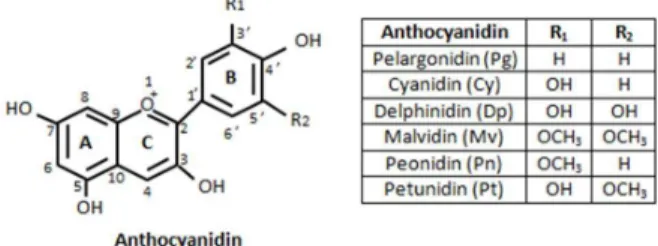
In 1988, it was demonstrated that anthocyanin are glycosylated derivatives of the 3,5,7,3΄-tetrahydroxyflavylium cation. Anthocyanin compounds contain two benzoyl rings A and B that are separated by a heterocyclic ring C (Figure 1). According to Bueno et al. (2012), the difference between anthocyanins consists in the number of hydroxyl groups, the nature, the number of sugar compounds, and the position of these attachments.
Figure 1. The flavylium cation (Mazza and Miniati, 1993)
Distribution of anthocyanins in fruits
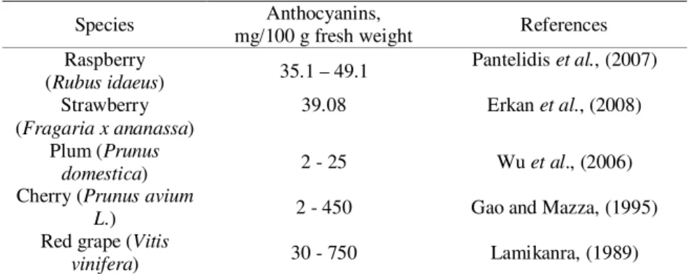
In 1990, scientists found that most anthocyanin fruits contain two aglycones, out of which cyanidin is being the most common. Anthocyanin content in fruits vary along with the concentration values that range from 0.25 mg/100 g fresh weight in pear peel, to >200 mg/100 g fresh weight in black fruits. Cultivars with bright red peel were found to contain 30 mg anthocyanins/100 g peel, whereas dark red to black cultivars where anthocyanins are located throughout the fruit contained 350– 450 mg/100 g fresh weight. At 4 mg/100 g fresh weight, anthocyanins levels in pink cultivars were about 10-100 fold lower compared to red and black fruits, respectively.
The anthocyanin content in different fruits is presented in the table below (Table 1).
Table 1. Anthocyanin content in different fruits (Szajdek and Borowska, 2008)
Species Anthocyanins,
mg/100 g fresh weight References Raspberry
(Rubus idaeus) 35.1 – 49.1
Pantelidis et al., (2007)
Strawberry (Fragaria x ananassa)
39.08 Erkan et al., (2008)
Plum (Prunus
domestica) 2 - 25 Wu et al., (2006)
Cherry (Prunus avium
L.) 2 - 450 Gao and Mazza, (1995)
Red grape (Vitis
vinifera) 30 - 750 Lamikanra, (1989)
Table 2. Anthocyanin profile in different fruits (Szajdek and Borowska, 2008)
Species Anthocyanin profile References
Raspberry (Rubus idaeus)
Cyanidin sophoroside, cyanidin 3-glucoside, cyanidin 3-glucorutinoside, cyanidin rutinoside, pelargonidin 3-sophoroside, pelargonidin 3-glucoside
Proteggente et al., (2002)
Strawberry (Fragaria x ananassa)
Pelargonidin glucoside, cyanidin 3-glucoside, pelargonidin 3-arabinoside,
pelargonidin 3-sukcynyloglucoside, cyanidin 3-sukcynyloglucoside
Skupien and Oszmianski, (2004)
Plum (Prunus domestica)
Cyanidin xyloside, cyanidin 3-glucoside, cyanidin 3-rutinoside, peonidin glucoside, peonidin
3-rutinoside
Usenik et al., (2009)
Cherry (Prunus avium L.)
Cyanidin glucoside, cyanidin 3-rutinoside, pelargonidin 3-3-rutinoside,
peonidin 3-rutinoside
Ballistreri et al., (2013)
Red grape (Vitis vinifera)
Delphinidin 3,5-diglucoside, cyanidin diglucoside, petunidin 3,5-diglucoside, peonidin 3,5-3,5-diglucoside,
malvidin 3,5-diglucoside
Huang et al., (2009)
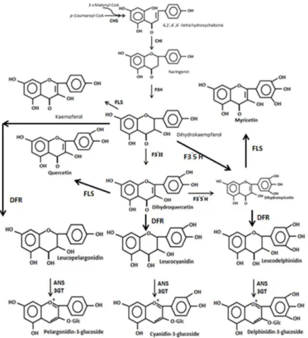
Biosynthesis of anthocyanins
study from 1994, the activity of dihydroflavonol 4-reductase (DFR) is required to synthesize the leucoanthocyanidins, that can be transformed to anthocyanidins in a reaction catalyzed by anthocyanidin synthase, which, then, undergo oxidation, dehydration, and glycosylation, to form the anthocyanins perlogonidin, cyanidin, and delphinidin.
According to the studies of Speciale et al. (2014) in the post-synthesis, cyanidin can be methylated on its 3' hydroxyl group to form peonidin, while delphinidin can be methylated on its 3' hydroxyl group to form petudinin or on both its 3' and 5' hydroxyl groups to form malvidin.
Figure 2. Anthocyanin biosinthesys pathway via the shikimic acid pathway (Holton and Cornish, 1995)
Stability of anthocyanin compounds
of the pigment, pH, temperature, light, copigments, metal ions, enzymes, oxygen, ascorbic acid, sugars, among others.
a) Molecular structure
It has been generally known since 1982 until today that the molecular structure of some anthocyanins is more stable than the others. Generally, increased hydroxylation decreases stability, whereas increased methylation increases it. Some determinations made by scientists in 1996 concluded that the colour of foods containing anthocyanins that are rich in pelargonidin, cyanidin, or delphinidin aglycones is less stable than that of food containing petunidin or malvidin aglycones.
b) pH
The changes of the pH values may lead to changes in the molecular structure of anthocyanins due to the ionic nature. At low pH values they are more stabile. According to the studies of the researcher Musoke, (2002) it is known that in aqueous media, four anthocyanin structures exist in equilibrium: flavylium cation, carbinol pseudobase, quinonoidal base and chalcone.
At the pH 1.0 the predominant species is the red-colored flavylium cation. It has been reported by scientists since 1998 that within the pH values of 2.0 and 4.0, the uncharged blue quinonoidal unstable species prevails, and if the pH is increased, an anionic quinonoidal species is formed. In 1993 it was reported that, at higher pH values (5.0 and 6.0), the carbinol pseudobase and chalcone structures are formed. Other researchers confirmed in 1998 that the unusual stability of acylated anthocyanins is at pH over 5.0.
c) Light
Even if the light is an important factor in anthocyanins biosinthesys, it is also known that light accelerates the degradation of anthocyanins, their colour being better maintained in the dark. This adverse effect has been demonstrated in several fruit juices and red wines. In 1996, researchers demonstrated that in fruit juices, acylated anthocyanins are more stable than unacylated derivatives. In addition, the absorbance of the juices tested was observed to increase after exposure to light. Further investigations of Yoshida et al. (2003a, b) showed that anthocyanins containing cinnamoyl derivatives are able to isomerize from trans to cis form, meaning colour intensification and resistance to the pyrilium ring hydration. d) Temperature
Bąkowska et al. (2003) said that high temperature values represent another important factor involved in the degradation of anthocyanins, wich leads to brown products. Recently, thanks to the studies of Cavalcanti et al., (2011) it has been demonstrated that anthocyanin content and antioxidant activity are better preserved at lower temperature values.
e) Oxigen
identified in some juices from berries. However it has been demonstrated that, at oxigen concentration values of 60-100 % for 0-7 days of storage in cold conditions, an increase in anthocyanin and phenolic content can be noticed.
f) Sugars
The same recent study of Cavalcanti et al. (2011) talks about a low stability of anthocyanin content due to sugar and its degradation products, and it was demonstrated that sugar concentration higher that 20 % has a negativ effect on anthocyanin content, while smaller concentration of sugar has an opposite effect. g) Ascorbic acid
It was suggested that an increase in the degradation of anthocyanins was a result of a condensation reaction between the anthoyanin molecules and the ascorbic acid. A mechanism proposed by Cavalcanti et al. (2011) in which the color degradation of anthocyanins is caused by the presence of ascorbic acid occurred due to the oxidative cleavage of the pyrilium ring by a free radical mechanism, in which ascorbic acid acts as an activator of molecular oxygen, producing free radicals. h) Enzymes
The most common enzymes involved in the degradation of anthocyanins are peroxidases and phenolases, such as phenol oxidases and polyphenol oxidases. Glycosidases are equally common. They can break the covalent bond between the aglycone of an anthocyanin and the glycosyl residue (Cavalcanti et al., 2011).
Extraction of anthocyanins
Anthocyanins are soluble in polar solvents and they are extracted from various plant materials by using solid–liquid extraction with solvents such as methanol, ethanol or water.
Anthocyanins are extracted with cold acidified solvents under mild conditions. The solvent system which denatures the cell membranes, simultaneously dissolving the anthocyanins and stabilizing them, is usually represented by methanol (or acetone, ethanol, acetonitrile). In the studies of Nicoue et al. (2007), the acid employed in the extraction of anthocyanins is usually acetic acid (≈ 7%) or trifluoroacetic acid (TFA ≈ 3%), whereas the organic solvent content varies from 50 to 100% of the mixture. Dai and Mumper (2010) discovered that the use of mineral acid can lead to the loss of attached acyl group.
Currently used methods for anthocyanin extraction are nonselective and result in solutions with large amounts of undesirable products such as sugars, acids, amino acids and proteins that require removal. For that, the crude extracts are purified with C18 cartridges previously activated with methanol, followed by water or 0.01 % aqueous HCl or 3 % formic acid. Anthocyanins were recovered from diluted fruit juice or wine by elution from a C18 cartridge with an aqueous eluent at a low pH (Corradini et al., 2011).
conventional extraction methods such as Soxhlet are still considered a reference method.
Non-conventional extraction techniques
In a research from 1998 it was declared that the most important issues in terms of conventional extraction are extended extraction time and high purity solvents, among others. The new extraction techniques already mentioned are considered green technologies, as they are in concordance with the standards required by the USA Environmental Protection Agency.
Ultrasound-assisted extraction (UAE)
It has been well known since the year 1996 that the extraction mechanism by ultrasound is based on two physical phenomena: diffusion across the cell wall and clearing the contents after the wall breaking. The most important factors for the action of ultrasound are temperature, pressure, frequency, and time of sonication. Pulsed-electric field extraction (PEF)
The pulsed electric field (PEF) is a useful technology for many scientists, used to improve processes like drying and extraction, etc. during the last decade. According to Puertolas et al. (2010), PEF extraction technique can operate in both mode, either continuous or batch.
Corralesa et al. (2008) found that the best extraction technology for the extraction of anthocyanin monoglucosides is by PEF. The use of a pulsed electric field treatment on the Merlot skin by Delsart et al. (2012) resulted in an increased extraction yield of polyphenols and anthocyanins.
Enzyme-assisted extraction (EAE)
According to a research from 1996, EAE is a novel and effective treatment that releases bounded compounds and increases the yield. Latif and Anwar (2009) discovered that there are two types of enzyme-assisted extraction: enzyme-assisted aqueous extraction (EAAE) and enzyme-assisted cold pressing (EACP).
EAE of phenolic compounds from grape pomace were tested and there was found a correlation between the content of total phenols and the degree of plant cell wall degraded by enzymes. Gomez-Garcia et al. (2012) extracted phenolic compounds from grape waste using an enzyme called novoferm and EAE had the strongest effect on phenolic release.
Microwave assisted extraction (MAE)
In 1994, scientists said about MAE that is a new extraction method that uses microwaves. MAE has several advantages that have been described since 2008 and these are quicker heating, reduced thermal gradients and equipment size, higher extract yield. This type of extraction method is faster than conventional extraction processes. It is a selective technique that extracts organic and organometallic compounds.
release bound phenolic acids from bran and flour fractions of sorghum and maize of different hardness by Chiremba et al. (2012).
Pressurized liquid extraction (PLE)
PLE method was first described in 1996. According to Nieto et al. (2010), this method is known by different names: pressurized fluid extraction (PFE), accelerated fluid extraction (ASE), enhanced solvent extraction (ESE), and high pressure solvent extraction (HSPE).
An advantage of PLE technique is that it requires small solvent amounts as a result of the combination between high pressure and temperature, which provides faster extraction yield.
According to Ibanez et al. (2012), PLE is also considered to be, like the other extraction methods, a green technology. PLE has been successfully applied to extract bioactive compounds from plant materials, thereby Erdogan et al. (2011) recovered individual phenolic compounds such as catechin, caffeic acid, chlorogenic acid, total phenolic contents and others from various parts of Anatolia propolis using the PLE method at optimum condition (40 ˚C, 1500 psi for 15 min). Supercritical fluid extraction (SFE)
A scientific study from 1999 showed that supercritical fluid possesses gas-like properties of diffusion, viscosity, and surface tension, liquid-like density and solvation power. According to the research of Ibanez et al. (2012), the most important factors which influence the extraction efficiency are: temperature, pressure, particle size and moisture content of feed material, extraction time, CO2 flow rate, and solvent-to-feed-ratio.
Separation and purification of anthocyanins from fruits
A research from 1980 highlighted that the extraction procedures are not selective for anthocyanins and because of that a cleanup and/or fractionation step is necessary to remove other materials present in the extract. For example, cross-flow ultrafiltration is used to eliminate sugars, present at higher concentration than the pigments in natural extracts. Another study realised later, in 1997, showed that ultrafiltration on cellulose acetate membranes removes proteins and polysaccharides, but some of the oligomeric pigments can form aggregates that could be retained by this membrane.
Chromatographic separation
Further purification and separation are carried out by chromatography that relies upon identical technologies and analytical methods, aiming at the isolation of pure substances from a mixture.
a) Paper and thin-layer chromatography
b) High-performance liquid chromatography (HPLC)
HPLC is the most used technique for the separation of anthocyanins, due to its high resolution. Reversed-phase C-18 columns are typically used, with different gradients of methanol or acetonitrile in water. The elution solvents used are acidified with formic or acetic acid (5–10%) such as to ensure that all anthocyanins are converted to flavylium cations, thereby improving the detection and the chromatographic resolution.
The best advantage of HPLC is the rate of separation at the analytical scale. Better than HPLC is the ultraperformance liquid chromatography (UPLC) because it uses an increased speed and resolution.
c) Capillary electrophoresis
Several advantages for seldom analytical separations of anthocyanins like small sample volume, low solvent consumption and short run times, are specific for the capillary electrophoresis (CE). The first method described in 1997 uses capillary zone electrophoresis with standard silica capillaries and borate running buffers at pH 8.0, resulting in anthocyanin degradation and a very poor sensitivity. Better resolution was obtained with capillary zone electrophoresis using acidic (apparent pH of 1.5) phosphate buffer containing 30 % acetonitrile, but the detection limit is rather high (25 mg/L).
Antocyanins identification and quantification
In chromatography, identification is classically based on the comparison of retention times or Rf with those of the reference compounds. The identification of anthocyanins requires a combination of spectrometric methods, the quantification of these compounds relying on this type of measurements.
a) UV–visible spectrophotometry
This method is essential for anthocyanin identification since the different structures show characteristic λmax. It has been known from different studies since 1990 that the nature of the sugar substituents does not modify the absorption spectrum. Acylation with cinnamic acids can be detected by the presence of a characteristic shoulder at the maximum absorbance wavelength of the acyl moiety (310 nm for p -coumaric acid, 320 nm for caffeic acid).
Quantification of anthocyanins is based on the measurement of their absorbance at their maximum absorbance wavelength in the visible range. This requires the establishment of calibration curves (or extinction coefficient), using anthocyanin standards. Total anthocyanins are measured after acidification (pH 1.0). Two methods are commonly known to be used for measuring and distinguishing anthocyanins from other pigments. The first one calculates anthocyanin concentration from the difference of absorbance values in the visible range at pH 4.5 and at pH 1.0:
The second method used measures pigments before and after sulfite bleaching and calculates the anthocyanin concentration from a calibration curve established using the same protocol on a standard anthocyanin. Both methods are based on the assumption that the color of genuine (monomeric) anthocyanins is modified by pH and sulfites, while that of the derived (polymeric) anthocyanins is not.
b) Mass spectrometry
MS separates ionized molecules on the basis of their mass-to-charge ratio (m/z). This implies that the molecular species of interest are charged and transferred into the gas phase by the MS ionization source and then separated according to their m/z by the mass analyzer. Anthocyanins from different sources are analyzed based on desorption and spray ionization techniques, associated to quadrupole (Q), time-of-flight (TOF), ion trap (IT), or Fourier transform ion cyclotron resonance (FT-ICR) analyzers. Fast atom bombardment mass spectrometry (FAB-MS) uses xenon or argon atoms to bombard the sample dissolved in a matrix (often glycerol). Matrix-assisted laser desorption ionization (MALDI), mostly associated with the TOF analyzer, is a soft ionization technique, in which the analyte, mixed with a matrix, is desorbed and ionized on laser irradiation. MALDI-TOF-MS is sensitive, fast and relatively tolerant to contaminants.
c) NMR spectroscopy
NMR spectroscopy is essential for the formal identification of anthocyanins and involves the complete assignment of all proton and carbon signals, based on the chemical shifts and coupling constants of the one-dimensional (1D) 1H and 13C spectra, and on the correlation of cross-peaks in 2D experiments. NMR spectra are usually recorded in deuterated solvents (deuterated dimethylsufoxide - DMSO-d6or methanol - CD3OD) to avoid the overlap of the solvent and analyte signals. Identification is normally performed on the flavylium cation, in acidified solvents, for example, with deuterated trifluoroacetic acid, but NMR can also be used to study the different anthocyanin forms.
d)Infrared, Resonance Raman and fluorescence absorption spectroscopies
Infrared (IR) and resonance Raman (RR) are two types of vibrational spectroscopy, providing qualitative and quantitative information about the vibrations of the chemical bonds of the molecules. The midinfrared, 4000–400 cm−1 (30–2.5 μm), based on the fundamental stretching and rotating vibrations of molecules, is of limited use for anthocyanin identification.
Autofluorescence of anthocyanins has not been explored. However, a recent paper of Poustka et al. (2007) has shown that anthocyanins exhibit autofluorescent properties when excited with the helium–neon laser at 543 nm and used this property to detect them in plants.
Bioactive properties and health promoting of anthocyanins
Antioxidant and antimicrobial activity
According to Yang et al. (2011), the antioxidant potential of anthocyanins depends on their chemical structure. This property is also influenced by the number of hydroxyl groups, the oxonium ion in the C ring, the hydroxylation, methylation, acylation and glycosylation pattern, among others.
In their studies, many scientists measure the antioxidant activity of anthocyanidins by using different assay methods. Using ferric reducing ability method, the 3-glucosides of delphinidin, petunidin and malvidin were observed to have 2-2.5 times higher antioxidant activity than the ascorbic acid. Using Trolox or oxygen radical-absorbing capacity (ORAC), the antioxidant capacity of anthocyanins was 3-6 times higher than that of the Trolox standard.
The antimicrobial activity of anthocyanins has been reviewed recently in the study conducted by Cisowska et al. (2011). The study revealed that there are different mechanisms by means of which anthocyanins can lead to microorganism toxicity. From the studies carried out by Lacombe et al. (2010), anthocyanins may cause cell deformation, destruction of their wall, and membrane condensation of cellular material with the presence of the cytoplasmic material and membrane debris outside the cells.
Despite these observations, Hidalgo et al. (2012) observed that incubating malvidin 3-O-glucoside with fecal bacteria resulted in mainly gallic, syringic, and p -coumaric acids. All the anthocyanins and their metabolites tested enhanced significantly the growth of Bifidobacterium spp. and Lactobacillus-Enterococcus
spp.
Cardiovascular properties
Due to a number of factors, cardiovascular diseases are in full development. Epidemiological studies of Levantesi et al. (2013) suggested that the consumption of red wine may be cardioprotective. The association between grape and phenolic compounds from wine, and coronary heart disease has been proved by the studies of different scientists, partly due to the presence of anthocyanin compounds. Anti-inflammatory properties
Inflammations occur due to cyclooxygenase (COX) enzymes that convert arachidonic acid to prostaglandins. A study from 2001 was able to determine the decrease of COX activities by 52-74%. Delphinidin and cyanidin against pelargonidin, peonidin, and malvidin have been shown to inhibit COX-2 expression. Hou et al. (2005) determined that only anthocyanins with the o -dihydroxyphenyl structure may have anti-inflammatory properties.
Neuronal properties
Kang et al. (2006) studied the neuroprotective effect of mulberry fruit extract with a content of cyanidin 3-O-glucoside on a brain injury mouse model with middle cerebral artery occlusion. A blueberry-rich diet tested in 2003 was demonstrated to have a positive impact on neuronal functions and in the protection against Alzheimer’s disease, preventing spatial memory deficits along with memory enhancement.
Eyesight properties
The effect on vision was among the first reported property of anthocyanins. In a study of Kramer (2004) it was suggested that by consuming fruit derived anthocyanins, the vision may be enhanced.
Antitumoral potential
The anticancer properties of anthocyanins that may be attributed to multiple mechanisms were established in a research of He and Giusti (2010), a study largely based on in vitro evidence. Based on his research studies, Zhang et al. (2008) discovered that anthocyanins isolated from strawberries can reduce the vitality of human oral, colon, and prostate cancer cells at a dose of 100 μg/mL. Jing et al. (2008) also observed that the most active against of all cancer cell lines was the anthocyanin fraction plus proanthocyanidin.
Anti-diabetic and anti-obesity effects
In a study from 2004, scientists found out that vegetables and fruits may trim the risk of obesity and diminish the incidence of type-2 diabetes. Ghosh and Konishi, (2007) observed that, through pancreatic β-cells, dietary constituents can regulate blood glucose levels or draw in insulin production in the case of type-2 diabetes. For example, pelargonidin 3-O-galactoside and its aglycone, pelargonidin, caused a 1.4-fold increase in insulin secretion at 4 mM glucose concentration, while other anthocyanins had only a marginal effect.
Conclusions
Anthocyanins are a group of water-soluble pigments found in various parts of plants. These pigments possess beneficial properties for human health, because of different biological activities and are able to provide protection against some human pathological conditions (cardiovascular diseases, cancer, inflammation, etc.).
The growing demand to extract plant bioactive compounds encourages the search for new extraction methods. The advanced chromatography methods and the environmental protection are two very important factors for the development of non-conventional extraction processes. Proper choice of standard methods influences the measurements of extraction efficiency. By far, the increasing economic significance of bioactive compounds could lead to various extraction methods in the future.
References
Alupului, A., Calinescu, I., and Lavric, V. 2012. Microwave extraction of active principles from medicinal plants. U.P.B.Science Bulletin, Series B, 74(2), 1454-2331.
Andersen, O.M., and Jordheim, M. 2006. The anthocyanins. In: Andersen, O. M., Markham, K.R. (eds.). Flavonoids Chemistry, Biochemistry and Applications. 471–551. CRC Press, Taylor and Francis, Boca Raton.
Asghari, J., Ondruschka, B., and Mazaheritehrani, M. 2011. Extraction of bioactive chemical compounds from the medicinal Asian plants by microwave irradiation. Journal of Medicinal Plants Research, 5(4), 495–506.
Bąkowska, A., Kucharska,A.Z., and Oszmiański, J. 2003. The effect of heating, UV irradiation, and storage stability of anthocyanin-polyphenol copigment complex. Food Chemistry, 81, 349–355.
Ballistreri, G., Continella A., Gentile A., Amenta M., and Fabroni S.R. 2013. Fruit quality and bioactive compounds relevant to human health of sweet cherry (Prunus avium L.) cultivars grown in Italy. Food Chemistry, 140, 630-638.
Bornsk, S.M., Ziberna, L., Polak, T., Vanzo, A., Ulrih, N.P., Abram, V., Tramer, F., and Passamonti, S. 2012. Bilberry and blueberry anthocyanin pathways act as powerful intracellular antioxidants in mammalian cells. Food Chemistry, 134(4), 1848-1884 Bueno, J.M., Saez-Plaza, P., Ramos-Escudero, F., Jimenez, A.M., Fett, R., and Asuero,
A.G. 2012. Analysis and Antioxidant Capacity of Anthocyanin Pigments. Part II: Chemical Structure, Color and Intake of Anthocyanins. Critical Reviews in Analytical Chemistry, 42(2), 126–151.
Cavalcanti, R.N., Santos, D.T., and Meireles, M.A.A. 2011. Non-thermal stabilization mechanisms of anthocyanins in model and food systems - An overview. Food Research International, 44(2), 499-509.
Chiremba, C., Rooney, L.W., and Trust, B.J. 2012. Microwave-assisted extraction of bound phenolic acids in bran and flour fractions from sorghum and maize cultivars varying in hardness. Journal of Chromatography Analysis, 1012(2), 119–128.
Cisowska, A., Wojnicz, D., and Hendrich, A.B. 2011. Anthocyanins as antimicrobial agents of natural plant origin. Natural Product Communications, 6(1), 149–56.
Corradini, E., Foglia, P., Giansanti, P., Gubbiotti, R., Samperi, R., and Laganà, A. 2011. Flavonoids: Chemical Properties and Analytical Methodologiesof Identification and Quantitation in Foods and Plants. Natural Product Research, 25(5), 469–495.
Corralesa, M., Toepflb, S., Butza, P., Knorrc, D., and Tauschera, B. 2008. Extraction of anthocyanins from grape by-products assisted by ultrasonics, high hydrostatic pressure or pulsed electric fields: a comparison. Innovative Food Science and Emerging Technologies, 9(1), 85–91.
De Pascual-Teresa, S. and Sanchez-Ballesta, M.T. 2007. Anthocyanins: from plant to health. Phytochemistry Reviews, 7(2), 28199.
Erdogan, S., Ates, B., Durmaz, G., Yilmaz, I., and Seckin, T. 2011. Pressurized liquid extraction of phenolic compounds from Anatolia propolis and their radical scavenging capacities. Food and Chemical Toxicology, 49(7), 1592–1597.
Erkan, M., Wang, S.Y., and Wang, C.Y. 2008. Effect of UV treatment on antioxidant capacity, antioxidant enzyme activity and decay in strawberry fruit. Postharvest Biology and Technology, 48, 163–171.
Gao, L., and Mazza, G. 1995. Characterization, quantitation, and distribution of anthocyanins and colorless phenolics in sweet cherries. Journal of Agricultural and Food Chemistry, 43, 343–346.
Ghafoor, K., Choi, Y.H., Jeon, J.Y., and Jo, I.H. 2010. Optimization of ultrasound-assisted extraction of phenolic compounds, antioxidants and anthocyanins from grape (Vitis vinifera) seeds. Journal of Agricultural and Food Chemistry, 57(11), 4988–4994. Ghafoor, K., Hui, T., and Choi, Y.H. 2012. Optimization of ultrasound-assisted extraction
of total anthocyanins from grape peel. Journal of Food Biochemistry, 35, 735–746. Ghosh, D., and Konishi, T. 2007. Anthocyanins and anthocyanin-rich extracts: role in
diabetes and eye function. Asia Pacific Journal of Clinical Nutrition, 16(2), 200–208. Gomez-Garcia, R., Martinez-Avila, G.C.G., and Aguilar, C.N. 2012. Enzyme-assisted
extraction of antioxidative phenolics from grape (Vitis vinifera L.) residues. 3 Biotech, 2(4), 297-300.
Hidalgo, M., Oruna-Concha, M.J., Kolida, S., Walton, G.E., Kallithraka, S., Spencer, J.P.E., and De Pascual-Teresa, S. 2012. Metabolism of anthocyanins by human gut microflora and their influence on gut bacterial growth. Journal of Agricultural and Food Chemistry, 60(15), 3882–3890.
Holton, T.A., and Cornish, E.C. 1995. Genetics and biochemistry of anthocyanin biosynthesis. Plant Cell, 7, 1071-1083.
Hou, D.X. 2005. Potential mechanisms of cancer chemoprevention by anthocyanins.
Current Molecular Medicine, 3(2), 149–159.
Ibanez, E., Herrero, M., Mendiola, J.A., and Castro-Puyana, M. 2012. Extraction and characterization of bioactive compounds with health benefits from marine resources: macro and micro algae, cyanobacteria, and invertebrates. In: Hayes, M. (ed.) Marine Bioactive Compounds: Sources, Characterization and Applications. 55–98, Springer, Madrid, Spain.
Joseph, J.A., Shukitt-Hale, B., and Lau, F.C. 2007. Fruit Polyphenols and Their Effects on Neuronal Signaling and Behavior in Senescence. Annals of the New York Academy of Science, 1100, 470-485.
Kang, T.H., Hur, J.Y., Kim, H.B., Ryu, J.H., and Kim, S.Y. 2006. Neuroprotective effects of the cyanidin-3-O-beta-d-glucopyranoside isolated from mulberry fruit against cerebral ischemia. Neuroscience Letters, 391(3), 122–126.
Kong, J.M., Chia, L., Goh, N., and Chia, T. 2003. Analysis and biological activities of anthocyanins. Phytochemistry. 69, 1939–1940.
Kramer, J.H. 2004. Anthocyanosides of Vaccinium myrtillus (bilberry) for night vision - a systematic review of placebo-controlled trials. Survey of Ophthalmology. 49, 618. Lacombe, A., Wu, V.C.H., Tyler, S., and Edwards, K. 2010. Antimicrobial action of the
Lamikanra, O. 1989. Anthocyanins of Vitis rotundifolia hybrid grapes. Food Chemistry, 33, 225–237.
Latif, S., and Anwar, F. 2009. Physicochemical studies of hemp (Cannabis sativa) seed oil using enzyme-assisted cold-pressing. European Journal of Lipid Science and Technology, 111(10), 1042–1048.
Levantesi, G., Marfisi, R., Mozaffarian, D., Franzosi, M.G., Maggioni, A., Nicolosi, G.L., Schweiger, C., Silletta, M., Tavazzi, L., Tognoni, G., and Marchioli, R. 2013. Wine consumption and risk of cardiovascular events after myocardial infarction: results from the GISSI-Prevenzione trial. International Journal of Cardiology, 163(3), 282–287. Martin, S., Favot, L., Matz, R., Lugnier, C., and Andriantsitohaina, R. 2003. Delphinidin
inhibits endothelial cell proliferation and cell cycle progression through a transient activation of ERK -1/-2. Biochemistry and Pharmacology, 65, 669.
Mazza, G. and Miniati, E. 1993. Anthocyanins in fruits, vegetables, and grains. CRC Press, Boca Raton, FL.
Musoke, B.E. 2002. The uses of plant colours (anthocyanin pigments) and methods to isolate them from plants., In: Materials of The World’s Women Congress, 2002, 212– 214, Makerere University, Uganda.
Nicoue, E.E., Savards, S., and Belkacemi, K. 2007. Anthocyanins in Wild Blueberries of Quebec: Extraction and Identification. Journal of Agriculture and Food Chemistry, 55(14), 5625–5635.
Nieto, A., Borrull, F., Pocurull, E., and Marce, R.M. 2010. Pressurized liquid extraction: a useful technique to extract pharmaceuticals and personal-care products from sewage sludge. Trends in Analytical Chemistry, 29(7), 752–764.
Pantelidis, G.E., Vasilakakis, Manganaris, G.A., and Diamantidis, G. 2007. Antioxidant capacity, phenol, anthocyanin and ascorbic acid contents in raspberries, blackberries, red currants, gooseberries and Cornelian cherries. Food Chemistry, 102, 777-783.
Poustka, F., Irani, N.G., Feller, A., Lu, Y., Pourcel, L., Frame, K., and Grotewold, E. 2007. A trafficking pathway for anthocyanins overlaps with the endoplasmic reticulum-tovacuole protein-sorting route in Arabidopsis and contributes to the formation of vacuolar inclusions. Plant Physiology, 145, 1323–1335.
Proteggente, A.R., Pannala, A.S., Paganga, G., Van Buren, L.,Wagner, E., Wiseman, S., Van De Put, F., and Dacombe, C. 2002. The antioxidant activity of regularly consumed fruit and vegetables reflects their phenolic and vitamin C composition. Free Radical Research, 36, 217–233.
Puertolas, E., Lopez, N., Saldana, G., Alvarez, I., and Raso, J. 2010. Evaluation of phenolic extraction during fermentation of red grapes treated by a continuous pulsed electric fields process at pilot-plant scale. Journal of Food Engineering, 119(3), 1063–1070. Shukitt-Hale, B., Kalt, W., Carey, A.N., Vinqvist-Tymchuk, M., McDonald, J., and Joseph,
J.A. 2009. Plum juice, but not dried plum powder, is effective in mitigating cognitive deficits in aged rats. Nutrition, 25(5), 567–573.
Speciale, A., Cimino, F., Saija, A., Canali, R., and Virgili, F. 2014. Bioavailability and molecular activities of anthocyanins as modulators of endothelial function. Genes & Nutrition, July, 9(4), 404.
Szajdek, A. and Borowska, E.J. 2008. Bioactive compounds and health-promoting properties of berry fruits: Review. Plant Foods for Human Nutrition, 63, 147–156. Tsuda, T. 2012. Dietary anthocyanin-rich plants: biochemical basis and recent progress in
health benefits studies. Molecular Nutrition and Food Research, 56(1), 159–170. Tsuda, T., Kato, Y., and Osawa, T. 2000. Mechanism for the peroxynitrite scavenging
activity by anthocyanins. Federation of European Biochemical Societies, 484, 207-2010.
Usenik, V., Stampar, F., and Veberic, R. 2009. Anthocyanins and fruit colour in plums (Prunus domestica) during ripening. Food chemistry, 114, 529-534.
Wu, X.L., Beecher, G.R., Holden, J.M., Haytowitz, D.B., Gebhardt, S.E., and Prior, R.L. 2006. Concentrations of anthocyanins in common foods in the United States and estimation of normal consumption. Journal of Agricultural and Food Chemistry, 54, 4069–4075.
Yang, M., Koo, S.I., Song, W.O., and Chun, O.K. 2011. Food matrix affecting anthocyanin bioavailability: review. Current Medicinal Chemistry, 18(2), 291–300.
Yoshida, K., Mori M., Kawachi M., Okuno R., Kameda K., and Kondo T. 2003a. A UV-B resistant polyacylated anthocyanin, HBA, from blue petals of morning glory.
Tetrahedron Letters, 44, 7875–7880.
Yoshida, K., Okuno R., Kameda K., Mori M., and Kondo T. 2003b. Influence of E, Z-isomerization and stability of acylated anthocyanins under the UV irradiation.
Biochemical Engineering Journal, 14, 163–169.