Analysis of the
Paracoccidioides brasiliensis
triosephosphate
isomerase suggests the potential for adhesin function
Luiz Augusto Pereira1, Soˆnia Nair B ´ao2, Moˆnica Santiago Barbosa1, Juliana Leal M. da Silva3, Maria Sueli S. Felipe4, Jaime Martins de Santana5, Maria Jos ´e S. Mendes-Giannini3& C ´elia Maria de Almeida Soares1 1
Laborat ´orio de Biologia Molecular, Instituto de Cieˆncias Biol ´ogicas, Universidade Federal de Goi ´as, Goiaˆnia, Goi ´as, Brazil;2Laborat ´orio de Microscopia, Universidade de Bras´ılia, Bras´ılia, DF;3Laborat ´orio de Micologia Cl´ınica, Universidade Estadual Ju´lio de Mesquita Filho, Araraquara, Sa˜o Paulo; 4Laborat ´orio de Biologia Molecular, Universidade de Bras´ılia; and5Laborat ´orio Interdisciplinar em Doenc¸a de Chagas, Universidade de Bras´ılia, Bras´ılia, DF, Brazil
Correspondence:C ´elia Maria de Almeida Soares, Laborat ´orio de Biologia Molecular, ICB2, Campus 2, Universidade Federal de Goi ´as, 74001-970, Goiaˆnia, Goi ´as, Brazil. Tel./fax:155 62 35211110; e-mail: [email protected]
Received 14 February 2007; revised 3 May 2007; accepted 14 June 2007.
First published online 23 August 2007.
DOI:10.1111/j.1567-1364.2007.00292.x
Editor: Jos ´e Ruiz-Herrera
Keywords
Paracoccidioides brasiliensis ; TPI; interaction with epithelial cells; infection.
Abstract
Paracoccidioides brasiliensisis an important fungal pathogen. The disease it causes, paracoccidioidomycosis (PCM), ranges from localized pulmonary infection to systemic processes that endanger the life of the patient.Paracoccidioides brasiliensis
adhesion to host tissues contributes to its virulence, but we know relatively little about molecules and the molecular mechanisms governing fungal adhesion to mammalian cells. Triosephosphate isomerase (TPI: EC 5.3.1.1) ofP. brasiliensis
(PbTPI) is a fungal antigen characterized by microsequencing of peptides. The protein, which is predominantly expressed in the yeast parasitic phase, localizes at the cell wall and in the cytoplasmic compartment. TPI and the respective polyclonal antibody produced against this protein inhibited the interaction of
P. brasiliensis to in vitro cultured epithelial cells. TPI binds preferentially to laminin, as determined by peptide inhibition assays. Collectively, these results suggest that TPI is required for interactions betweenP. brasiliensisand extracellular matrix molecules such as laminin and that this interaction may play an important role in the fungal adherence and invasion of host cells.
Introduction
The pathogenic fungus Paracoccidioides brasiliensis causes paracoccidioidomycosis (PCM), one of the most important systemic mycoses in Latin America. The severe nature of the disease and the occurrence of sequelae, frequently causing pulmonary dysfunction and other disabilities, render it a pathogen of considerable medical importance. The fungus is thermally dimorphic, grows as a yeast-like structure in the host tissue and as mycelium in the saprobic condition. The mycelial phase produces infective propagules that lodge in the host alveoli, and adhere and invade the alveolar cells and/or the basal lamina (Franco & Montenegro, 1994).
The attachment of microorganisms to a biological surface is a complex process involving specific interactions between adhesins and complex receptors on host tissues (Finlay & Cossart, 1997). During their evolution, fungal pathogens have acquired the ability to exploit cell surface matrix components as ligands for attachment (Rostand & Esko, 1997). The extracellular matrix of the alveolar basal lamina
is a complex mixture of molecules, including fibronectin, laminin, and collagens. Although the matrix is not available for binding in normal cells, the components can be exposed after tissue damage resulting from either inflammatory process or lytic activity by toxins of the microorganism (Pattiet al., 1994).
The adhesion of P. brasiliensis to cells is seen as an important determinant of pathogenesis. The fungal proteins functioning as adhesins inP. brasiliensisare just now coming to light. Some fungal proteins including a glycoprotein of 43 kDa (gp43), a 30 kDa protein, proteins of 19 and 32 kDa and glyceraldehyde-3-phosphate dehydrogenase (GAPDH) have been identified and found to mediate adhesion to extracellular matrix components and human cells (Vicentini
et al., 1994; Andreotti et al., 2005; Gonz´ales et al., 2005; Barbosaet al., 2006; Mendes-Gianniniet al., 2006).
analysis; it was originally described in our laboratory as a fungal antigen reactive with sera of PCM patients and sequences from N-terminal and internal peptides were obtained (Fonseca et al., 2001). Subsequently, the cognate cDNA was obtained and the recombinant protein was over-produced in a heterologous system (Pereiraet al., 2004).
It has been reported that pathogen-derived TPI plays an important role in host–parasite interaction. The immuniza-tion of experimental animals with monoclonal antibodies to TPI, with the recombinant protein, as well as with plasmid DNA, led to a significant protection against schistosomiasis (Harn, 1987; Miaoet al., 1998; Zhuet al., 2006). Recently, the enzyme was described as a novel binding protein of the integrinaIIb cytoplasmic domain (Liuet al., 2006).
Little is known about molecules related to fungal host-interaction, such as adhesion molecules and the molecular mechanisms governingP. brasiliensisadhesion to mamma-lian cells. With this in mind, we investigated the role of TPI as a putative novel molecule involved in the fungus interac-tion with the host. This work describes the producinterac-tion of an antirecombinant TPI (rPbTPI) polyclonal antibody that recognizes the native protein in yeast and mycelium, as well as recognizing the recombinant molecule. Immunoelectron microscopy was employed to define the TPI subcellular localization inP. brasiliensis. ThePbTPI was detected in the cytoplasm and in the cell wall of the yeast phase of P. brasiliensis. TPI binds preferentially to laminin, as deter-mined by peptide inhibition assays. Treatment ofP. brasi-liensis yeast cells with the anti-TPI polyclonal antibody promoted inhibition of interaction of P. brasiliensis to epithelial cells. Likewise, the treatment ofin vitrocultured epithelial cells with the recombinant protein blocked bind-ing ofP. brasiliensis.
All the data indicate that TPI present on the surface of P. brasiliensis is able to interact with host extracellular matrix proteins. This fungal cell-wall characterized protein could be crucial for the initial fungal adherence and inva-sion, thus promoting the fungal infection.
Materials and methods
Paracoccidioides brasiliensis isolates growth conditions and differentiation assays
Paracoccidioides brasiliensis Pb01 strain (ATCC-MYA-826) has been investigated by our laboratory and was cultivated for 7 days in semisolid Fava Netto’s medium at 361C in the yeast form and at 221C for its mycelia phase, as previously described (Barbosa et al., 2006). The differentiation was performed in the same medium above, without agar, by changing the temperature of the culture from 22 to 361C for the mycelium to yeast transition, as described (Moreira
et al., 2004). The cells were previously grown in liquid
medium for 18 h before changing the incubation tempera-ture, which was maintained for 15 days. Aliquots were taken at different time intervals and processed for further analysis.
Heterologous expression ofP. brasiliensis TPI, recombinant protein purification and antibody production
The production and purification of the recombinant TPI was performed as described (Pereiraet al., 2004). Briefly, the cDNA was cloned into the EcoRI/XhoI sites of pGEX-4T-3 (GE Healthcare). The recombinant TPI was expressed in the soluble form by the bacteria and the protein was purified by affinity chromatography under nondenaturing conditions. The recombinant protein was used to generate specific rabbit polyclonal serum. The rPbTPI (300mg) was injected into rabbit with Freud’s adjuvant, three times at 2-week intervals. The obtained serum, containing specific anti-TPI polyclonal antibodies, was sampled and stored at 201C.
Preparation of fungal cell extracts
Yeast and mycelial protein crude extracts were obtained as described (Barbosaet al., 2006). Two types of extracts were produced: (1) a total cell homogenate and (2) a cell-free homogenate. For the preparation of total cell homogenate, mycelium and yeast cells were frozen and exhaustively ground with mortar and pestle in the presence of protease inhibitors. The mixture was centrifuged (12 000g) at 41C
for 10 min, and the supernatant was used. The cell-free extracts of the yeast cells were obtained as described (Barbosaet al., 2006). In brief, yeast cells of P. brasiliensis
(300 mg) were resuspended in 1.0 mL of 10 mM phosphate-buffered saline (PBS), pH 7.2, and vortexed for 30 s. The cells were centrifuged (5600g) for 1 min, and the
super-natant was collected and used for further analysis. The protein content of samples was determined.
Determination of TPI specific activity
The enzymatic activity was determined by coupling the conversion of dihydroxyacetone phosphate to glyceralde-hyde 3-phosphate, as described (Plaut & Knowles, 1972). The increase in the absorbance at 340 nm caused by the reduction of NAD was monitored. One activity unit (U) is defined as the conversion of 1mmol substrate min 1at 251C. The reaction mixture was composed of 100 mM triethanola-mine buffer, pH 7.6, 5 mM EDTA, 0.2 mM NAD, 20mg mL 1
glyceraldehyde-3-phosphate dehydrogenase, 1 mM dihy-droxyacetone phosphate, and 20mL (at a concentration of
extinction coefficient of the product (e340= 6220 M cm 1). Chemicals used in the enzymatic assay were from Sigma Chemical Co. (St Louis, MO).
Western-blot analysis
SDS-polyacrylamide gel electrophoresis (SDS-PAGE) was carried out and the proteins were electrophoretically trans-ferred to a nylon membrane, according to standard proto-cols. TPI was detected with the polyclonal antibody (1 : 1000 diluted). The reaction was revealed with 5-bromo-4-chloro-3-indolylphosphate/nitroblue tetrazolium (BCIP/NBT).
Ultrastructure of the yeast cells and immunocytochemistry of TPI
For the ultrastructural and immunocytochemistry studies, we employed the protocols previously described in Barbosa
et al. (2006). After fixation of the yeast cells, ultrathin sections were stained with 3% (w/v) uranyl acetate and lead citrate.
For ultrastructural immunocytochemistry studies, the ultrathin sections were incubated for 1 h with the polyclonal antibody raised against the recombinant TPI (diluted 1 : 100) and for 1 h at room temperature with the labeled secondary antibody rabbit IgG, Au-conjugated (10 nm average particle size; 1 : 20 dilution). The grids were stained as described above and observed with a Jeol 1011 transmission electron microscope (Jeol, Tokyo, Japan). Controls were obtained.
Affinity ligand assays
Far-Western assays were carried out as previously described (Guichetet al., 1997; Barbosaet al., 2006). Recombinant TPI was submitted to SDS-PAGE and blotted onto nitrocellulose membranes, which were assayed for laminin and fibronectin binding as follows. The membranes were incubated with 30mg laminin mL 1 or 30mg fibronectin mL 1, for 90 min.
The blots were sequentially incubated with rabbit antibodies antilaminin or antifibronectin (1 : 100 diluted) and with peroxidase-labelled goat antirabbit immunoglobulin (1 : 1000 diluted). The reactive bands were developed with hydrogen peroxide and diaminobenzidine. Controls were obtained.
Binding assays of recombinant PbTPI toin vitro
cultured A549 pneumocytes and Vero cells
Type II pneumocyte line A549 from the American Type Culture Collection (ATCC CCL185, Manassas, VA) and African green monkey kidney cells (Vero cells, ATCC CCL81) were used in the experiments. Briefly, the A540 pneumocytes and Vero cells were maintained in Dulbecco’s modified Eagle’s medium (DMEM) and Medium 199 (Sig-ma), respectively, both supplemented with 10% (v/v) heat-inactivated fetal calf serum. Monolayers of cells were
in-cubated with 50mg mL 1ofP. brasiliensisrecombinant TPI at 361C for 5 h. Distilled water was added for 4 h and the cell lysates were fractionated by electrophoresis (12% SDS-PAGE). Proteins in the gel were electrotransferred to mem-branes. Western blot analysis was performed as described before. Negative controls were obtained by analyzing the supernatant of lysed cells not preincubated with the recom-binant TPI, as well as by coating the cell culture flask with 50mg mL 1of TPI.
Interference of TPI and polyclonal antibody in interaction ofP. brasiliensis toin vitro cultured cells
A549 pneumocytes and Vero cells were incubated for 1 h at 361C with the recombinant TPI at 50mg mL 1 in 10 mM
PBS. After washing the cells in the respective culture mediums 1108yeast cells ofP. brasiliensiswere added to the cultures. Incubation was performed for 5 h at 361C, as previously described (Barbosa et al., 2006). In parallel, 1108yeast cells ofP. brasiliensiswere incubated for 1 h at 361C with the polyclonal antibody anti-TPI (1 : 1000 di-luted). Cells were washed and allowed to interact with the A549 pneumocytes or Vero cells. Three types of control experiments were performed: (1) with yeast cells not pre-incubated with the recombinant TPI protein; (2) with yeast cells not preincubated with the anti-TPI antibody and (3) with yeast cells preincubated just with bovine serum albu-min (BSA), at 50mg mL 1. The number ofP. brasiliensisyeast cells interacting with thein vitrocultured epithelial cells was determined, as described (Esquenaziet al., 2003; Mendes-Gianniniet al., 2004). Results are presented as the means of counts performed three times with SDs included.
Inhibitory effects of synthetic peptides on binding of TPI to pneumocytes
Gly–Ser–Arg (YIGSR) from laminin were used in the experiments. As a negative control, TPI was omitted and replaced by PBS.
Statistical analysis
Results are expressed as the meanSE of the mean of three independent experiments. Statistical analysis was performed usingANOVA (F-test followed by Duncan test). P-values of 0.05 or less were considered statistically significant.
Results
Production of polyclonal antibody and analysis of TPI accumulation inP. brasiliensis
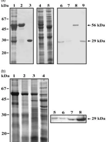
The cDNA encoding the P. brasiliensis TPI (GenBank accession number AY250089) was overexpressed into bac-terial cells. After induction with IPTG, a 56-kDa fusion recombinant protein was detected in the bacterial lysates (Fig. 1a, lane 2). The fusion protein was cleaved by the addition of thrombin protease (Fig. 1a, lane 3). As observed, highly purified protein was obtained that migrated on SDS-PAGE as a single species of 29 kDa. The purified recombi-nant TPI was used to produce rabbit polyclonal antibody. The protein total extracts ofP. brasiliensisyeast and myce-lium (Fig. 1a, lanes 4 and 5, respectively) were visualized after Coomassie blue staining. Those samples, including the recombinant TPI before and after thrombin cleavage, were blotted onto membranes and reacted to the polyclonal antibody (Fig. 1a, lanes 6–9). As demonstrated, a single band of 29 kDa was detected in extracts of both yeast and mycelium (Fig. 1a, lanes 6 and 7). Recombinant TPI in the bacterial lysates and purified recombinant TPI were also recognized as a single band by the polyclonal antibody (Fig. 1a, lanes 8 and lane 9, respectively). No cross-reactivity to the rabbit preimmune serum was evidenced with the samples (data not shown).
Total cellular extracts from mycelium, mycelium in tran-sition to yeast at days 1, 7 and 15 after the temperature shift, were taken and analyzed by one-dimensional gel electro-phoresis (Fig. 1b, lanes 1–4, respectively). The samples were run in parallel and transferred to membrane to react with the polyclonal antibody (Fig. 1b, lanes 5–8). The native protein is weakly accumulated in the mycelia phase (Fig. 1b, lane 5) and its expression is progressively increased during the transition to yeast (Fig. 1b, lanes 6–8).
Detection of TPI protein by immunoelectron microscopy ofP. brasiliensis yeast cells
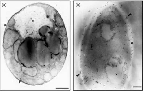
Immunocytochemistry experiments were performed to de-fine the cellular localization of the TPI protein in yeast cells.
Electron microscopy of conventionally embedded cells re-vealed the ultrastructure of theP. brasiliensisyeast form (Fig. 2a). In yeast cells processed by the postembedding method, Fig. 1.Recognition ofP. brasiliensisTPI by the rabbit polyclonal antibody and protein expression during fungal dimorphic transition. (a) Recombi-nant and native TPI are recognized by the polyclonal antibody.Escherichia coliXL1-Blue cells harbouring the pGEX-4T-3-TPI plasmid were grown at 301C to an A260 nmof 0.6 and harvested after (lane 1) a 2 h incubation with 0.1 mM IPTG. Lane 2 – the affinity-isolated recombinant TPI. Lane 3 – the recombinant fusion protein cleaved by thrombin. Lane 4 – protein extracts from yeast (30mg), Lane 5 – protein extracts from mycelium
(30mg). The proteins were fractionated by one-dimensional gel electro-phoresis and stained using Coomassie blue. Lanes 6–9 – Western blot analysis of the native and recombinant TPI. The same samples and quantities of proteins were fractionated (12% SDS-PAGE) and transferred to membrane. Lane 6 – protein extracts from yeast. Lane 7 – protein extracts from mycelium. Lane 8 –E. colitransformed with pGEX-4T-3-TPI total extract; Lane 9 – the recombinant fusion protein cleaved by thrombin. The blots were reacted to the polyclonal anti-TPI antibody. Molecular size markers are indicated. Arrows indicate the recombinant fusion protein and the native TPI. (b) Analysis of the expression of TPI during the transition from mycelium to yeast inP. brasiliensis. The proteins (30mg) were fractionated by one-dimensional gel electrophoresis and
gold particles were present in cytoplasm and the cell wall (Fig. 2b). Control samples not exposed to the poly-clonal antibody, as well as sample incubated with the rabbit preimmune serum, prior to the incubation with the gold-conjugated antibody were free of label (data not shown).
Enzymatic activity of native and recombinant TPI ofP. brasiliensis
As the TPI ofP. brasiliensiswas found to be localized both at the cell wall and internally in the cytoplasm, we attempted to evaluate the enzymatic activity of the native protein in total cellular extracts of yeast and mycelium, as well as in the cell-free extract of yeast cells, which corresponds to the most superficial components of the cell wall. Also the recombi-nant protein was assayed for its enzymatic activity. Table 1 contains the results of these experiments. The TPI specific activity was substantially higher in the yeast cells when compared with mycelia, in agreement with the higher amount of the protein in the parasitic phase, as demon-strated by Western blot analysis. Additionally, the cell-free extract fraction exhibited a high specific activity of TPI, corroborating the high amount of gold particles found in the fungal cell wall by the immunoelectron microscopy experiments. The recombinant molecule presented the high-est activity in the thigh-ested samples.
Binding of recombinant TPI to extracellular matrix proteins and toin vitro cultured A549 pneumocytes and Vero cells
The ability of the recombinant TPI of P. brasiliensis to bind laminin and fibronectin was determined by
far-Western blotting assays (Fig. 3a). The recombinant protein manifests the ability to bind to laminin (Fig. 3a, lane 2) and fibronectin (Fig. 3a, lane 3). The positive control was developed with the anti-TPI polyclonal antibody (Fig. 3a, lane 1). Negative controls were obtained by incubating the recombinant molecule with just the secondary anti-body or in the absence of the extracellular matrix proteins (data not shown). An additional control demonstrated the specificity of the binding of TPI to the extracellular matrix proteins, as no reactivity between BSA and the extracellular matrix proteins was demonstrated and the polyclonal antibody to TPI did not present cross-reactivity to BSA (data not shown). The adhesin characteristic of TPI was also evaluated by interaction of the recombinant protein with pneumocytes and Vero cells (Fig. 3b and c, respec-tively). The purified protein behaved as an adhesin, binding to thein vitrocultured cells (Fig. 3b and c, lane 2). Negative and positive controls were developed, respectively, with pneumocytes A549 and Vero cells not incubated with the recombinant TPI (Fig. 3b and c, respectively, lane 3), as well Fig. 2.Immunoelectron microscopy detection
of TPI inP. brasiliensisyeast cells by postembed-ding methods. (a) Transmission electron micro-scopy ofP. brasiliensisyeast cells; nucleus (n), intracytoplasmic vacuoles (v) mitochondria (m). The plasma membrane (arrow) and cell wall (w) are also shown. (b) Gold particles (arrowheads) are observed at the fungus cell wall (w) and in the cytoplasm (double arrowheads). Bars, 1mm
(a), 0.2mm (b).
Table 1. Analysis of the enzymatic activity of the native and recombinantP. brasiliensisTPI
Protein source
PbTPI specific activity (U mg1protein)
Yeast ‘cell free’ extract 2.020.0075
Yeast 1.90.0059
Mycelium 0.60.0038
Purified recombinant TPI (rPbTPI) 18.80.0015
One activity unit (U) is defined as the conversion of 1mmol sub-strate min1 at 25
as with the recombinant protein added to the cell cul-ture flask, in the absence of the cells monolayer (Fig. 3b and c, lanes 1).
Inhibitory effects of TPI and polyclonal antibody onP. brasiliensis interaction to cells and
competitive assay with TPI and peptides during interaction with pneumocytes
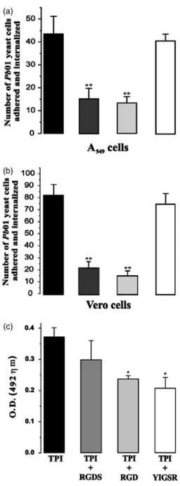
Yeast cells were assayed for the interaction with in vitro
cultured pneumocytes and Vero cells, as shown in Fig. 4a and b, respectively. Paracoccidioides brasiliensis yeast cells were treated with the antibody anti-TPI prior to interaction with thein vitrocultured cells; pneumocytes and Vero cells were pretreated with recombinant TPI prior to the interac-tion with P. brasiliensis yeast cells. As demonstrated, the treatment of pneumocytes with recombinant TPI resulted in 65% inhibition of the adherence and internalization of Fig. 3. Interaction of the rPbTPI to extracellular matrix components and
to thein vitrocultured A549 pneumocytes and Vero cells. (a) The rPbTPI (0.5mg) was subjected to SDS-PAGE and electroblotted. The membranes were reacted with laminin (lane 2) and fibronectin (lane 3) and subse-quently incubated with the rabbit IgG antilaminin and antifibronectin antibodies, respectively. Use of peroxidase-conjugated antirabbit IgG revealed the reactions. The positive control was obtained by incubating the recombinant protein with the anti-TPI polyclonal antibody (lane 1). (b and c) Cultured pneumocytes (b) and Vero cells (c) were incubated with 50mg of recombinant TPI for 5 h at 361C. The cells were lysed, the supernatant was fractionated by SDS-PAGE and proteins were trans-ferred to membranes. Detection was performed by incubation with rabbit anti-TPI polyclonal antibody and subsequent reaction with alkaline phosphatase-coupled anti-rabbit IgG. The reaction was developed with BCIP/NBT. Positive control, recombinant protein transferred to the membranes (lane 1); supernatant of pneumocytes or Vero cells after incubation of the cells with the recombinant TPI (lane 2); supernatant of pneumocytes and Vero cells not incubated with the recombinant TPI (lane 3).
Fig. 4. Interaction assay data and competitive assay withPbTPI and peptides during the interaction with pneumocytes. (a and b) Histograms showing the interaction (adhesion plus internalization) ofP. brasiliensis
yeast cells to pneumocytes A549 and Vero cells, respectively. Prior to each assay, the yeast cells were treated for 1 h with the anti-TPI polyclonal antibody (1 : 1000 diluted). In another set of experiments the A549 and Vero cells were pretreated for 1 h with 50mg mL 1of the rPbTPI. The
interaction ofP. brasiliensisto pneumocytes and Vero cells was analyzed after 5 h (a and b, respectively). Black bars, control; dark grey bars, epithelial cells treated with the recombinant TPI; light grey bars, Para-coccidioides brasiliensisyeast cells treated with the anti-TPI polyclonal antibody; white bars, cells treated with BSA. The values represent the meanSD of three independent experiments performed in triplicate. Asterisks denote values statistically different from control atPo0.0001.
Vertical bars indicate SD. (c) Pneumocyte A549 cells were incubated with TPI (10mg mL1); TPI (10mg mL 1) and the synthetic peptides of
P. brasiliensis to those cells. In addition, the treatment of
P. brasiliensis yeast cells with the polyclonal antibody re-sulted in 69% inhibition of the adherence and internaliza-tion to pneumocytes (Fig. 4a). Similar results were obtained when Vero cells were infected with P. brasiliensis. The treatment of the epithelial cells with the recombinant TPI resulted in 73% inhibition of fungal adherence and inter-nalization. We observed an 85% inhibition of adhesion/ internalization with the treatment of the yeast cells with the polyclonal antibody (Fig. 4b).
As laminin and fibronectin bind to immobilized TPI we attempted to evaluate whether peptides of those molecules had the ability to inhibit TPI binding toin vitrocultured pneumocytes. Competitive binding was performed with synthetic peptides corresponding to the adhesive recogni-tion sequences of fibronectin and laminin, as shown in Fig. 4c. The peptide RGDS from fibronectin exhibited a small inhibitory effect (P40.05) on the binding of TPI to
in vitrocultured pneumocytes. Synthetic peptide RGD, from fibronectin and laminin, inhibited the interaction of TPI with the cultured pneumocytes by 36.0% (Po0.05), as shown. The peptide YIGSR, from laminin, showed an inhibitory effect of 44.1% (Po0.05).
Discussion
In the present study we have investigated the role of TPI as an adhesin putatively involved in host cell binding. The fungal cell wall is the initial site of interaction between fungal cells and their host and thus is a key structure for entry and infection. Although not formally considered an intracellular pathogen,P. brasiliensiscan enter epithelial cells (Hannaet al., 2000; Mendes-Gianniniet al., 2000). Endocy-tosis of this fungus requires intact epithelial cell microfila-ments and microtubules and triggers host-cell apoptosis (Mendes-Gianniniet al., 2004). Although the invasion of nonphagocytic cells is likely central to the pathogenesis of the disease, there is a paucity of knowledge about the fungal surface proteins that induce invasion, as well as the host-cell receptors to the fungal adhesins.
In addition to a cytoplasmic location, the TPI of
P. brasiliensisis present at the fungal cell wall. Despite its external location, the protein lacks an N-terminal signal peptide, as previously demonstrated (Fonsecaet al., 2001; Pereiraet al., 2004). Evidence has been accumulating using as current models Saccharomyces cerevisiae and Candida albicans, clearly showing that many proteins that lack an N-terminal peptide also reach the cell surface. Of special note, many of those surface proteins lacking the N-terminal signal peptide are also found in the cytoplasm, where they perform well-known functions (Nombelaet al., 2006). Some of those nonconventionally secreted proteins are involved in binding to host components (Gozalbo et al., 1998). In regard to
P. brasiliensis, we have demonstrated that two adhesins, glyceraldehyde-3-phosphate dehydrogenase (GAPDH) (Barbosa et al., 2006) and TPI, described here, do not present canonical sequences to surface trafficking. In addi-tion to its probable binding to host-cell components med-iating fungal adhesion and invasion, TPI exhibits enzymatic activity at the cell wall, as demonstrated in this work.
Adhesion encoding genes are not constitutively expressed, but in general are activated by diverse environmental triggers (Cheng et al., 2005). We demonstrated that the expression of TPI is developmentally regulated inP. brasi-liensis, with expression increasing as the fungus adopts the pathogenic yeast-like morphology. Data on TPI enzymatic activity corroborate the Western blot result.
The ability ofP. brasiliensis to colonize host tissues may be facilitated by fungal surface proteins with high affinity to extracellular matrix molecules, and the outcome of such colonization depends largely on the receptor/ligand interac-tions between the host cells and the fungus. As it has been proposed that initial infection ofP. brasiliensisoriginates in the lung following inhalation of airborne conidia (Franco
et al., 1994), the fungal ability to initiate infection may be due to the adhesion of its spores to both extracellular matrix molecules and lung epithelial cells. The TPI can be involved in the interaction of P. brasiliensis with the extracellular matrix components laminin and fibronectin, as inferred by the adhesion experiments of those molecules with immobi-lized TPI. The recombinant TPI and the polyclonal antibody raised against the molecule were able to interfere with the interaction of P. brasiliensis to in vitro cultured epithelial cells. Competitive assays with the RGD peptide, part of the laminin and fibronectin molecules, reduced by 36.0% the adhesion of TPI to pneumocytes. On the other hand, YIGSR derived from the laminin reduced the binding by 44.1% and RGDS did not reduce significantly the adhesion of TPI to pneumocytes. On the basis of those findings we can spec-ulate that laminin mainly mediates the adhesion of TPI to the epithelial cells.
Recognition and binding to the host cells is a key step in the pathogenesis of many fungi, and consequently the characterization of novel adherence molecules and identifi-cation of the molecular basis ofP. brasiliensisattachment to host cells remain important objectives.
Acknowledgements
invaluable discussion and for the critical review of this manuscript.
References
Andreotti PF, Monteiro da Silva JL, Baila˜o AM, Soares CMA, Benard G, Soares CP & Mendes-Giannini MJ (2005) Isolation and partial characterization of a 30-kDa adhesin from
Paracoccidioides brasiliensis.Microbes Infect7: 875–881. Barbosa MS, Bao SN, Andreotti PF, de Faria FP, Felipe MS, dos
Santos Feitosa L, Mendes-Giannini MJ & Soares CMA (2006) Glyceraldehyde 3-phosphate dehydrogenase of
Paracoccidioides brasiliensisis a cell surface protein, involved in fungal adhesion to extracellular matrix proteins and
interaction with cells.Infect Immun74: 382–389.
Cheng G, Wozniak K, Wallig MA, Fidel PL Jr, Trupin SR & Hoyer LL (2005) Comparison betweenCandida albicansagglutinin-like sequence gene expression patterns in human clinical specimens and models of vaginal candidiasis.Infect Immun73: 1656–1663. Esquenazi D, de Souza W, Alviano CS & Rozental S (2003) The
role of surface carbohydrates on the interaction of
microconidia ofThichophyton mentagrophyteswith epithelial cell.FEMS Immunol Med Microbiol30: 113–123.
Finlay BB & Cossart P (1997) Exploitation of mammalian host cell functions by bacterial pathogens.Science276: 718–725. Fonseca CA, Jesu´ıno RSA, Felipe MSS, Cunha DA, Brito W &
Soares CMA (2001) Two-dimensional electrophoresis and characterization of antigens fromParacoccidioides brasiliensis.
Microbes Infect3: 535–542.
Franco M & Montenegro MR (1994) Pathology. Paracoccidioido-mycosis(Franco M, Lacaz C, Restrepo A & Del Negro G, eds), pp. 131–147. CRC Press, Boca Raton, FL.
Gonz´ales A, G ´omez BL, Diez S, Hern´andez O, Restrepo A, Hamilton AJ & Cano LE (2005) Purification and partial characterization of aParacoccidioides brasiliensisprotein with capacity to bind to extracellular matrix proteins.Infect Immun
73: 2486–2495.
Gozalbo D, Gil-Navarro I, Azorin I, Renau-Piqueras J, Martinez JP & Gil ML (1998) The cell wall-associated glyceraldehyde-3-phosphate dehydrogenase ofCandida albicansis also a fibronectin and laminin binding protein.Infect Immun66: 2052–2059.
Guichet A, Copeland JW, Erdelyi M, Hlousek D, Zavorszky P, Ho J, Brown S, Percival-Smith A, Krause HM & Ephrussi A (1997) The nuclear receptor homologue Ftz-F1 and the
homeodomain protein Ftz are mutually dependent cofactors.
Nature385: 548–552.
Hanna SA, Monteiro da Silva JL & Giannini MJ (2000) Adherence and intracellular parasitism ofParacoccidioides brasiliensisin vero cells.Microbes Infect2: 877–884.
Harn DA (1987) Immunization with schistosome membrane antigens.Acta Trop Suppl12: 46–49.
Liu QY, Corjay M, Feuerstein GZ & Nambi P (2006) Identification and characterization of triosephosphate isomerase that specifically interacts with the integrinaIIb cytoplasmic domain.Biochem Pharmacol72: 551–557. Manque PM, Eichinger D, Juliano MA, Juliano L, Araya JE &
Yoshida N (2000) Characterization of the cell adhesion site of
Trypanosoma cruzimetacyclic stage surface glycoprotein gp82.
Infect Immun68: 478–484.
Mendes-Giannini MJS, Taylor ML, Bouchara JBet al. (2000) Pathogenesis II: fungal responses to host responses: interaction of host cells with fungi.Med Mycol38: 113–123.
Mendes-Giannini MJ, Hanna SA, da Silva JL, Andreotti PF, Vincenzi LR, Benard G, Lenzi HL & Soares CP (2004) Invasion of epithelial mammalian cells byParacoccidioides brasiliensis
leads to cytoskeletal rearrangement and apoptosis of the host cell.Microbes Infect6: 882–891.
Mendes-Giannini MJS, Andreotti PF, Vicenzi LR, da Silva JLM, Lenzi LH, Benard G, Zancop´e-Oliveira RM, Guedes LM & Soares CP (2006) Binding of extracellular matrix proteins toParacoccidioides brasiliensis.Microbes Infect8: 1550–1559.
Miao YX, Liu SX & McManus DP (1998) Isolation of native, biochemically purified triosephosphate isomerase from a Chinese strain ofSchistosoma japonicumand its protective efficacy in mice.Parasitol Int47: 195–199.
Moreira SFI, Baila˜o AM, Barbosa MS, Jesuino RSA, Felipe MSS, Pereira M & Soares CMA (2004) Monofunctional catalase P of
Paracoccidioides brasiliensis: identification, molecular cloning and expression analysis.Yeast21: 173–182.
Nombela C, Gil C & Chaffin L (2006) Non-conventional protein secretion in yeast.Trends Microbiol14: 15–21.
Patti JM, Allen BL, Mc Gavin MJ & Hook M (1994) MSCRAMM-mediated adherence of microorganisms to host tissues.Annu Rev Microbiol48: 585–617.
Pereira LA, Pereira M, Felipe MSS, Zancop´e-Oliveira RM & Soares CMA (2004) Proteomic identification, nucleotide sequence, heterologous expression and immunological reactivity of the triosephosphate isomerase ofParacoccidioides brasiliensis.Microbes Infect6: 892–900.
Plaut B & Knowles JR (1972) pH-dependence of the triose phosphate isomerase reaction.Biochem J129: 311–320. Rostand KS & Esko JD (1997) Microbial adherence to and invasion through proteoglycans.Infect Immun65: 1–8. Vicentini AP, Gesztesi JL, Franco MF, de Souza W, Moraes JZ,
Travassos LR & Lopes JD (1994) Binding ofParacoccidioides brasiliensisto laminin through surface glycoprotein gp43 leads to enhancement of fungal pathogenesis.Infect Immun62: 1465–1469.