Research Article
An Evaluation of 3-Rhamnosylquercetin, a Glycosylated
Form of Quercetin, against the Myotoxic and Edematogenic
Effects of sPLA
2
from
Crotalus durissus terrificus
Daniela de Oliveira Toyama,
1Henrique Hessel Gaeta,
2Marcus Vinícius Terashima de Pinho,
2,3Marcelo José Pena Ferreira,
4Paulete Romoff,
4Fábio Filippi Matioli,
5Angelo José Magro,
5Marcos Roberto de Mattos Fontes,
5and Marcos Hikari Toyama
21Centro de Ciˆencias Biol´ogicas e da Sa´ude (CCBS), Universidade Presbiteriana Mackenzie, 01302-907 S˜ao Paulo, SP, Brazil 2Campus Experimental do Litoral Paulista, UNESP, Laborat´orio de Biologia Molecular e Pept´ıdeos, BIOMOLPEP,
11330-900 S˜ao Vicente, SP, Brazil
3Programa de P´os-Graduac¸˜ao em Farmacologia, Faculdade de Ciˆencias M´edicas, UNICAMP, 13083-970 Campinas, SP, Brazil 4Escola de Engenharia, Universidade Presbiteriana Mackenzie, 01302-907 S˜ao Paulo, SP, Brazil
5Departamento de F´ısica e Biof´ısica, Instituto de Biociˆencias, UNESP, 18618-970 Botucatu, SP, Brazil
Correspondence should be addressed to Marcos Hikari Toyama; marcoshikaritoyama@gmail.com
Received 11 October 2013; Revised 9 December 2013; Accepted 9 December 2013; Published 18 February 2014
Academic Editor: Kota V. Ramana
Copyright © 2014 Daniela de Oliveira Toyama et al. his is an open access article distributed under the Creative Commons Attribution License, which permits unrestricted use, distribution, and reproduction in any medium, provided the original work is properly cited.
his paper shows the results of quercitrin efects on the structure and biological activity of secretory phospholipase (sPLA2) from
Crotalus durissus terriicus, which is the main toxin involved in the pharmacological efects of this snake venom. According to our mass spectrometry and circular dichroism results, quercetin was able to promote a chemical modiication of some amino acid
residues and modify the secondary structure ofC. d. terriicussPLA2. Moreover, molecular docking studies showed that quercitrin
can establish chemical interactions with some of the crucial amino acid residues involved in the enzymatic activity of the sPLA2,
indicating that this lavonoid could also physically impair substrate molecule access to the catalytic site of the toxin. Additionally,
in vitroandin vivoassays showed that the quercitrin strongly diminished the catalytic activity of the protein, altered its Vmax and
Km values, and presented a more potent inhibition of essential pharmacological activities in theC. d. terriicussPLA2, such as its
myotoxicity and edematogenic efect, in comparison to quercetin. hus, we concluded that the rhamnose group found in quercitrin
is most likely essential to the antivenom activities of this lavonoid againstC. d. terriicussPLA2.
1. Introduction
At present, phospholipase A2s (PLA2s) (EC 3.1.1.4) can be classiied into various groups and subgroups according to a complex molecular taxonomy. Several PLA2s have recently been isolated and characterized. One of the most investigated groups of PLA2s includes the secretory phospholipase A2s (sPLA2), which are primarily found in the venom of several animals. he sPLA2s exhibit well-established functions in the digestion of dietary phospholipids, although they also have
important functions in the host’s defense against bacterial infections, and they are involved in pathological processes such as atherosclerosis and cancer [1, 2]. Moreover, mam-malian genomes encode several types of sPLA2-binding pro-teins, indicating that sPLA2s may have enzyme-independent activities related to their ability to bind to cellular target proteins [3]. Several recent studies have shown that snake venom sPLA2s present a mechanism of action that is very similar to that of human sPLA2s [4, 5], and some secre-tory phospholipase A2s puriied from humans can induce Volume 2014, Article ID 341270, 11 pages
pharmacological events similar to those of snake venom phospholipase A2[6].
hus, there is great interest in using snake venom sPLA2s as molecular target model to evaluate and investigate for natural compounds that potentially inhibit the activities of phospholipase A2homologous molecules in other organisms [7–9]. his approach could be especially useful for develop-ing better comprehension of several inlammatory diseases, considering the role of sPLA2s in the acute inlammation process and the fact that their uncontrolled production can contribute to the exacerbation of these pathological processes [10–12]. In this regard, the search for new molecules capable of signiicantly reducing the enzymatic activity of sPLA2and decreasing the production of arachidonic acid through this route is very important from a therapeutic standpoint [13,14]. Various natural compounds have the potential to inhibit or negatively modulate the activities of PLA2s and other enzymes involved in the cascade of arachidonic acid, conse-quently presenting a potential method for reducing and con-trolling the inlammatory process. he compounds known as lavonoids present remarkable anti-inlammatory activity; these molecules can inhibit the enzymatic activity of PLA2s and other enzymes involved in the arachidonic acid pathway, and they can reduce the synthesis of some inlammatory intermediates [15–17]. he most common natural lavonoid is quercetin (Q), which is generally found in its glycosylated forms as quercitrin (Qn) or rutin (quercetin rutinoside).
Although some studies indicate that Q has a more pro-nounced efect in downregulating the inlammatory response relative to Qn, other studies highlight a signiicant anti-inlammatory efect from both glycosides (Qn and ruti-noside) in experimental colitis models in rats and other bio-logical essays [17,18]. hus, the main objective of this work is to clarify, from a structural point of view, the efects of quercitrin’s anti-inlammatory properties and the inluence of its structural properties on the edema and myonecrosis induced by sPLA2puriied fromC. d. terriicus.
In this study, we performed experimental and theoretical procedures including chromatography, circular dichroism, molecular docking, and otherin vitroandin vivobiological essays to evaluate the efects of Q and Qn onC. d. terriicus sPLA2. he results obtained from these experiments showed that Qn is a more efective inhibitor of importantC. d. terri-icussPLA2biochemical and pharmacological activities than Q, indicating that the deoxy sugar rhamnose group is most likely involved in the anti-inlammatory and antimyotoxic properties presented by the glycosylated molecular form of quercetin (Q).
2. Material and Methods
2.1. Materials. he venom fromCrotalus durissus terriicus
(C. d. terriicus) was kindly donated by the Butantan Institute (S˜ao Paulo, Brazil). he solvents, chemicals, and reagents used for protein puriication and characterization (HPLC grade or higher) were acquired from Sigma-Aldrich Chemi-cals (3050 Spruce St., St. Louis, MO 63103, USA), Merck (One Merck Drive, Whitehouse Station, NJ, USA), and Bio-Rad
(USA). Male Swiss mice (20–25 g) were obtained from the Multidisciplinary Center for Biological Research (CEMIB) of the State University of Campinas (UNICAMP). he animals were maintained under standard conditions (22 ± 2∘C; 12 h light/dark cycle), with food and water availablead libitum. All animal experiments were performed in accordance with Brazilian laws for the Care and Use of Laboratory Animals, and the protocols were approved by the Committee of Ethics from UNICAMP number 2898-1.
2.2. Puriication of Quercitrin. Quercitrin (Qn) was puriied from the leaves of Baccharis microdonta DC. that were collected in Campos do Jord˜ao, SP, in June 2008. A voucher specimen has been deposited at the Herbarium of Prefeitura Municipal de S˜ao Paulo (PMSP) under number 8980. Dried and powdered leaves (241 g) were defatted with n-hexane and subsequently extracted with methanol at room tem-perature. Following its concentration under a vacuum, the crude MeOH extract (97.5 g) was suspended in MeOH : H2O (1 : 1), and successively partitioned with hexanes (6.55 g), CH2Cl2(6.69 g), EtOAc (11.83 g) and n-BuOH (18.87 g). Part of the EtOAc phase (8.0 g) was dissolved with hot methanol, resulting in a precipitate (1.70 g) and a soluble fraction (6.11 g). he soluble portion was then subjected to gel iltration on Sephadex LH-20 eluted with MeOH to make 10 fractions (A1–A10). Fraction A5 (108.8 mg) was subjected to HPLC puriication to obtain the Qn lavonoid, which was identiied on the basis of its UV, ESI-MS, and NMR data in comparison with data reported in the literature [19].
2.3. Puriication of Phospholipase A2. To purify theC. d. terri-icussPLA2, whole venom was irst fractionated as previously described by [20]. Dried venom (45 mg) was dissolved in ammonium bicarbonate bufer (0.2 M, pH 8.0) and clariied by centrifugation (4,500×g, 1 min). he supernatant was injected into a molecular exclusion HPLC column (Superdex 75,1×60 cm, Pharmacia), and the chromatographic run was performed with a low rate of 0.2 mL/min for the elution of fractions. he absorbance was monitored at 280 nm. he separated crotoxin-like fraction was immediately lyophilized. he lyophilized fraction was then subjected to reverse-phase chromatography using a�-Bondapak C18 column (0.39× 30 cm) with a low rate of 1 mL/min for fraction elution. he absorbance was monitored at 280 nm. Aterwards, this fraction was eluted by using a nonlinear gradient with bufer A (0.1% of triluoroacetic acid in Milli-Q water) and bufer B (acetonitrile 66% in bufer A). he inal fraction was the C. d. terriicussPLA2, and its purity was evaluated by tricine SDS-PAGE and mass spectrometry on a MALDI-TOF mass spectrometer as previously described by [21].
puriiedC. d. terriicussPLA2solution (1 mg/mL). he mix-ture was then incubated for 90 min at room temperamix-ture, and 200�L aliquots were loaded into a preparative reverse-phase column to separate the treated enzyme (sPLA2: Q and sPLA2: Qn). Following column equilibration with HPLC bufer A (aqueous 0.1% TFA), the samples were eluted by using a discontinuous gradient of HPLC bufer B (66.6% of acetonitrile in 0.1% TFA) at a constant low rate of 1.0 mL/ min. he chromatographic run was monitored at 214 nm.
2.5. Circular Dichroism Spectroscopy. he secondary
struc-ture can be determined by CD spectroscopy in the “far-UV” spectral region (190–250 nm). At these wavelengths the chromophore is the peptide bond, and the signal arises when it is located in a regular, folded environment. he CD spectrum of a protein in the “near-UV” spectral region (250– 350 nm) is sensitive to certain aspects of tertiary structure of proteins. At these wavelengths the chromophores are the aromatic amino acids and disulide bonds, and the CD signals they produce are sensitive to the overall tertiary structure of the protein.
In this study, we used both assay types to evaluate the sec-ondary structure and monitor shits in the tertiary structure of native sPLA2and sPLA2s that were chemically modiied by quercitrin. To determine the protein secondary structure, sPLA2, sPLA2: Q, and sPLA2: Qn were dissolved in 10 mM sodium phosphate bufer (pH 7.4), and the inal protein con-centrations were adjusted to 8.7 mM. his protein solution was then subjected to centrifugation at 4,000×g for 5 min, and the resulting supernatant was transferred to a 1 mm path-length quartz cuvette. Circular dichroism spectra within a wavelength range of 185–300 nm were acquired in-house with a J720 spectropolarimeter (Jasco Corp., Japan) by using a bandwidth of 1 nm and a response time of 1 s. Data collection was performed at room temperature with a scanning speed of 100 nm/min. Nine scans were obtained for each sample, and all spectra were corrected by subtracting bufer blanks. he near-UV CD spectrum (>250 nm) of the samples provided information on the tertiary protein structure. he signals obtained in the range of 250–300 nm were caused by the absorption, dipole orientation, and the nature of the sur-rounding environment around the phenylalanine, tyrosine, cysteine (or S-S disulide bridges), and tryptophan residues in the protein. In this study, the CD HPLC detector from Jasco Corp., Japan, was used to enable the scanning of sPLA2, sPLA2: Q, and sPLA2: Qn peaks.
2.6. Molecular Docking. For quercitrin (Qn) in silico design and docking simulations, the Avogadro v.0.9.4 ( http://avo-gadro.openmolecules.net/) program was used to generate the in silicomodel and improve its overall structure through a steepest-descent algorithm for energy minimization based on the MMF94 force ield. All docking simulations between the Qn model and theC. d. terriicussPLA2crystallographic structure (PDB ID 2QOG) [22] were executed with the GOLD v.5.0.1 (CCDC Sotware Limited, Cambridge, UK) program [23]. he docking site was deined within a 10 ˚A radius around the His48 residue located at the catalytic site
of monomers A and C of theC. d. terriicussPLA2 crystallo-graphic structure. Additionally, other cavities on the protein surface were tested to identify other potential docking sites. he N�1 atoms from the catalytic histidine of the C. d. terriicussPLA2crystallographic model were protonated, and the simulations generated approximately 1000 docking solu-tions to provide a representative population. he remaining docking parameters were deined according to the GOLD v.5.0.1 default settings. he docking solutions between Qn and theC. d. terriicussPLA2structural model were scored and rescored by using the GoldScore itness function and the number of H-bonds between the protein and the Qn, respectively. he GoldScore itness is the sum of the protein-ligand bond energy, protein-protein-ligand van der Waals (vdw) energy, ligand internal vdw energy, ligand torsional strain energy, and the ligand intramolecular hydrogen bond energy. his sum represents the amount of docked complex, but it can be excessively high in the case of weak bonds. herefore, the number of H-bonds between the protein and the Qn was also observed when choosing the best docking solutions [23].
2.7. Enzymatic Assay of sPLA2. sPLA2activity was measured by following the protocols described in [24] for a 96-well plate assay using 4-nitro-3-octanoyloxy-benzoic acid (NABA or NOB, manufactured by BIOMOL, USA) as the substrate. Enzyme activity, which was expressed as the initial velocity of the reaction (Vo), was calculated on the basis of the increase in absorbance ater 20 min. All assays were performed by using� = 12and the absorbance at 425 nm was measured by using a SpectraMax 340 multiwell plate reader (Molecular Devices, Sunnyvale, CA). Ater the addition of sPLA2(20�g), the reaction mixture was incubated for 40 min at 37∘C, and the absorbance was read at 10 min intervals. he efect of the substrate concentration on enzyme activity was deter-mined by measuring the absorbance increase ater a 20 min incubation in Tris-HCl bufer, pH 8.0, at 37∘C. All assays were performed in triplicate, and the absorbance at 425 nm was measured by using a SpectraMax 340 multiwell plate reader (Molecular Devices, Sunnyvale, CA). he remaining enzymatic assay was conducted as described above. Q and Qn were dissolved in 1% DMSO.
2.8. Paw Edema. A paw edema assay was performed by using
2.9. Evaluation of Myonecrosis. he liberation of creatine kinase (CK) from damaged muscle cells was determined by recording the enzyme activity in mouse plasma by using the CK-NAc kit (http://www.laborlab.com.br/, Laborlab, Brazil) as described in [25]. Native sPLA2and sPLA2were previously treated with both Q and Qn. hese samples were injected into the let gastrocnemius muscle of male Swiss mice (18–20 g;
� = 5). he right gastrocnemius muscle was injected with
50�L of 0.5 mg/mL sPLA2 samples. Control mice received an equal volume of 0.15 M NaCl. Ater 3 h, the mice were anesthetized, and blood was collected from the abdominal vena cavainto tubes containing heparin as an anticoagulant. he plasma was stored at 4∘C for a maximum of 12 h before the assay. he amount of CK was then determined with 40�L of plasma, which was incubated for 3 minutes at 37∘C with 1.0 mL of the reagent according to the kit protocol. he resulting activity was expressed in U/L.
2.10. Statistical Analysis. Results are reported as the means± SEM of replicated experiments. he signiicance of difer-ences between means was assessed by an analysis of variance followed by Dunnett’s test when several experimental groups were compared to the control group. he conidence limit for signiicance was 5%.
3. Results
3.1. Puriication of Chemically Treated sPLA2 and sPLA2.
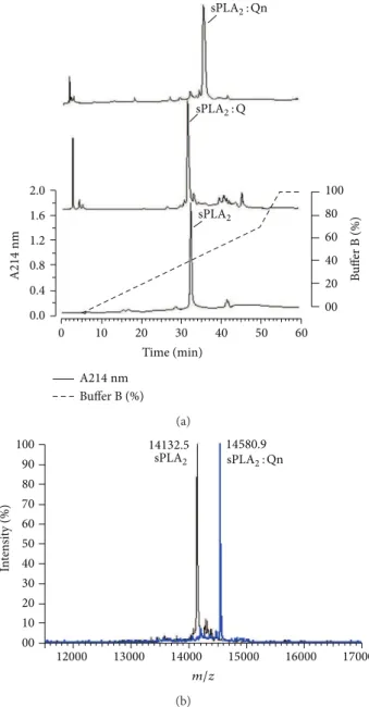
Figure1(a)shows the chromatography proiles of the eluted native sPLA2, sPLA2: Q, and sPLA2: Qn. he retention times of native sPLA2, sPLA2: Q, and sPLA2: Qn were 32.5, 31.8, and 33.5 minutes, respectively. All sPLA2, sPLA2: Q, and sPLA2: Qn samples were lyophilized and stored for future analysis. Figure1(b)shows the mass spectrometry proile of native sPLA2and sPLA2: Q, which was the same as that found by [21]; this inding shows that the methods used here were stable, and they generated reliable and accurate data. Further-more, the analysis result of sPLA2mass spectrometry: Qn was 14580.90, so this mass is the product of sPLA2incubation with Qn. hus, Figure1(b)only shows the results of sPLA2: Qn, and the results of the incubation of the product of sPLA2: Q were presented in the Figure1(b).
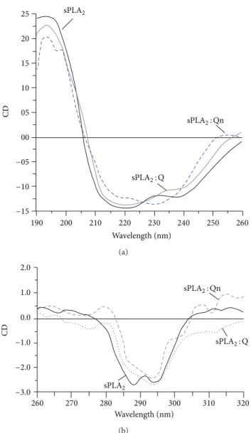
3.2. Circular Dichroism Analysis. Figure2shows the circu-lar dichroism proile of the native sPLA2, sPLA2: Qn, and sPLA2: Q, which were subjected to the same test conditions. he far-UV region (ultraviolet) ranging between 190 and 260 nm was used to reveal important features of its secondary structure. he results are shown in Figure 2(a), indicating that Qn was able to induce some secondary modiications in native sPLA2in comparison with quercetin (Q). In addition, Qn was able to induce a signiicant change in the random coil region of native sPLA2. he near-UV CD spectrum (>250 nm) of protein provides information on the tertiary structure. he signals obtained in the 250–300 nm region are caused by absorption, dipole orientation, and the nature of the environment surrounding the phenylalanine, tyrosine, cysteine (or S-S disulide bridges), and tryptophan amino
2.0
1.6
1.2
0.8
0.4
0.0
0 10 20 30 40 50 60
Time (min)
100
80
60
40
20
00
A
214
nm
B
uf
er B (%)
Bufer B (%)
sPLA2
A214nm
sPLA2: Qn
sPLA2: Q
(a)
100
90
80
70
60
50
40
30
20
10
00
12000 13000 14000 15000 16000 17000
m/z
In
ten
si
ty (%)
14132.5 14580.9
sPLA2 sPLA2: Qn
(b)
Figure 1: Puriication and chemical modiication of secretory
phospholipase A2(sPLA2). A fractionation of the whole venom was
performed by reverse-phase HPLC (C5 column, 0.10 cm×25 cm)
using a nonlinear concentration gradient of bufer to obtain a
high-purity protein. (a) shows a comparative proile of native sPLA2,
sPLA2: Q, and sPLA2: Qn when subjected to reverse-phase HPLC.
(b) shows the MALDI-TOF mass spectrometry analysis of native
sPLA2 and sPLA2: Qn, indicating the diference in the molecular
mass corresponding to one molecule of bound quercitrin.
acids. Figure 2(b) shows the UV CD spectrum of native sPLA2, sPLA2: Q, and sPLA2: Qn, and from these results, the previous native sPLA2treatment with Qn induced more evident tertiary shits than native sPLA2.
3.3. Molecular Docking of sPLA2 with Compounds. In
25
20
15
10
05
00
−05
−10
−15
CD
190 200 210 220 230 240 250 260
Wavelength (nm) sPLA2
sPLA2: Qn
sPLA2: Q
(a)
CD
Wavelength (nm) 2.0
1.0
0.0
−1.0
−2.0
−3.0
260 270 280 290 300 310 320
sPLA2
sPLA2: Qn
sPLA2: Q
(b)
Figure 2: he far-UV (ultraviolet) CD spectrum of proteins can reveal important characteristics of their secondary structure. (a)
shows the results of CD spectra from native sPLA2, sPLA2: Q, and
sPLA2: Qn. Data from 185–280 nm are shown. he CD spectra are
expressed in theta machine units in millidegrees. he near-UV
CD spectrum (>250 nm) of proteins provides information on the
tertiary structure. he signals obtained in the 250–300 nm region are caused by the absorption, dipole orientation, and the nature of the surrounding environment around the phenylalanine, tyrosine, cysteine (or S-S disulide bridges), and tryptophan amino acids. (b)
shows the near-UV CD spectrum of the native sPLA2, sPLA2: Q, and
sPLA2: Qn.
model [22], and they were used to analyze the probable pref-erential orientation of the ligands (Q and Qn) in a complex with theC. d. terriicussPLA2. Based on the docking scores, this computational analysis showed that Qn has a higher ainity for the active site ofC. d. terriicus sPLA2 than Q. he Avogadro v.0.9.4 (http://avogadro.openmolecules.net/) program was used to generate an in silico model of Qn and improve its overall structure through a steepest-descent algorithm for energy minimization based on the MMF94
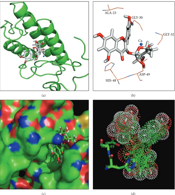
force ield. All docking simulations between the Qn model and theC. d. terriicussPLA2crystallographic structure (PDB ID 2QOG) [22] were performed with the GOLD v.5.0.1 (CCDC Sotware Limited, Cambridge, UK) program [23]. he docking site was deined by a 10 ˚A radius around the His48 residue, which was located at the catalytic site of the A and C monomers of theC. d. terriicussPLA2crystallographic structure. Additionally, other cavities on the protein surface were also tested to identify other potential docking sites. he N�1 atoms from the catalytic histidine in the C. d. terriicussPLA2crystallographic model were protonated, and the simulations generated approximately 1000 docking solu-tions to provide a representative population. he remaining docking parameters were deined according to the GOLD v.5.0.1 default settings. he docking solutions between Qn and theC. d. terriicussPLA2structural model were scored and rescored by using the GoldScore itness function. As shown in Figure3, the main interactions between the ligand and the protein involve amino acid residues Asp49, His48, and Gly30 and the Ca2+ion.
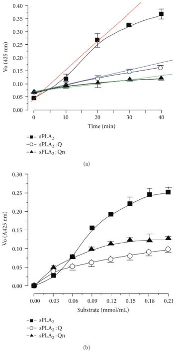
3.4. Enzymatic Assays. All enzymatic assays yield a product that is linear over a short period of time at an initial rate ater the beginning of the enzyme activity (when performed under appropriate conditions). he linear slope indicates that the rate of the enzymatic reaction and the increase in product formation are proportional to the enzyme reaction. As the reaction proceeds, the substrate is consumed and the acceleration decreases. Figure 4(a) shows the time-course efect of an enzymatic reaction. he native sPLA2exhibited a linear rate increase over a 20 min reaction and the sPLA2: Q and sPLA2: Qn experienced a reduction in enzymatic activity of approximately57 ± 4% and63 ± 12%, respectively, in the same time period (Figure4(a)). In fact, there is no statistically signiicant diference in Figure4(a)between both inhibitors at 20 minutes of enzyme kinetic experiments. he data in Figure4(a)suggest a trend towards greater Qn inhibition over Q. he results of Figure4(a)show that the saturation of the active site of sPLA2 in the presence of Qn already occurs ater 40 minutes whereas the sPLA2 incubated with Q, the active site of sPLA2is saturated ater 30 minutes. hese results suggest that the inhibition proile of Q to Qn is diferent and that these compounds have slightly diferent inhibition capabilities of sPLA2 when it is puriied from the Crotalus
durissus terriicus venom, and the inhibitions induced by
Q or Qn were statistically similar. he sPLA2 of Crotalus
durissus terriicus has been characterized as an allosteric
enzyme in the presence of 4-nitro-3-(octanoyloxy)benzoic acid (NOBA or NOB), which is a chromogenic substrate speciic for phospholipase A2[25–27]. Figure4(b)shows the substrate efects on the sPLA2activity, and the native sPLA2 exhibited a Vmax value of0.254 ± 0.09and a Km value of
0.08 ± 0.002, whereas sPLA2: Qn and sPLA2: Q had Vmax
values of 0.12 ± 0.03 and 0.10 ± 0.03, and Km values of
0.04 ± 0.002and0.051 ± 0.004, respectively.
3.5. Pharmacological Assays. he native sPLA2 had a
(a)
ALA-23
GLY-32
GLY-30
Ca
ASP-49
HIS-48
(b)
(c) (d)
Figure 3: Structural representation of Q bound to sPLA2from docking simulations. (a) shows a cartoon representation of the sPLA2structure.
quercetin is shown in a stick representation, and the Ca2+ion is represented as a blue sphere. (b) shows the quercetin molecule and its main
amino acid interactions. (c) shows a surface representation of sPLA2bound to a quercetin molecule (stick representation). (d) shows the dot
representation of the Quercetin molecule in the bound position. Two amino acid residues (Asp49 and His48) from sPLA2are represented as
sticks.
a swelling value of0.27 ± 0.06mL (� = 5, and∗� < 0.05)
and0.32 ± 0.04mL (� = 5, and∗� < 0.05) for this time
interval. Within the same time interval, sPLA2: Q showed maximum edema of0.18 ± 0.04mL (� = 5,∗� < 0.05) and
0.28 ± 0.05mL (� = 5,∗� < 0.05) in the same time interval.
Furthermore, sPLA2: Qn showed maximum edema values of
0.18 ± 0.05mL (� = 5, ∗� < 0.05) and 0.023 ± 0.05mL
(� = 5,∗� < 0.05), respectively. hese results showed that
both Q and Qn signiicantly inhibit sPLA2enzyme activity, and the inhibition by Qn was two times higher than that of Q (Figure5(a)). Figure5(b)shows the myotoxic activity induced by native sPLA2, sPLA2: Q, and sPLA2: Qn. he extent of the damage caused by sPLA2to skeletal muscles was
assessed by quantifying the CK levels, which are widely used as an indirect marker of muscle damage. For trials with snake toxins, CK is used as a marker to assess the damage to skeletal muscles in the presence of snake venom. hree hours ater the native sPLA2injection, the CK value was 1,230 ± 270U/L
(� = 5,∗� < 0.05). For the sPLA2: Q and sPLA2: Qn, the
serum CK levels were780 ± 120U/L (� = 5,∗� < 0.05) and
680 ± 69(� = 5,∗� < 0.05), respectively.
In addition to trials with sPLA2s that had been chemically treated with both lavonoids, assays in which the animals were pretreated with 100�L (0.3 mM/mL, IP injection,� = 5and
∗� < 0.05) of Q and Qn were also performed. Figure6(a)
0.40
0.35
0.30
0.25
0.20
0.15
0.10
0.05
0.00
Vo
(
42
5
nm)
0 10 20 30 40
Time (min) sPLA2
sPLA2: Qn
sPLA2: Q
(a)
0.30
0.25
0.20
0.15
0.10
0.05
0.00
Vo
(
A
42
5
nm)
0.00 0.03 0.06 0.09 0.12 0.15 0.18 0.21
Substrate (mmol/mL) sPLA2
sPLA2: Qn
sPLA2: Q
(b)
Figure 4: (a) shows the results of the enzymatic activity assays that were performed by using a synthetic chromogenic substrate for
PLA2 (NOBA). he reaction was monitored at 425 nm. sPLA2: Q
and sPLA2: Qn exhibited a signiicant decrease in activity when
compared to native sPLA2. (b) shows the efect of the substrate
concentration on enzyme activity in the presence of native sPLA2,
sPLA2: Q, and sPLA2: Qn.
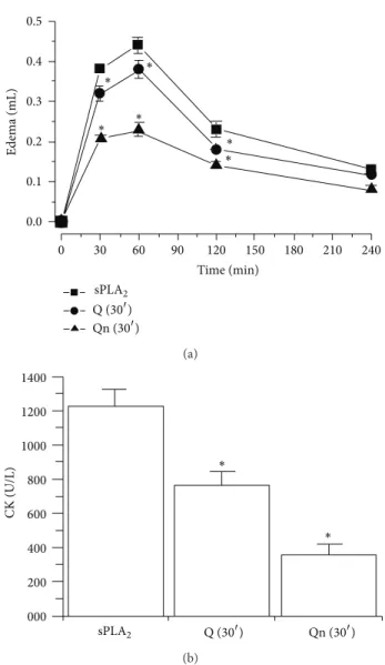
he results of the edema assay for animals receiving 0.9% saline (100�L, IP injection,� = 5and∗� < 0.05) were0.38 ±
0.06mL (� = 5and∗� < 0.05) and0.44 ± 0.05mL (� = 5and
∗� < 0.05) at 30 min and 60 min into the edema time-course
experiment ater the injection of native sPLA2, respectively. he animals that received Q (30�) had0.28 ± 0.03mL edema values at 30 min (� = 5,∗� < 0.05) and0.38 ± 0.08mL at 60 min (� = 5,∗� < 0.05). he animals treated with Qn
0.4
0.3
0.2
0.1
0.0
0 30 60 90 120 150 180 210 240
Time (min)
Edema (mL)
∗ ∗ ∗
∗
sPLA2
sPLA2: Qn
sPLA2: Q
(a)
∗
∗ 1000
800
600
400
200
000
CK (U/L)
sPLA2 sPLA2: Q sPLA2: Qn
(b)
Figure 5: (a) shows the results of paw edema that was induced
ater the injection of sPLA2, sPLA2: Q, and sPLA2: Qn into the right
paws of Swiss mice. Measurements were made ater 30, 60, 120, and 240 min, and all the edema results expressed in (a) were obtained by subtracting the saline injection values. (b) shows the myonecrosis levels as evaluated by CK levels in Swiss mice. Fity micrograms of
native sPLA2, sPLA2: Q, and sPLA2: Qn at a inal concentration of
0.5 mg/mL were injected into the gastrocnemius muscle. he results are expressed as units of enzymatic activity per liter (U/L). Error bars
indicate the SEM.∗� < 0.05compared to native sPLA2.
(30�) had swelling times of0.21 ± 0.04mL at 30 min (� = 5,
∗� < 0.05) and0.23 ± 0.09mL at 60 min (� = 5,∗� < 0.05).
∗
∗ ∗
∗
∗ ∗
Q (30)
Qn (30)
0.5
0.4
0.3
0.2
0.1
0.0
0 30 60 90 120 150 180 210 240
Time (min)
Edema (mL)
sPLA2
(a)
∗ ∗
CK (U/L)
1400
1200
1000
800
600
400
200
000
Q (30) Qn (30)
sPLA2
(b)
Figure 6: (a) shows the results from paw edema in the animals that
were injected with quercitrin (Qn 30�) and quercetin (Q 30�) 30 min
before sPLA2administration into the right paw of Swiss mice. he
control group received a saline injection prior to the administration
of sPLA2. Measurements were made ater 30, 60, 120, and 240 min,
and all edema results expressed in (a) were obtained ater subtracting the edema values from the saline injection. (b) shows the results of
paw edema in animals that were injected with quercitrin (Qn 30�)
or quercetin (Q 30�) 30 min before the administration of sPLA2.
he control group received saline. Myonecrosis was evaluated on
the basis of CK levels ater 50 mg of native sPLA2 was injected at
a inal concentration of 0.5 mg/mL into the gastrocnemius muscle. he results are expressed as units of enzymatic activity per liter
(U/L). Error bars indicate the SEM.∗� < 0.05compared to native
sPLA2.
terriicus. Qn exhibited a neutralizing efect that was two
times higher than the efect induced by Q.
4. Discussion
Quercetin (Q) is considered one of the most abundant natural lavonoids and it is mainly found in fruits and other foods.
Quercetin is typically consumed in its glycosylated form as quercitrin (Qn), but multiple studies carried out with the aglycone form demonstrated its potent anti-inlammatory efect. However, thein vivo efectiveness of this compound has been questioned. he results of experiments onin vivo models of inlammation showed that Qn was more efective in reducing inlammation in comparison to Q, which showed better results in thein vitroassays [18,28,29].
herefore, to shed some light on the inhibitory role of Qn in the inlammatory process, the efect of this lavonoid was evaluated in a typical sPLA2 puriied from the venom ofC. d. terriicusby using several experimental and theoret-ical methods, including chromatography, circular dichroism, molecular docking, and otherin vitroandin vivobiological assays. Chromatography showed that binding to Q did not change the retention time of sPLA2 relative to sPLA2: Qn samples, which exhibited a longer retention time than the native sPLA2 and sPLA2: Q. his inding suggests that Qn may have caused structural changes in sPLA2, as also indi-cated by the results from circular dichroism and luorescence scanning assays. hese structural changes may be caused by the molecular interactions of Qn with the C. d. terriicus sPLA2, which could involve hydrogen bonding, hydrophobic and electrostatic interactions between Qn and the amino acid residues Gly 30, Gly 32, His 48, and Asp 49 and the Ca2+ion as suggested by the molecular docking results. Indeed, crystal complexes of porcine pancreatic phospholipase A2/berberine (PDB ID 4DBK),Daboia russelii pulchellasPLA2/berberine (PDB ID 2QVD, [30]), and acidicBothrops jararacussusPLA2 (BthA-I)/p-bromophenacyl bromide presented similar lig-and/protein interactions to those of theC. d. terriicussPLA2/ Qn docking complex, that is, involving amino acids from the Ca2+-binding loop (e.g., Gly 30) and catalytic site (e.g., Asp 49 and His 48).
he results of the enzyme kinetic studies show that the inhibition induced by quercitrin (Qn) is not the same as that observed for quercetin (Q), and this inding is apparent ater 40 minutes (Figure 4(a)). his diference in the inhibitory capacity of Q and Qn against the sPLA2 from Crotalus
durissus terriicus is supported by the results shown in
Figure4(b), which demonstrate the kinetic behavior of native sPLA2, of sPLA2with quercetin, and sPLA2with quercitrin. he diference between the lavonoids is most likely caused by the presence of a rhamnose sugar in Qn in accordance with the docking studies presented in Figure3, which shows the insertion of Qn in the hydrophobic channels of sPLA2. Rhamnose appears to inhibit the substrate’s access to the sPLA2catalytic site.
the sPLA2that was pretreated with Q or Qn indicate that the calcium loop region may be involved in the molecular inter-action between the sPLA2 fromCrotalus durissus terriicus and the receptors. In previous studies, Lambeau et al. used a Ca2+loop mutant derived from sPLA2that was isolated from venom to demonstrate the importance of this loop in the sPLA2interaction with the M-type receptor [31].
sPLA2 from venom has been found to interact with a variety of mammalian sPLA2-binding proteins, such as N-and M-type receptors, 14-3-3 proteins N-and calmodulin, pen-traxins and associated proteins, crocalbin, pulmonary sur-factant proteins, KDR VEGF receptor 2, and factor Xa [32]. Furthermore, Rouault et al. also demonstrated that not only is the calcium binding loop region involved in binding to the receptor, but the interfacial binding domain is also involved. hus, the stereochemical inhibition from when the substrate was binding to the active site of sPLA2 (as induced by Qn) could explain the diferent degrees of inhibition for quercetin (Q) and quercitrin (Qn).
Catalytically active sPLA2can induce various biological and pathological efects, as in the sPLA2 present in snake venom. Generally, PLA2causes these biological, physiologi-cal, and pathological activities through its enzymatic activity, which result in the increased production of arachidonic acid, which is the rate-limiting step in the generation of eicosanoids and platelet activating factors. his efect is caused by increased levels of intracellular arachidonic acid that stimulate the activity of cyclooxygenase 2 [33–35] and induce an increase in free radical peroxides and pro inlam-matory cytokines. he increased levels of hydrogen peroxide may therefore lead to an increase in the lipid peroxidation levels, which can lead to cell membrane lesions such as those in skeletal muscle cells. Furthermore, sPLA2can reportedly increase the mobilization of internal calcium through an indi-rect mechanism. his mobilization may lead to the activation of calpain, a member of a cytoplasmic protease family that can stimulate the activity of xanthine oxidase. his activity can lead to an increase in the concentration of molecular oxygen and may further exacerbate cellular injury [36,37]. Several studies showed that Q and Qn are potent antioxidants. Results obtained by other authors demonstrated that the protective efect of these two lavonoids may be caused by their ability to neutralize the cytotoxic action of free radicals [38,39]. he diference in the levels of protective or neutralizing efects observed between Q and Qn treatments may be caused by the presence of rhamnose because the only diference between Q and Qn is the presence of this sugar.
According to Lespade et al., [40], Kim et al., [41] glycosy-lation may increase the antioxidant properties of lavonoids [40,41]. Moreover, Qn confers better protection than Q in some cases by protecting cells from ROS generation as well as ROS side efects [42]. he results in Figure 6show that pretreating animals with Q and Qn can greatly reduce the toxic activity of sPLA2. hese results also suggest that the action of these compounds occurs at the intracellular level and involves the neutralization of ROS and ROS side efects such as the activation and enhancement of the inlammation cascade. he presence of rhamnose in Qn is crucial to its
protective activity against sPLA2fromCrotalus durissus terri-icusin bothin vitroandin vivostudies, which indicates that quercitrin (Qn) is more efective than quercetin (Q) at the cellular level. Qn inhibits the interfacial binding domain of the sPLA2fromCrotalus durissus terriicusfrom interacting with its receptor.
Conflict of Interests
he authors have no conlict of interests to disclose.
Acknowledgments
he authors are grateful to the Coordenadoria de Aperfeic¸oa-mento de Pessoal de N´ıvel Superior (CAPES), the Fundo Mackenzie de Pesquisa, the Conselho Nacional de Desen-volvimento Cient´ıico e Tecnol´ogico (CNPq), and the Fun-dac¸˜ao de Amparo `a Pesquisa do Estado de S˜ao Paulo (FAPESP) for their inancial support (FAPESP Proc. nos. 2011/06704-4, 2012/06502-5, and 2013/12077-8) and to the Instituto Nacional para Pesquisa em Toxinas (INCT-Tox).
References
[1] D. J. Rigden, L. W. Hwa, S. Marangoni, M. H. Toyama, and I.
Polikarpov, “he structure of the D49 phospholipase A2
pira-toxin III fromBothrops pirajaireveals unprecedented structural
displacement of the calcium-binding loop: possible relationship
to cooperative substrate binding,”Acta Crystallographica D, vol.
59, part, 2, pp. 255–262, 2003.
[2] F. H. R. Fagundes, R. Apar´ıcio, M. L. Dos Santos et al., “A
catalyt-ically inactive Lys49 PLA2 isoform fromBothrops jararacussu
venom that stimulates insulin secretion in pancreatic beta cells,”
Protein and Peptide Letters, vol. 18, no. 11, pp. 1133–1139, 2011.
[3] G. Lambeau and M. H. Gelb, “Biochemistry and physiology
of mammalian secreted phospholipases A2,”Annual Review of
Biochemistry, vol. 77, pp. 495–520, 2008.
[4] M. Murakami, Y. Taketomi, H. Sato, and K. Yamamoto,
“Secreted phospholipase A2revisited,”Journal of Biochemistry,
vol. 150, no. 3, pp. 233–255, 2011.
[5] M. Murakami and G. Lambeau, “Emerging roles of secreted
phospholipase A2enzymes: an update,”Biochimie, vol. 95, no.
1, pp. 43–50, 2013.
[6] B. S. Vishwanath, A. A. Fawzy, and R. C. Franson,
“Edema-inducing activity of phospholipase A2 puriied from human
synovial luid and inhibition by aristolochic acid,”
Inlamma-tion, vol. 12, no. 6, pp. 549–561, 1988.
[7] A. Razpotnik, I. Kriˇzaj, J. ˇSribar et al., “A new phospholipase A2
isolated from the sea anemone Urticina crassicornis—its
pri-mary structure and phylogenetic classiication,”FEBS Journal,
vol. 277, no. 12, pp. 2641–2653, 2010.
[8] R. M. Ximenes, R. S. Alves, T. P. Pereira et al., “Harpalycin 2 inhibits the enzymatic and platelet aggregation activities of
PrTX-III, a D49 phospholipase A2 from Bothrops pirajai
venom,”BMC Complementary and Alternative Medicine, vol. 12,
article 139, 2012.
[9] F. A. Marangoni, L. A. Ponce-Soto, S. Marangoni, and E. C.
puriication and pharmacological and structural
characteriza-tion of new PLA2Bleu TX-III,”BioMed Research International,
vol. 2013, Article ID 941467, 9 pages, 2013.
[10] G. U. Meduri and C. R. Yates, “Systemic
inlammation-asso-ciated glucocorticoid resistance and outcome of ARDS,”Annals
of the New York Academy of Sciences, vol. 1024, pp. 24–53, 2004. [11] R. D. Di Villa Bianca, C. Coletta, E. Mitidieri et al., “Hydrogen sulphide induces mouse paw oedema through activation of
phospholipase A2,”British Journal of Pharmacology, vol. 161, no.
8, pp. 1835–1842, 2010.
[12] A. K. Mahalka and P. K. Kinnunen, “Class speciic peptide
inhibitors for secretory phospholipases A2,”Biochemical and
Biophysical Research Communications, vol. 436, no. 2, pp. 349– 353, 2013.
[13] V. D. Mouchlis, E. Barbayianni, T. M. Mavromoustakos, and G. Kokotos, “he application of rational design on phospholipase
A2inhibitors,”Current Medicinal Chemistry, vol. 18, no. 17, pp.
2566–2582, 2011.
[14] M. Toyama, S. D. Rodrigues, D. O. Toyama et al.,
“Phospholi-pases A2protein structure and natural products interactions in
development of new pharmaceuticals,” inProtein Structure, E.
Faraggi, Ed., 2012.
[15] R. J. Nijveldt, E. Van Nood, D. E. C. Van Hoorn, P. G. Boelens, K. Van Norren, and P. A. M. Van Leeuwen, “Flavonoids: a review of probable mechanisms of action and potential applications,”
American Journal of Clinical Nutrition, vol. 74, no. 4, pp. 418– 425, 2001.
[16] P. Rathee, H. Chaudhary, S. Rathee, D. Rathee, V. Kumar, and K. Kohli, “Mechanism of action of lavonoids as anti-inlammatory
agents: a review,”Inlammation and Allergy, vol. 8, no. 3, pp.
229–235, 2009.
[17] M. Comalada, D. Camuesco, S. Sierra et al., “In vivoquercitrin
anti-inlammatory efect involves release of quercetin, which
inhibits inlammation through down-regulation of the NF-�B
pathway,”European Journal of Immunology, vol. 35, no. 2, pp.
584–592, 2005.
[18] F. S´anchez De Medina, B. Vera, J. G´alvez, and A. Zarzuelo, “Efect of quercitrin on the early stages of hapten induced
colonic inlammation in the rat,”Life Sciences, vol. 70, no. 26,
pp. 3097–3108, 2002.
[19] J. Mendez, A. R. Bilia, and I. Morelli, “Phytochemical inves-tigations of Licania genus. Flavonoids and triterpenoids from
Licania pittieri,”Pharmaceutica Acta Helvetiae, vol. 70, no. 3, pp.
223–226, 1995.
[20] S. C. B. Oliveira, F. V. Fonseca, E. Antunes et al., “Modulation
of the pharmacological efects of enzymatically-active PLA2by
BTL-2, an isolectin isolated from the Bryothamnion triquetrum
red alga,”BMC Biochemistry, vol. 9, no. 1, article 16, 2008.
[21] C. A. Cotrim, S. C. B. De Oliveira, E. B. S. Diz Filho et al., “Quer-cetin as an inhibitor of snake venom secretory phospholipase A2,”Chemico-Biological Interactions, vol. 189, no. 1-2, pp. 9–16, 2011.
[22] D. P. Marchi-Salvador, L. C. Corrˆea, A. J. Magro, C. Z. Oliveira, A. M. Soares, and M. R. M. Fontes, “Insights into the role of oligomeric state on the biological activities of crotoxin: crystal
structure of a tetrameric phospholipase A2 formed by two
isoforms of crotoxin B from Crotalus durissus terriicus venom,”
Proteins, vol. 72, no. 3, pp. 883–891, 2008.
[23] G. Jones, P. Willett, and R. C. Glen, “Molecular recognition of receptor sites using a genetic algorithm with a description of
desolvation,”Journal of Molecular Biology, vol. 245, no. 1, pp. 43–
53, 1995.
[24] R. M. Ximenes, M. M. Rabello, R. M. Ara´ujo et al.,
“Inhi-bition of neurotoxic secretory phospholipases A2 enzymatic,
edematogenic, and myotoxic activities by harpalycin 2, an
isolavone isolated fromHarpalyce brasilianabenth,”
Evidence-Based Complementary and Alternative Medicine, vol. 2012, Article ID 987517, 9 pages, 2012.
[25] D. D. O. Toyama, E. B. D. S. Diz Filho, B. S. Cavada et al., “Umbelliferone induces changes in the structure and
pharma-cological activities of Bn IV, a phospholipase A2isoform isolated
fromBothrops neuwiedi,”Toxicon, vol. 57, no. 6, pp. 851–860, 2011.
[26] E. B. S. Diz Filho, S. Marangoni, D. O. Toyama et al., “Enzymatic
and structural characterizationof new PLA2 isoform isolated
from white venom ofCrotalus durissus ruruima,”Toxicon, vol.
53, no. 1, pp. 104–114, 2009.
[27] M. H. Toyama, D. G. de Oliveira, L. O. Beriam, J. C. Novello, L. Rodrigues-Simioni, and S. Marangoni, “Structural, enzymatic
and biological properties of new PLA2 isoform fromCrotalus
durissus terriicusvenom,”Toxicon, vol. 41, no. 8, pp. 1033–1038, 2003.
[28] C. F. Lin, Y. L. Leu, S. A. Al-Suwayeh, M. C. Ku, T. L. Hwang, and J. Y. Fang, “Anti-inlammatory activity and percutaneous absorption of quercetin and its polymethoxylated compound
and glycosides: the relationships to chemical structures,”
Euro-pean Journal of Pharmaceutical Sciences, vol. 47, no. 5, pp. 857– 864, 2012.
[29] X. Dai, Y. Ding, Z. Zhang, X. Cai, and Y. Li, “Quercetin and quercitrin protect against cytokine-induced injuries in RINm5F
�-cells via the mitochondrial pathway and NF-�B signaling,”
International Journal of Molecular Medicine, vol. 31, no. 1, pp. 265–271, 2013.
[30] D. N. Chandra, G. K. Prasanth, N. Singh et al., “Identiication of
a novel and potent inhibitor of phospholipase A2in a medicinal
plant: crystal structure at 1.93 ˚A and Surface Plasmon
Reso-nance analysis of phospholipase A2complexed with berberine,”
Biochimica et Biophysica Acta, vol. 1814, no. 5, pp. 657–663, 2011. [31] G. Lambeau, P. Ancian, J.-P. Nicolas et al., “Structural elements
of secretory phospholipases A2 involved in the binding to
M-type receptors,”he Journal of Biological Chemistry, vol. 270, no.
10, pp. 5534–5540, 1995.
[32] M. Rouault, C. Le Calvez, E. Boilard et al., “Recombinant production and properties of binding of the full set of mouse
secreted phospholipases A2 to the mouse M-type receptor,”
Biochemistry, vol. 46, no. 6, pp. 1647–1662, 2007.
[33] W. K. Han, A. Sapirstein, C. C. Hung, A. Alessandrini, and J.
V. Bonventre, “Cross-talk between cytosolic phospholipase A2�
(cPLA2�) and secretory phospholipase A2(sPLA2) in hydrogen
peroxide-induced arachidonic acid release in murine mesangial
cells: sPLA2 regulates cPLA2�activity that is responsible for
arachidonic acid release,”he Journal of Biological Chemistry,
vol. 278, no. 26, pp. 24153–24163, 2003.
[34] M. Chalimoniuk, “Secretory phospholipase A2 and its role in
oxidative stress and inlammation,”Postepy Biochemii, vol. 58,
no. 2, pp. 204–208, 2012.
[35] H. Nakamura, K. Yasufuku, T. Makiyama, I. Matsumoto, H. Fujino, and T. Murayama, “Arachidonic acid metabolism via
cytosolic phospholipase A2�induces cytotoxicity in
niemann-pick disease type C cells,”Journal of Cellular Physiology, vol. 227,
no. 7, pp. 2847–2855, 2012.
[36] H. Gissel and T. Clausen, “Excitation-induced CA2+ inlux and
skeletal muscle cell damage,”Acta Physiologica Scandinavica,
[37] H. Gissel, “he role of CA2+ in muscle cell damage,”Annals of the New York Academy of Sciences, vol. 1066, pp. 166–180, 2005. [38] M. A. Aderogba, E. K. Okoh, and T. O. Idowu, “Evaluation of the antioxidant activity of the secondary metabolites from
Piliostigma reticulatum (DC.) Hochst,” Journal of Biological
Sciences, vol. 5, no. 2, pp. 239–242, 2005.
[39] H. K. Sandhar, B. Kumar, S. Prasher, P. Tiwari, M. Salhan, and P. Sharma, “A review of phytochemistry and pharmacology of
lavonoids,”Internationale Pharmaceutica Sciencia, vol. 1, no. 1,
pp. 25–41, 2011.
[40] L. Lespade and S. Bercion, “heoretical investigation of the efect of sugar substitution on the antioxidant properties of
lavonoids,”Free Radical Research, vol. 46, no. 3, pp. 346–358,
2012.
[41] B. H. Kim, J. S. Choi, E. H. Yi et al., “Relative antioxidant activi-ties of quercetin and its structurally related substances and their
efects on NF-�B/CRE/AP-1 signaling in murine macrophages,”
Molecules and Cells, vol. 35, no. 5, pp. 410–420, 2013.
[42] Y. Yin, W. Li, Y. O. Son et al., “Quercitrin protects skin from
UVB-induced oxidative damage,”Toxicology and Applied
Submit your manuscripts at
http://www.hindawi.com
Pain
Research and Treatment
Hindawi Publishing Corporation
http://www.hindawi.com Volume 2014
World Journal
Hindawi Publishing Corporation
http://www.hindawi.com Volume 2014
Hindawi Publishing Corporation http://www.hindawi.com
Volume 2014
Toxins
Journal of
Vaccines
Journal of
Hindawi Publishing Corporation
http://www.hindawi.com Volume 2014
Hindawi Publishing Corporation
http://www.hindawi.com Volume 2014
Antibiotics
Toxicology
Journal of
Hindawi Publishing Corporation
http://www.hindawi.com Volume 2014
Stroke
Research and Treatment
Hindawi Publishing Corporation
http://www.hindawi.com Volume 2014
Drug Delivery
Journal ofHindawi Publishing Corporation
http://www.hindawi.com Volume 2014 Hindawi Publishing Corporation
http://www.hindawi.com Volume 2014 Advances in Pharmacological Sciences
Tropical Medicine
Hindawi Publishing Corporationhttp://www.hindawi.com Volume 2014
Medicinal ChemistryInternational Journal of
Hindawi Publishing Corporation
http://www.hindawi.com Volume 2014
Addiction
Journal ofHindawi Publishing Corporation
http://www.hindawi.com Volume 2014
Hindawi Publishing Corporation
http://www.hindawi.com Volume 2014
BioMed
Research International Emergency Medicine International
Hindawi Publishing Corporation
http://www.hindawi.com Volume 2014
Hindawi Publishing Corporation
http://www.hindawi.com Volume 2014
Diseases
Hindawi Publishing Corporation
http://www.hindawi.com Volume 2014
Anesthesiology Research and Practice
Scientifica
Hindawi Publishing Corporation
http://www.hindawi.com Volume 2014
Journal of
Hindawi Publishing Corporation
http://www.hindawi.com Volume 2014
Pharmaceutics
Hindawi Publishing Corporation
http://www.hindawi.com Volume 2014