The
Chlamydia psittaci
Genome: A Comparative Analysis
of Intracellular Pathogens
Anja Voigt1., Gerhard Scho¨fl1., Hans Peter Saluz1,2
*
1Leibniz-Institute for Natural Product Research and Infection Biology, Jena, Germany,2Friedrich Schiller University, Jena, Germany
Abstract
Background:Chlamydiaceaeare a family of obligate intracellular pathogens causing a wide range of diseases in animals and humans, and facing unique evolutionary constraints not encountered by free-living prokaryotes. To investigate genomic aspects of infection, virulence and host preference we have sequenced Chlamydia psittaci, the pathogenic agent of ornithosis.
Results:A comparison of the genome of the avianChlamydia psittaciisolate 6BC with the genomes of other chlamydial species,C. trachomatis,C. muridarum,C. pneumoniae,C. abortus,C. felisand C. caviae, revealed a high level of sequence conservation and synteny across taxa, with the major exception of the human pathogen C. trachomatis. Important differences manifest in the polymorphic membrane protein family specific for theChlamydiaeand in the highly variable chlamydial plasticity zone. We identified a number ofpsittaci-specific polymorphic membrane proteins of the G family that may be related to differences in host-range and/or virulence as compared to closely relatedChlamydiaceae. We calculated non-synonymous to synonymous substitution rate ratios for pairs of orthologous genes to identify putative targets of adaptive evolution and predicted type III secreted effector proteins.
Conclusions:This study is the first detailed analysis of theChlamydia psittacigenome sequence. It provides insights in the genome architecture ofC. psittaciand proposes a number of novel candidate genes mostly of yet unknown function that may be important for pathogen-host interactions.
Citation:Voigt A, Scho¨fl G, Saluz HP (2012) TheChlamydia psittaciGenome: A Comparative Analysis of Intracellular Pathogens. PLoS ONE 7(4): e35097. doi:10.1371/journal.pone.0035097
Editor:David M. Ojcius, University of California Merced, United States of America
ReceivedDecember 1, 2011;AcceptedMarch 8, 2012;PublishedApril 10, 2012
Copyright:ß2012 Voigt et al. This is an open-access article distributed under the terms of the Creative Commons Attribution License, which permits unrestricted use, distribution, and reproduction in any medium, provided the original author and source are credited.
Funding:This work was supported by the Jena School of Microbial Communication (JSCM), the Bundesministerium fu¨r Bildung und Forschung (BMBF), and the Thu¨ringer Aufbaubank (TAB). The funders had no role in study design, data collection and analysis, decision to publish, or preparation of the manuscript.
Competing Interests:This work was supported by the ThU¨ringer Aufbaubank. This does not alter the authors’ adherence to all the PLoS ONE policies on sharing data and materials, as detailed online in the guide for authors.
* E-mail: [email protected]
.These authors contributed equally to this work.
Introduction
Chlamydia psittaci is the pathogenic agent of ornithosis or psittacosis, a primarily avian respiratory disease with sizeable impact on poultry farming and bird breeding economic returns [1]. It belongs to the family Chlamydiaceae, a small group of extremely successful obligate intracellular pathogens that efficient-ly colonize mucosal surfaces and thrive within a wide variety of animal hosts, including humans [1].
Although birds are the primary targets ofC. psittaci infections [2], transmissions from birds to humans have been reported, especially where humans come into close contact with infected birds on a regular basis, as in the case of veterinarians, poultry farmers, or bird breeders [3–5]. Moreover, C. psittaci has been isolated from a variety of other mammalian hosts, including cattle and other ruminants, horses, and pigs [1,6].
Other medically important members of the familyChlamydiaceae
are the human-specificC. trachomatis and the wide-host-rangeC. pneumoniae. Worldwide,C. trachomatisis a leading cause of sexually transmitted bacterial diseases and ocular infections (trachoma), potentially leading to blindness [7].C. pneumoniaeis transmitted by
respiratory droplets and the causative agent of an atypical pneumonia and other acute respiratory illnesses [8].
Currently, within Chlamydiaceae a total of nine species are organized in the single genusChlamydia:C. trachomatis,C. muridarum,
C. pneumoniae,C. pecorum,C. suis,C. abortus,C. felis,C. caviae, andC. psittaci. All Chlamydiaceae need to infect eukaryote host cells for replication. Despite major differences in host range, tissue tropism, and disease pathology they all share a characteristic biphasic developmental cycle unique among prokaryotes [9,10]. The infectious chlamydial form, the elementary body (EB), enters the eukaryotic cell and becomes internalized in a vacuole in the cytoplasm of the host cell. In this so called inclusion, the EB differentiates into a non-infectious, metabolically active form, the reticulate body (RB). The RB multiplies by binary fission [1,10]. Depending on the strain, two to three days after infection the RBs transform back into EBs, which get released by lysis of the host cell or exocytosis [9].
As a consequence of this life style, the arguably most important driving force for the evolution of these pathogens is the interaction with their peculiar ecological niche, the inclusion within the cytoplasm of a eukaryotic host. Specific genomic manifestations of
the obligate intracellular lifestyle have long been recognized, e.g. a strong trend towards reduced genome sizes, a dramatical reduction of effective population size relative to environmental bacteria making purifying selection comparatively less efficient, extreme sequence divergence in proteins that mediate the interaction with the host environment such as outer surface proteins and secretion systems (reviewed in [11]). Hence, a comparison of genes involved either directly or indirectly in interactions with the host cell is most likely to shed light on the evolution of the intracellular life style of theChlamydiaceae, and their adaptation to different eukaryote hosts.
Currently, relatively little is known regarding the chlamydial factors involved in virulence, host interaction, or host specificity. Genes for which functions in relation to niche adaptation have been implicated are mainly (i) the polymorphic membrane proteins (pmps), a large family of proteins probably unique to the phylumChlamydiae[12,13], and considered to be important in adhesion of the EB to the host cell, molecular transport, and cell wall associated functions [14]; (ii) genes located in the chlamydial ‘‘plasticity zone’’, a distinct region close to the terminus of replication, characterized by unusually high levels of inter- and intraspecific polymorphism [15]; and (iii) the so called type III effector proteins, a highly variable class of proteins potentially secreted into the host cell by the molecular machinery of the type III secretion (T3S) apparatus.
The T3S apparatus and the effectors translocated by it form a sophisticated mechanism of bacterial pathogenesis found in a number of Gram-negative bacteria. It is characterized by the direct translocation of effector proteins into the host cytoplasm to mediate colonization and parasitation of susceptible hosts [16,17]. T3S effector proteins display little sequence homology across species. Although the N-terminal regions of T3S effectors show unusual amino acid compositions (e.g. [18]) no unambiguous common motif among different T3S signal sequences has been established, making the computational prediction of putative T3S effectors a difficult challenge [18,19].
Whole-genome comparison between phylogenetically distant chlamydial species that parasitize a range of host species, vary in their host specificity and pathogenicity can provide a foundation from which to comprehend factors involved in chlamydial niche adaptation. For this study we sequenced the genome of the pathogenic avian type strainChlamydia psittaci6BC. Meanwhile two additional C. psittaci genome sequences have become available [20,21]. This study represents the first detailed analysis of theC. psittaci genome sequence, however. Our results show a typical chlamydial genome with a coding capacity of 967 CDSs. The
comparative analysis of theC. psittacigenome with those of other
Chlamydiaceae confirms the exceptional roles of the family of polymorphic membrane proteins and the chlamydial plasticity zone as source of most interspecies variation. The prediction of putative type III secreted effectors and a genome-wide analyses of non-synonymous to synonymous substitution rate ratios yield a number of novel candidate genes likely involved in host-pathogen interactions and adaptive divergence betweenC. psittaciand their relatives.
Results and Discussion
Genome sequence of the avian isolate 6BC ofC. psittaci
Chlamydia psittaci6BC possesses a single circular chromosome of 1.172 Mb and a plasmid of 7553 bp. The bacterial chromosome is predicted to contain 967 coding sequences (CDSs) and the plasmid is predicted to harbour eight coding sequences. 26% of the CDSs are annotated as encoding hypothetical products. The general features of theC. psittacigenome in comparison to other sequenced chlamydial genomes are summarized in Table 1.
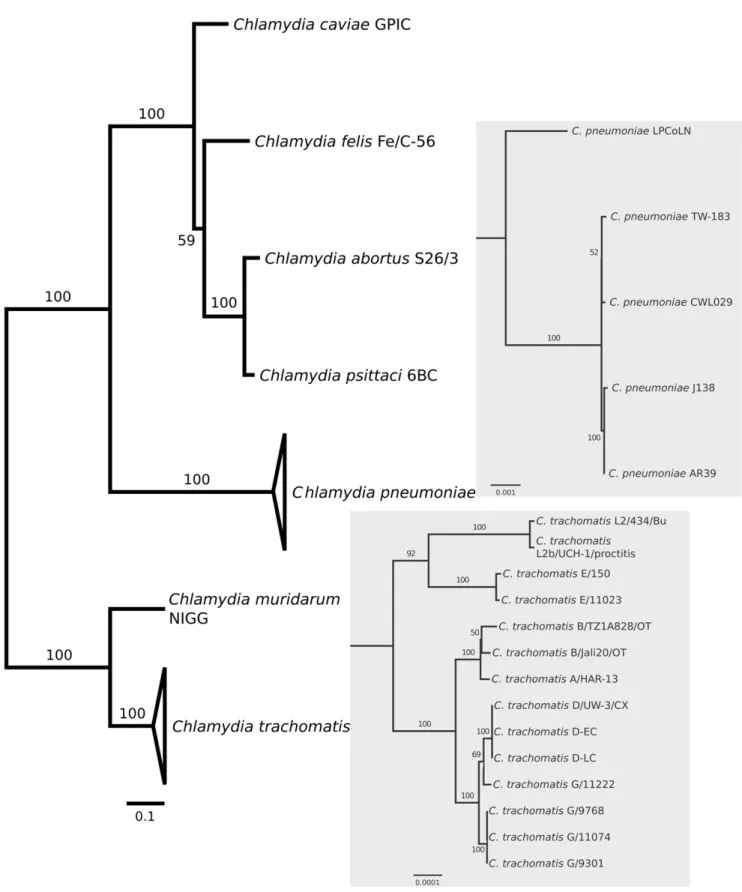
A phylogenetic tree was reconstructed for all species within
Chlamydiaceae for which full-length genomic sequences have become available, including intraspecific variants (Figure 1). The inferred topology is consistent with previous phylogenies of the
Chlamydiaceae(e.g. [22,23]), and shows the close relationship ofC. psittaciwith the three chlamydial species originally considered as the ‘‘mammalian’’Chlamydia psittaci abortion, feline, and Guinea pig strains (i.e.,C. abortus,C. felis, andC. caviae; here referred to as ‘‘psittaci-group’’).
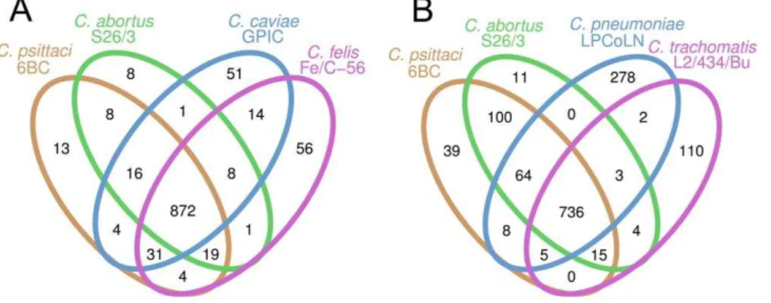
A comparison of the genomes of thepsittaci-group shows that all four species are highly conserved in terms of size, in the number of coding sequences and in nucleotide composition (Table 1). The degree of conservation is illustrated by a comparison of the coding capacity of these genomes: 872 of the predicted CDSs ofC. psittaci
are shared among all four species of thepsittaci-group (with a total CDS count ranging from 967 to 1005), 82 are divergent, and only 13 genes are unique toC. psittaci(Figure 2A). Notably, six of the 13
C. psittacigenes identified as having no significant homology to any other chlamydial species encode polymorphic membrane proteins (pmps) of the G family, most others encode hypothetical proteins with unknown functions (Table 2). Four of these unique genes (CPSIT_0306, CPSIT_0429, CPSIT_0605, and CPSIT_0846) are predicted type III secreted effector proteins (Table 3 and Table S1). Three of these genes are located in the hypervariable chlamydial plasticity zone (CPSIT_603, CPSIT_605, and CPSIT_607, Figure 3), a region at the terminus of replication
Table 1.Summary ofChlamydiaceaegenome features.
C. psittaci6BC C. abortusS26/3 C. felisFe/C-56 C. caviaeGPIC C. pneumoniaeLPCoLN
C. trachomatisL2/434/ Bu
Genome size [bp] 1,171,660 1,144,377 1,166,239 1,173,390 1,241,020 1,038,842
No. of CDSs 967 932 1005 998 1097 874
Coding density [%] 89.39 87.62 91.22 89.42 89.44 89.09
Average gene size [bp]
1083 1075 1059 1051 1012 1059
% G+C content 39.06 39.87 39.38 39.22 40.55 41.33
% G+C of CDSs 39.46 40.24 39.95 39.82 41.26 41.66
tRNA 38 38 38 38 38 37
rRNA operons 1 1 1 1 1 2
Figure 1. Phylogenomic relationships among sequenced chlamydial genomes.The maximum-likelihood tree is based on 100 randomly chosen conserved orthologous genes. Bootstrap values are displayed at the branches. The panels show the within-species phylogenetic relationships of the sequenced genomes ofChlamydia pneumoniae(upper panel) andC. trachomatis(lower panel).
doi:10.1371/journal.pone.0035097.g001
Comparative Genome Analysis ofChlamydia psittaci
that contains an array of chlamydial niche-specific genes putatively important in host-specific interactions [24].
When the more divergent speciesC. pneumoniaeandC. trachomatis
were included in the comparison, 736 of the predicted CDSs (ranging from 874 to 1097) were common to all Chlamydiaceae
(Figure 2B). 64 genes were shared exclusively by C. psittaci, C. abortus, andC. pneumoniae, all members of the recently deprecated genusChlamydophila[23,25] (Figure 2B).
Whole-genome comparisons with the other published Chlamyd-iaceaegenomes show that theC. psittaci6BC genome is essentially syntenic to sequences from C. abortus, C. felis, andC. pneumoniae
(Figure 4).
A high degree of genomic rearrangement, however, becomes apparent with respect toC. trachomatis(Figure 4). Major deviations from synteny and/or sequence conservation are found in a hypervariable region near the replication terminus that has been termed ‘‘plasticity zone’’ (PZ) [15] and among the family of polymorphic membrane protein (pmp) genes [26] (Figure 4). These regions are suspected to harbour key genomic correlates to species-specific adaptation to different environmental niches, differential tissue tropism and differences in virulence and
pathogenicity [27]. Additionally, putative virulence factors, e.g. proteins mediating the chlamydial attachment to the host cell, or those related to the chlamydial inclusion membrane and development, may play a crucial role in niche adaptation, such as members of Inc/Tmh protein family (inclusion membrane proteins and transmembrane head proteins) and type III secreted effector proteins (T3SE) [15,28].
It is interesting to note, that a recent revision of the taxonomy of theChlamydiaceaesaw the genusChlamydiaretained as the sole genus in the family [25], thus deprecating the usage of the genus
Chlamydophilaintroduced by Everett et al. in 1999 [29]. However, the phylogenetic analysis based on concatenated protein sequences (Figure 1), the numbers of shared orthologous genes (Figure 2B), and the level of synteny between genomes (Figure 4) all clearly do support the separation of Chlamydiaceae into two groups that correspond to the former genera, Chlamydia and Chlamydophila. Thus, irrespective of whether by any formal criteriaChlamydophila
can be considered a genus, the label does remain useful as a moniker for an evolutionarily distinct branch within the
Chlamydiaceae.
Plasticity zone and polymorphic membrane proteins
The size and organisation of the plasticity zone differs substantially among Chlamydiaceae (Figure 3). This high level of genetic diversity is thought to correspond to rapid evolution of a set of putative virulence factors which have accumulated in chlamydial PZs. Thus, a number of the genes contained in the chlamydial PZs have been linked to host-pathogen interactions/ pathogenesis, e.g., a MAC/perforin domain gene [30], a cytotoxin gene similar to the EHEC adherence factor and clostridial large cytotoxins [24,31], tryptophan biosynthesis genes [31,32], or phospholipase D family enzymes [33].
In C. psittaci6BC the PZ spans about 29 kb and encodes 16 genes. It has less gene content than the respective plasticity zones ofC. caviaeandC. felis(22 and 29 genes) but is larger than the PZ of
C. abortus(11 genes). These differences arise becauseC. psittaciand
C. abortus lack the complete tryptophan biosynthesis operon (trpABFCDR, kynU, prsA) present in the C. felis and C. caviae
genomes. In contrast toC. abortus, C. psittacishares a putatively functional 10074 bp EHEC-like adherence factor (CPSIT_0606), a 2466 bp MAC/perforin domain gene (CPSIT_0608), and a
guaAB-addcluster serving purine nucleotide interconversion withC. caviaeandC. felis(Figure 3). Three of the hypothetical proteins in
Figure 2. Venn diagrams showing the numbers of predicted CDSs that are unique or shared among two or more taxa. A.Number of shared genes among the closely related C. psittaci, C. abortus,C. caviae, andC. felis; B. number of shared genes among the wider range of
Chlamydiaceae. Pseudogenes were scored as absent in this analysis. doi:10.1371/journal.pone.0035097.g002
Table 2.Predicted CDSs unique toC. psittaci6BC.
CDS Product description
CPSIT_0309 polymorphic membrane protein, G family
CPSIT_0310 polymorphic membrane protein, G family
CPSIT_03111 polymorphic membrane protein, G family
CPSIT_03121 polymorphic membrane protein, G family
CPSIT_03161 polymorphic membrane protein, G family
CPSIT_0330 putative outer membrane protein
CPSIT_04291 hypothetical protein
CPSIT_0603 conserved hypothetical protein
CPSIT_0605 hypothetical protein
CPSIT_0607 hypothetical protein
CPSIT_0661 conserved domain protein
CPSIT_0668 polymorphic membrane protein, G family
CPSIT_08461 putative TMH-family membrane protein
1predicted T3S effector protein.
Table 3.Comparison of predicted type III secreted proteins ofC. psittaci6BC with orthologs in otherChlamydiaceae.
C. psittaci6BC
ORF SVM value Annotation C. abortusS26/3C. felisFe/C-56 C. caviaeGPICC. pneumoniaeLPCoLN C. trachomatisL2/434/Bu
CPSIT0357 1.957 hypothetical protein - 678* 325* -
-CPSIT0192 1.655 orthologous toC. trachomatisTARP 167* 837* 170* 1089* 0716*
CPSIT0429 1.309 hypothetical protein - - - -
-CPSIT0580 1.224 putative inner membrane protein 522* 472* 536* -
-CPSIT0074 1.221 conserved hypothetical serine rich protein 063* 942* 062* -
-CPSIT0422 1.152 conserved hypothetical protein - 618* 390 -
-CPSIT0397 1.146 conserved hypothetical protein 357* 640* 367* 0938* 0528
CPSIT0463 1.109 putative inner membrane protein 412* 582 426 -
-CPSIT0421 1.087 conserved hypothetical protein 376* 619* 389* -
-CPSIT0532 1.087 inclusion membrane protein B 477* 516* 491 0799* 0484*
CPSIT0997 1.080 putative inner membrane protein 910* 073* 941* 0230* 0828*
CPSIT0606 1.061 adherence factor 550 442 558* -
-CPSIT0245 1.054 carbohydrate isomerase 215* 788* 219* 1041* 0656
CPSIT0757 1.050 dihydrodipicolinate reductase 680* 303* 715* 0474* 0618
CPSIT0594 1.005 inclusion membrane protein A 536* 458* 550* -
-CPSIT0220 0.971 cyclodiphosphate synthase 191* 812* 195* 01062 0693
CPSIT0431 0.952 putative membrane protein - 610 397* -
-CPSIT0846 0.896 putative TMH-family/IncA-family protein 766 - - -
-CPSIT0844 0.864 putative TMH-family/IncA-family protein 764* 218 797* -
-CPSIT0689 0.858 conserved hypothetical protein 618 - 647 -
-CPSIT0933 0.846 putative membrane protein 852 129* - 0291*
-CPSIT0314 0.829 polymorphic membrane protein, G family 283* 719* - -
-CPSIT0749 0.769 conserved hypothetical protein 673* - 707 -
-CPSIT0296 0.746 hypothetical serine rich protein 264* 738* 270* -
-CPSIT0785 0.737 conserved hypothetical serine rich protein 706* 277* 739* 0448* 0238*
CPSIT0853 0.707 putative membrane protein 773* 214* 803* -
-CPSIT0602 0.706 conserved hypothetical protein 546 448 - -
-CPSIT0962 0.704 flagellar biosynthesis/type III secretory pathway 876* 106* 908* 0265* 0087
CPSIT0490 0.677 conserved hypothetical serine rich protein 437* 556* 451* 0841* 0338*
CPSIT1042 0.648 deoxyribonucleotide triphosphate pyrophosphatase 952* 031* 982* 0180 0869
CPSIT0828 0.635 DNA recombination protein 747* 234* 779* 0407 0197
CPSIT0760 0.573 hypothetical membrane protein 683* 300* 718 -
-CPSIT0656 0.552 putative integral membrane protein 588* 388* 616 0664* 402*
CPSIT0974 0.549 trigger factor 887* 095* 919* 0254* 0076
CPSIT0313 0.545 polymorphic membrane protein, G family 282* 721* 284* 0958
-Comparative
Genome
Analysis
of
Chlamydia
psittaci
PLoS
ONE
|
www.plos
one.org
5
April
2012
|
Volume
7
|
Issue
4
|
theC. psittaciPZ (CPSIT_0603, CPSIT_0605, CPSIT_0607) are predicted to be type III secreted effector proteins.
Among the recognizable putative toxin genes of the PZ, a full-length version of a chlamydial MAC/perforin domain gene was present in theC. psittaci,C. felis,C. pneumoniaekoala LPCoLN and
C. trachomatisisolates. A MAC/perforin domain protein was absent from theC. abortusgenome [27], and showed frame disruptions in
C. caviaeand theC. pneumoniaehuman isolates [34] (Figure 3). The
C. psittaci MAC/perforin also shows a MIR (protein nannosyl-transferase, IP3R and RyR) domain indicative of a possible ligand transferase function. It shares this feature only with the more distant orthologs inC. pneumoniaekoala LPCoLN andC. trachomatis, but notC. felis[34].
Many eukaryotic MAC/perforin proteins function as mem-brane perforation proteins [35], and are known to play important roles in plant and animal immune response to bacterial infections [36]. The functional role of the MAC/perforin domain genes in
Chlamydiae is unclear. It has been suggested that proteins with MAC/perforin structures might help in evading the host immune response through structural mimicry, e.g. if the assembly of a host MAC/perforin complex is prevented by pathogen-derived MAC/ perforin molecules present on the pathogen’s cell surface [37,38]. It is likely that the chlamydial MAC/perforin genes were obtained by horizontal gene transfer from an eukaryotic source [39,40].
A full-length lymphostatin/EHEC adherence factor is present only inC. psittaci,C. felis, andC. caviae(Figure 3) as well as inC. muridarum(three orthologs [15]) andC. pecorum[41]. This cytotoxin is generally absent from the human-specific pathogens C. trachomatis (except for gene fragments [42,43]) and C. pneumoniae
[15,34].
The second major source of diversity among chlamydial genomes is the group of polymorphic membrane proteins. The pmps are a large protein family likely unique to the Chlamydiae
[12,13]. Pmps are characterized by an unusually high level of mutational change within and across species, suggesting relatively fast evolutionary rates and high selective pressures potentially associated with adaptation to different hosts or immune responses [34,44,45]. They are present in varying numbers ranging from nine inC. trachomatisandC. muridarum[46] to 21 putative CDSs in
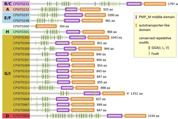
C. pneumoniae[34,47] andC. psittaci(this study; Figure 5). The pmps group phylogenetically into six basic subfamilies (A, B/C, D, E/F, G/I, and H; Figure 6; [46]). Of these subfamilies, family G/I is the largest and the most rapidly evolving with numerous evolutionary recent independent events of gene duplication and loss in the various chlamydial lineages (Figure 6). The tendency to a proliferation of G/I family pmps is especially pronounced among the species belonging to the former genusChlamydophila (i.e. the
psittaci-group, C. pneumoniae, andC. pecorum). While there are only two G/I pmps present inC. trachomatisandC. muridarum, there are 14 pmp G/I family genes present in the C. psittaci genome (Figure 7).
InC. psittaci6BC, one pmp gene is predicted to be truncated on the N-terminal side (CPSIT_0314). Similar to otherChlamydiaceae, a number of pmps harbour long poly-G tracts. Interestingly, while these poly-G stretches appeared to be in frame in the sequence generated by us, in a parallel sequencing effort onC. psittaci6BC [21] the three pmp genes corresponding to CPSIT_0305, CPSIT_0312, and CPSIT_0666 are found to contain frameshifts in these long homopolymeric tracts. Whether this is a sequencing artefact or represents rapid change due to slippage mutations is unclear.
Despite their overall low amino acid and nucleotide similarities, all pmps share a unique domain structure. They contain a C-terminal autotransporter-like domain, a central pmp middle
domain and a varying number of the Chlamydia-specific short tetrapeptide motifs GGA(I, L, V) and FxxN on the N-terminal side [14,44,48] (compare Figure 5). In C. psittaci the numbers of conserved tetrapeptide motifs range from two to 18 for GGA(I, L, V), and from four up to 23 for FxxN. On average 9 FxxN and 4.8 GGA(I, L, V) motifs are found per pmp gene. These numbers are similar to other chlamydial species:C. trachomatis(13.6 and 6.5) and
C. pneumoniae(11.3 and 5.0) [44]. Importantly, it has recently been shown that at least two copies of these repetitive tetrapeptide motives are essential for chlamydial adhesion to the host cell [49].
Putative type III secreted effector proteins
Like a variety of other Gram-negative pathogens,C. psittaciuses a conserved type III secretion machinery as a basic mechanism of virulence determination, that allows transporting specific proteins known as type III secreted effectors (T3SE) into the cytoplasm of their host cells [17,50,51]. Many of the chlamydial T3S effectors are targeted to the inclusion membrane that encapsulates the pathogen inside their host cell [52]. Hence, these effectors are thought to play a crucial role for the modification of the chlamydial environment and for the survival ofChlamydiaein their inclusion vacuole [15].
Figure 3. Comparison of the plasticity zone ofC. psittaci6BC,C. abortusS26/3,C. felisFe/C-56,C. caviaeGPIC,C. pneumoniaeLPCoLN and AR39, andC. trachomatisL2/434/Bu.Genes are labelled with the published locus tag numbers. Colour-coded genes are discussed in the text. Pseudogenes are marked byY.
doi:10.1371/journal.pone.0035097.g003
Figure 4. Global genome comparison between theC. psittaci6BC,C. abortusS26/3,C. felisFe/C-56,C. pneumoniaeLPCoLN, andC. trachomatisL2/434/Bu genomes.The figure shows orthologous matches visualized using genoPlotR (compare Methods). The grey tick marks above and below the sequence lines represent the predicted CDSs on the plus strand and the minus strand of the genomes, respectively. Colour-marked are (blue) members of the polymorphic membrane protein family (pmp) and (green) the position of the plasticity zone (PZ). The red lines connecting genome lines represent direct orthologous matches. The blue lines represent reversed matches. Darker colours correspond to a higher bit scores.
doi:10.1371/journal.pone.0035097.g004
Comparative Genome Analysis ofChlamydia psittaci
To predict potential T3S effector proteins in the genomes ofC. psittaci6BC,C. abortusS26/3,C. felisFe/C-56,C. caviaeGPIC,C. pneumoniae LPCoLN, and C. trachomatis L2/434/Bu we used a support vector machine (SVM) classifier developed by Wang et al. [18] that is based on T3S-specific features extracted from the N-terminal amino acid composition profile of proteins and the prediction software EffectiveT3 [19] (see Methods section).
Chlamydia psittaci CDSs predicted by these methodologies to encode T3S effector proteins and their orthologs in the other investigated species are presented in Table 3 (and Tables S1, S2, S3, S4, S5, S6, S7). Using a decision threshold of 0.5, 40 CDSs are classified as T3S effectors by the SVM algorithm (Table 3) and 68 CDSs are identified by EffectiveT3 (Table S7). 15 CDSs are identified by both approaches (Table 3).
As can be expected, many of the proteins classified as T3S effectors in C. psittaci are homologs to experimentally verified effector proteins from other species. CPSIT_0192, for instance, is orthologous to the important C. trachomatis T3S effector Tarp (translocated actin-recruiting protein) [53]. Tarp orthologs are present in all examined chlamydial species (Table 3). CPSIT_0192 has 100% query coverage and 91% sequence identity toC. abortus
CAB167 and possesses three Chlamydia-specific domains of unknown function (DUF1547) and an actin-binding I/LWEQ domain. Query coverage and sequence identity to C. trachomatis
Tarp CTL0716 are 62% and 37%, respectively. This highlights the high degree of variability in these genes among the
Chlamydiaceae. Even within C. trachomatis variation in the Tarp
sequence has been reported [43]. Despite significant sequence differences to C. trachomatis, both, the C. psittaci and the C. trachomatisTarp are expressed late in the developmental cycle and may have the same function [54,55].
An important family of T3S effectors tightly associated with the inclusion membrane are the Inc proteins. Members of this family show little general sequence similarity, but share a conspicuous bilobed hydrophobic domain of 60–80 amino acid residues [56]. An enrichment for coiled-coil regions typical for eukaryotic organisms has recently been described for putative Incs [12].
The C. trachomatis genome contains seven characterized Inc proteins (Inc A to G). The high sequence diversity in the Inc protein family makes the occurrence of most Inc proteins largely strain-specific. Thus, in C. psittaci, of three characterized Inc proteins A, B, and C only Inc B (CPSIT_0532) is conserved enough to be identified as an ortholog to C. trachomatis Inc B (CTL0484) by reciprocal BLAST.
InC. psittaciboth Inc A and B were classified as T3S effectors by both prediction approaches (Table 3). For both proteins, type III secretion has also been experimentally confirmed inC. psittaciand
C. pneumoniae[54,57]. Based on high immunological activity Inc A was the first Inc protein identified [58]. Inc B modulates host immune responses and might be involved in inclusion develop-ment and prevention of early lysosomal fusion [59]. TheC. psittaci
Inc C (CPSIT_0531) has not been classified as a T3SE by the SVM approach (but it is recognized as a T3SE by EffectiveT3; Table S7). This is likely explained by the complete lack of
sequence homology in the N-terminal region with respect to theC. trachomatis Inc C (CTL0485; query coverage 53%, sequence identity 50%), which is an experimentally verified T3S effector.
Another cluster of genes putatively belonging to the larger Inc protein family and presumably playing similar roles, are the transmembrane head proteins (TMH) [27]. Transmembrane head proteins are characterized by a paired N-terminal transmembrane domain (IncA) followed by alpha-helical coiled-coil domains and show levels of sequence similarity significantly lower than the genome average [27].
In C. psittaci the tmh locus encodes 8 CDSs (CPSIT_0841, CPSIT_0842, CPSIT_0843, CPSIT_0844, CPSIT_0846, CPSIT_0848, CPSIT_0850, CPSIT_0851), all of which harbour an N-terminal IncA domain. The TMH proteins CPSIT_0844 and CPSIT_0846 were classified as possible T3S effectors by both prediction approaches and are orthologous toC. abortusCAB764 and CAB766, respectively (Table 3)
The comparison withC. felisandC. caviaesuggest that the genes encoding the above proteins have arisen from a duplication event in the common ancestor of C. psittaciand C. abortus. The feline
Figure 6. Phylogenetic relationship of chlamydial pmp-family proteins.The maximum-likelihood tree is based on alignments of the conserved PMP_M middle domain and autotransporter domain. Species included in the tree areC. psittaci6BC (CPS),C. abortusS26/3 (CAB),C. felis
Fe/C-56 (CF) ,C. caviaeGPIC (CCA),C. pneumoniaeLPCoLN (CPK),C. muridarumNigg (TC) andC. trachomatisL2/434/Bu (CTL). Pmps cluster into 6 major subfamilies previously designated A (orange), B/C (purple), D (red), E/F (blue), G/I (yellow), and H (green) [44]. Bootstrap values are displayed at the branches. Pseudogenes are marked byY.
doi:10.1371/journal.pone.0035097.g006
Comparative Genome Analysis ofChlamydia psittaci
ortholog CF0218 was shown to be distributed throughout the chlamydial inclusion bodies and confirmed to be immunogenic [60], but has not been classified as a T3S effector by our approach. Besides a number of experimentally confirmed T3S effectors, some proteins with functional annotations that suggest a role in host-pathogen interactions and/or pathogenicity have been classified as T3S effectors. Among this group are a number of genes belonging to the pmp G family (CPSIT_0313, CPSIT_0314 [predicted by SVM], CPSIT_0311, CPSIT_0312, CPSIT_0316 [predicted by EffectiveT3]) and four of the 16 genes located in the plasticity zone. Thus, the adherence factor (CPSIT_0606) located in the PZ (Figure 3) is predicted as T3SE with a high SVM score and by EffectiveT3. Although the adherence factor has orthologs inC. caviaeGPIC,C. abortus S26/3 (only a small gene remnant showing 2% query coverage, but 93% sequence identity) andC. felisFe/C-56 (Table 3), the adherence factor is only predicted to be type III secreted for C. psittaci and C. caviae orthologs. The adherence factors ofC. felis(CF0442) andC. caviae(CCA_00558) show in comparison to the psittacine adherence factor a query coverage of 90%, and a DNA sequence identity of 45% and 44%, respectively, suggesting a high evolutionary turn over.
Selection pressure on chlamydial genomes
The basic measure of selective pressure acting on protein coding sequences is thedN=dS-ratio. Generally, low values ofdN=dS(i.e.,
values,1) are indicative of purifying selection acting on a given protein coding gene, while values.1 are usually interpreted as evidence for positive selection. Theoretically, the strength of purifying selection depends on the effective population size and the specific mutation and recombination rates of the compared lineages. Smaller effective population sizes and less recombination will lead to relatively largerdN=dS-values [61].
To characterize differences in selective pressure among chlamydial lineages on a genome-wide scale, we constructed the distributions ofdN=dS for pairs of chlamydial species over their
respective sets of orthologous genes. In agreement with previous findings [62,63], the shapes of these distributions were highly similar and best fitted by a log-normal distribution (Figure 8).
Figure 8. Distributions of dN=dS for orthologous genes from
pairs of chlamydial genomes. A. Distributions for comparisons among the closely related speciesC. psittaci,C. abortus,C. felis, andC. caviae. B. Distributions for comparisons among the more distantly related species C. psittaci and C. caviae vs. C. pneumoniae and C. trachomatis, respectively. As a point of reference the comparison C. psittacivs.C. abortusis included in both plots. Probability density curves were estimated by Gaussian-kernel smoothing.
doi:10.1371/journal.pone.0035097.g008
Figure 7. Organization of the pmp-family proteins compared betweenC. psittaci6BC,C. abortusS26/3,C. felisFe/C-56,C. pneumoniae
LPCoLN, andC. trachomatisL2/434/Bu.Arrows indicate the gene orientation. The colour code denotes the membership to a pmp-subfamily. CDSs are designated by the numeric part of the published locus tags. Orthologous genes as inferred from the phylogenetic analysis shown in Figure 6, are connected by grey bars. Pseudogenes are marked byY.
Median dN=dS-values fell in the range between 0.06 and 0.1
(Figure 9) and are indicative of the strong evolutionary constraints typical for the compact genomes of prokaryotes [63].
It has been observed that dN=dS correlates negatively with
evolutionary distance, i.e. the smaller the distance between genomes the larger the estimates of dN=dS, thus leading to an
overestimation of positive selection [64]. Such a pattern is not apparent among the chlamydial lineages compared here (Figure 9). In fact, with the notable exception of C. psittaci vs. C. abortus, pairwise comparisons between the closely related lineages within the ‘‘C. psittaci-group’’ show dN=dS-distributions shifted towards
lower values, indicative of higher levels genomic conservation, than comparisons across larger genomic distances (compare Figures 8A vs. 8B and Figure 9). In addition to exhibiting less genomic constraint than other comparisons within the same group of lineages, the C. psittaci/C. abortus comparison also shows the highest overall variance in dN=dS-ratios (interquartile
range = 0.106; interquartile ranges for all other comparisons range from 0.057 to 0.080)
The median dN=dS-values for pairwise comparisons among
chlamydial lineages range between 0.066 and 0.096, and fall thus in the upper third of the range for global median dN=dS-values
typically reported for prokaryotes (0.01–0.1 [63]). This is in line with a trend that the weakest purifying selection pressures are seen in obligate parasites, and is probably explained by their relatively small effective population sizes, frequent bottlenecks and low recombination rates [65].
Although no clear phenotypic correlates are apparent, the variability in purifying selection pressure affecting the evolution of different chlamydial lineages may thus also be a reflection of differences in their effective population sizes and/or the frequency of bottlenecks associated with differences in their life styles (e.g. host preferences or differences in pathogenicity may influence the numbers of infected carriers and thus the effective populations sizes of the pathogens).
Using an (arbitrary) cut-off value of a gene-wide dN=dS-ratio
greater than 0.75 for at least one of the nucleotide substitution models, theC. psittaci/C. abortuscomparison is the only one to give a list of potential candidate genes under positive selection (Table 4).
Metabolic pathways
The C. psittaci 6BC genome encodes for all central metabolic pathways such as the glycolytic pathway and the tricarboxylic acid (TCA) cycle. The TCA cycle of obligate intracellular pathogens varies from complete, in e.g. Coxiella burnetii [66] and Rickettsia prowazekii[67] to absent, in e.g. Mycoplasma [68]. Like all other chlamydial species [69], C. psittaci6BC lacks a number of core components of the tricarboxylic acid cycle, namely citrate synthase, aconitase, and isocitrate dehydrogenase. How Chlamyd-iaceaecompensate for these deficiencies is not clear.
Chlamydiaceae also vary in the completeness of the biotin pathway. LikeC. abortusS26/3,C. felisFe/C56, andC. pneumoniae
LPCoLN, theC. psittaci6BC genome contains all genes necessary for the production of biotin from pimeloyl-CoA (Figure S1). In contrast,C. trachomatisL2/434/Bu has lost several genes from this pathway such as adenosylmethionine-8-amino-7-oxononanoate aminotransferase bioA, dethiobiotin synthetase bioD, and biotin synthasebioB(Figure S1). AlsoC. muridarumandC. caviaeexhibit an incomplete biotin gene cluster [27].
Biotin is an essential cofactor involved in many pathways [70]. The phylogenetic distribution of the deficiencies in the biotin biosynthesis pathway within Chlamydiaceae suggests at least two independent events of gene loss (one in the common ancestor ofC. trachomatisandC. muridarum, and one inC. caviae). This correlates with differences in host specificity. While for C. trachomatis, C. muridarum, andC. caviaeonly one (or, in the case ofC. muridarumtwo closely related) host species has been reported, the host range is markedly broader for the other species [71]. Intracellular organisms generally are prone to loss of function of metabolic genes due to a relaxation of selective constraints in their metabolite-rich environment [72]. This trend may, however, be
Figure 9. Dependence of mediandN=dS on the genetic distance between genomes estimated as the median non-synonymous
substitution rate,dN.
doi:10.1371/journal.pone.0035097.g009
Comparative Genome Analysis ofChlamydia psittaci
exacerbated if a restricted host range leads to a reduced effective population size, and thus to a less efficient selection against deleterious mutations.
Differences between the chlamydial species also exist in the purine and pyrimidine pathways (Figures S2 and S3). The genome of C. psittaci 6BC contains a gene cluster consisting of IMP dehydrogenaseguaB, GMP synthaseguaA, and adenosine deam-inase add relevant for purine interconversion (Figure S2). These genes are also present inC. felis,C. caviae,C. pneumoniaeAR39 (with aguaBpseudogene, however), andC. muridarum[24], but lack from
C. abortus(only aguaBpseudogene),C. pneumoniaeLPCoLN, andC. trachomatis L2/434/Bu [27,34]. With respect to pyrimidine interconversion, the genomes of C. psittaci 6BC and all other
Chlamydiaceae encode the conversion of UMP to CTP (uridylate kinasepyrH, nucleoside diphosphate kinasendk, and CTP synthase
pyrG, Figure S3). With the exception ofC. trachomatisL2/434/Bu all Chlamydiaceae examined here also encode orotate phosphor-ibosyltransferase pyrE (Figure S3). Only C. pneumoniae encodes uridine kinase udk, responsible for the conversion of uridine or cytidine into uridine monophosphate or cytidine monophosphate (UMP/CMP). Also these patterns suggest multiple independent events of loss of function, possibly due to a reduction of selective constraints on metabolic genes, but lack any clear correlation to known differences in host adaptation.
Several studies maintain thatChlamydiaceae do not import host dNTPs for DNA synthesis, but convert NTPs to dNTPs [73–75]. BothC. psittaci6BC andC. trachomatisL2 can obtain all NTPs from the host cell [75–77]. In C. trachomatis nucleoside phosphate transporters Npt1 and Npt2 are present [74,78]. Npt1 mediates the exchange of host ATP and bacterial ADP, and Npt2 transports NTPs into the bacterium. An ATP/ADP translocase that enables the RBs to supply themselves with ATP from the host cell, has also been reported forC. psittaci6BC [79]. Our genomic data support this finding. TheC. psittaci6BC genome encodes for an ATP/ADP translocase (CPSIT_0474) with 79% sequence identity and 93% query coverage compared toC. trachomatisNpt1.
Chlamydial species differ, however, in their requirements with respect to the availability of external sources of NTPs or precursors. While all chlamydial species investigated here are able
to synthesize CTP from UTP, only C. psittaci and C. felis can potentially also interconvert ATP and GTP, because only these two species encode a completeguaAB-addcluster. In other words, allChlamydiaceaedeficient in theguaAB-addcluster have to import ATP, GTP, and UTP or precursors from the host cell [78,80].C. psittaciandC. feliscrucially only depend on an external source of UTP and either ATP or GTP or the respective precursors.
Uniquely amongChlamydiaceae,C. pneumoniaepossesses a uridine kinaseudk(EC 2.7.1.48), converting uridine or cytidine to UMP or CMP [34]. This potentially makesC. pneumoniaeindependent from an external source of UTP if it can take up its precursors, i.e. uridine or cytidine.
Interestingly, it has recently been shown, that a cytosolic 59 -nucleotidase can have phosphotransferase activity in addition to hydrolase activity [81,82]. For the chlamydial isolates examined in this study, a 59-nucleotidase (EC 3.1.3.5) has been predicted. If the phosphotransferase activity extends to the chlamydial 59 -nucleo-tidases, it potentially allows a conversion of (deoxy)guanosine, xanthosine, inosine, (deoxy)adenosine, uridine, and cytidine into their respective monophosphates (Figure S2 and S3). If these precursor molecules can be obtained from the host cell the guaAB-addcluster is rendered redundant, decreasing the selection pressure to maintain functional copies of these genes. However, whether
Chlamydiaceae have the ability to obtain NMP precursors from external sources at all is contentious. While Tribby and Moulder [83] asserted thatC. psittaciCal10 incorporates adenine, guanine, their (deoxy)ribonucleosides, later studies [75,76,84] found that only precursors which the host cell has converted to nucleotides can successfully be incorporated.
Conclusions
With the sequencing of the genome of Chlamydia psittaci the complete genomic sequences for all species but one (C. suis) of the
Chlamydiaceae has become available. The comparative study of these genomes provides important insights into evolutionary history of this group of closely related intracellular pathogens and allows the identification of genomic differences that may account for the observed variation in virulence, pathogenicity, and host specificity among the species. In this study we made use of the
Table 4.Genes potentially under positive selection betweenC. psittaciandC. abortus.
CCCL CDS1 CDS2 meandN=dSratio product description
CPSIT_0350 CAB314 0.974233 putative serine-rich exported protein
CPSIT_0844 CAB764 0.957362 putative TMH-family/IncA-family protein
CPSIT_0161 CAB139 0.911909 putative lipoprotein
CPSIT_0604 CAB549 0.904697 conserved hypothetical protein (plasticity zone)
CPSIT_0469 CAB416 0.821031 putative exported protein
CPSIT_0036 CAB032 0.741458 conserved hypothetical protein
CPSIT_0034 CAB030 0.697462 conserved hypothetical protein
CPSIT_0336 CAB302 0.680659 conserved hypothetical protein
CPSIT_0390 CAB351 0.671977 putative inner membrane protein
CPSIT_0876 CAB793 0.669905 hypothetical protein
CPSIT_0841 CAB760 0.598583 putative TMH-family/IncA-family protein
CPSIT_0685 CAB614 0.595976 co-chaperonin GroES
CPSIT_0545 CAB491 0.582204 hypothetical protein
CPSIT_0207 CAB180 0.537288 small cystein-rich outer membrane protein
CPSIT_0513 CAB460 0.484486 putative lipoprotein
C. psittacigenome to focus on the most prominent genomic regions outside of the well-conserved chlamydial core genome: the polymorphic membrane proteins, the chlamydial plasticity zone, and the type III secreted effector proteins. We have shown that the genetic differences ofC. psittaciwith respect to otherChlamydiaceae
includes an array of unique pmp genes of the G/I subfamily, the lack of a tryptophan operon in the plasticity zone (similar to its sister taxonC. abortus), the presence of an uninterrupted adherence factor and a MAC/perforin in the plasticity zone, and a number of candidate type III secreted effectors some of which are not present in all Chlamydiaceae or have not been classified as T3SEs in all species. In addition, a number of genes with functional annotations indicative of a role in host-pathogen interactions show some indication of recent positive selection after the split of theC. psittaci
and C. abortuslineages. Further investigation of these genes may provide insights in what enables some species to exploit a wide range of hosts while others seem restricted few closely related host species, or whether these genes indeed may account for differences in virulence and pathogenicity.
Methods
Chlamydial genomes
The avian Chlamydia psittaci isolate 6BC (GenBank accession number CP002549) was sequenced de novo by a combination of Roche 454 pyrosequencing, Illumina and Sanger sequencing to, on average, 487-fold sequence coverage, assembled and annotated as described in [85].
So far complete genomic sequences of seven other chlamydial species have been published:C. trachomatis[46],C. muridarum[15],
C. pecorum [41],C. pneumoniae[15,47,86],C. caviae[24],C. abortus
[27], and C. felis [87]. For a phylogenetic analysis based on complete genomes, sequences and annotations for the following publicly available chlamydial species were obtained from NCBI:
Chlamydia muridarum Nigg (GenBank: AE002160), Chlamydia trachomatis (14 strains: AM884176, CP000051, FM872308, FM872307, CP002052, CP002054, AE001273, CP001886, CP001890, CP001930, CP001887, CP001889, CP001888, AM884177),Chlamydia abortusS26/3 (CR848038),Chlamydia caviae
GPIC (AE015925), Chlamydia felis Fe/C-56 (AP006861), and
Chlamydia pneumoniae (5 strains: AE002161, AE001363, BA000008, AE009440, CP001713).
Comparative analyses where mostly restricted to the following subset of the above genomes: C. trachomatis L2/434/Bu (AM884176), C. pneumoniae LPCoLN (CP001713),C. felis Fe/C-56,C. abortusS26/3,C. caviaeGPIC, andC. psittaci6BC.
Comparative analyses of genome content
For the identification of species- and genus-specific orthologous genes, an all-vs.-all comparison of the translated coding sequences (CDSs) of 6 chlamydial genomes (see above) was performed using BLAT v34 [88]. The BLAT-identified bidirectional hits were filtered, keeping only those with an expect score less than10{3
and a cumulative match size of at least one-third of the query sequence length. Where query sequences yielded multiple matches, the match with the highest bit score was retained. Best reciprocal hits by these criteria were considered orthologs for the purpose of this study. A custom R script was written to construct multi-genome match tables and to generate four-way Venn diagrams. Discrepancies from mismatches between putative orthologs in the multi-genome comparison arose in 13 cases and were resolved manually by checking local synteny. To visualize the conservation of genomic context among theChlamydiaceae, a
whole-genome synteny plot based on best reciprocal BLAT hits was constructed using the genoPlotR package [89].
Metabolic pathway reconstruction forC. psittaciwas performed with ASGARD v1.5.3 [90], using the KEGG database [91] as a source for pathway definitions.
For constructing the global phylogeny of theChlamydiaceae, we retrieved a set of 478 orthologous genes conserved across all 24 available chlamydial genomes by all-vs.-all BLAT-comparisons of the CDSs as implemented in the orthology mapping software mercator (http://www.biostat.wisc.edu/ cdewey/mercator/). From this set of orthologs, we aligned a random sample of 100 genes with MAFFT v6.717b [92] using the L-INS-i option. The concatenated alignment, spanning 121,258 positions with a total of 58,523 informative sites was employed to reconstruct an unrooted phylogeny by maximum likelihood inference, using PHYML v3.0 [93] under a general-time reversible (GTR) model with six rate categories. To avoid long-branch attraction, intra- and interspecies phylogenies were estimated separately. Base frequencies, transi-tion/transversion ratios, and the gamma distribution parameter
(a) were estimated from the data. Topological robustness was assessed by 100 non-parametric bootstrap replicates.
Comparative analysis of the polymorphic membrane protein family
Comparative genomics and phylogenetic estimation were used to characterize evolutionary changes affecting the chlamydial polymorphic membrane protein (pmp) family. Predicted pmp sequences were extracted fromC. psittaci6BC,C. abortusS26/3,C. caviaeGPIC,C. felisFe/C-56,C. pneumoniaeLPCoLN,C. trachomatis
L2/434/Bu, and C. muridarum Nigg by searching all translated putative genes for the pmp-specific C-terminal autotransporterb -barrel domain and the conserved PMP_M middle domain [27] motifs using the Pfam HMM database. Interrupted pmp genes (in
C. felis) and annotated pseudogenes (C. abortus andC. pneumoniae) were reconstructed in silico for phylogenetic and comparative analyses.
Due to the inter- and intraspecific divergence of pmp-family proteins and following [27], the phylogenetic analysis of pmp genes was based on alignments of the conserved PMP_M middle domain and the C-terminal autotransporter domain alone. Multiple protein alignments were constructed with MAFFT v6.717b [92] using the L-INS-i option and the BLOSUM80 substitution matrix. A maximum likelihood tree was reconstructed using PHYML [93] under the WAG [94] model of protein evolution. Amino-acid frequencies and the gamma distribution parameterawere estimated from the data.
Test for positive selection and type III secreted proteins
To characterize the nature and strength of selective pressures affecting protein sequences, pairs of orthologous genes between the closely related genomes ofC. psittaci,C. abortus,C. felis, andC. caviae
as well as between the more distantly related genomes ofC. psittaci,
C. caviae and C. pneumoniae, C. trachomatis were identified as bidirectional best hits in an all-against-all BLAT search as described above. Amino acid sequences were aligned using the Needleman-Wunsch global alignment algorithm and the BLOSOM62 substi-tution matrix as implemented in R, and subsequently translated back to the corresponding DNA sequences. Ratios of the rates of non-synonymous to synonymous nucleotide substitutions per site (dN=dS), averaged over the entire alignment, were estimated using
KaKs_Calculator 2.0 [95]. We calculateddN=dS-ratios under four
of the candidate models of codon substitutions implemented in the software (c-NG,c-LWL,c-MLWL, andc-YN), and used dN=dS
-values averaged over all models as an estimate of the selective Comparative Genome Analysis ofChlamydia psittaci
pressure that affect the compared genomes after their divergence from their most recent common ancestor.
In silicoprediction of type III secreted (T3S) effector proteins was performed using BPBAac [18]. Briefly, BPBAac uses a Bi-profile Bayes (BPB) approach to feature extraction from training datasets [96] to extract T3S effector features from the position-specific N-terminal amino acid composition (Aac) profile of sets of validated T3S proteins and non-T3S proteins. Bi-Profile Bayes allows representing both the positive and the negative information contained in each peptide sequence in a single posterior probability vector. The posterior probability vectors derived from the training data sets are then used to train a machine learning algorithm, called support vector machine (SVM). Fundamentally, the SVM is a binary classifier that, given two datasets, learns to distinguish between them and to predict the classification of previously unseen samples. The robustness of the classification is expressed by SVM decision values (scores), which indicate the distance in feature space of data points to the nearest point on the decision boundary. Following the practice of [18], we shift the decision threshold from 0 to 0.5 to minimize the number of false positives reported as candidate type III secreted proteins. Additionally, T3S effector proteins were predicted using Effecti-veT3 (http://effectors.org) [19]. This approach relies on a taxonomically universal and conserved type III secretion signal sequence in the N-terminus [19]. For prediction we used the standard EffectiveT3 classification module and a cut-off score of 0.9999.
Supporting Information
Figure S1 Biotin biosyntesis pathways in C. psittaci 6BC.The genomes ofC. psittaci6BC,C. abortusS26/3,C. felisFe/ C-56, andC. pneumoniaeLPCoLN encode for all enzymes needed to convert pimeloyl-CoA to biotin. These include 8-amino-7-oxononanoate synthase bioF, adenosylmethionine-8-amino-7-ox-ononanoate aminotransferase bioA, dethiobiotin synthetase bioD, and biotin synthasebioB. The genome ofC. trachomatisL2/434/Bu encodes only the first step.
(TIF)
Figure S2 Purine biosynthesis pathway of C. psittaci 6BC.The genomes ofC. psittaci6BC andC. felisFe/C-56 encode a guaB/A-add cluster (dehydrogenaseguaB, GMP synthaseguaA, adenosine deaminaseadd) for the conversion of AMP, IMP, and GMP, while C. abortus S26/3, C. pneumoniae LPCoLN, and C. trachomatisL2/434/Bu lack this gene cluster. The scheme has been modified after the KEGG PATHWAY database ( www.genome. jp/kegg/pathway.html ). Dashed arrows indicate predicted reaction directions supported by enzyme profiles available from the KEGG ENZYME database ( http://www.genome.jp/kegg/ kegg3.html ).
(TIF)
Figure S3 Pyrimidine biosynthesis pathway ofC. psit-taci6BC.(A) A scheme of the pyrimidine biosynthesis pathway of
C. psittaci 6BC, C. abortus S26/3, C. felis Fe/C56, C. pneumoniae
LPCoLN, andC. trachomatisL2/434/Bu modified after the KEGG PATHWAY database ( www.genome.jp/kegg/pathway.html ). OnlyC. pneumoniaeencodes uridine kinaseudk(EC 2.7.1.48). Only
C. trachomatisL2/434/Bu lacks orotate phosphoribosyltransferase
pyrE(EC 2.4.2.10). (B) Partial view of the pyrimidine biosynthesis pathway including gene designations:pyrB, aspartate carbamoyl-transferase; pyrC, dihydroorotase; pyrD, dihydroorotate dehydro-genase;pyrE, orotate phosphoribosyltransferase;pyrF, orotidine 5-phosphate decarboxylase;pyrH, uridylate kinase; ndk, nucleoside diphosphate kinase;pyrG, CTP synthase. Dashed arrows indicate predicted reaction directions supported by enzyme profiles available from the KEGG ENZYME database ( http://www. genome.jp/kegg/kegg3.html ).
(TIF)
Table S1 Predicted type III secreted effectors in
Chlamydia psittaci6BC.
(DOC)
Table S2 Predicted type III secreted effectors in
Chlamydia trachomatisL2/434/Bu.
(DOC)
Table S3 Predicted type III secreted effectors in
Chlamydia abortusS26/3.
(DOC)
Table S4 Predicted type III secreted effectors in
Chlamydia felisFe/C-56.
(DOC)
Table S5 Predicted type III secreted effectors in
Chlamydia pneumoniaeLPCOLN.
(DOC)
Table S6 Predicted type III secreted effectors in
Chlamydia caviaeGPIC.
(DOC)
Table S7 Type III secreted effectors in Chlamydia
psittaci 6BC predicted by EffectiveT3 ( http://www.
effectors.org/)
(DOC)
Acknowledgments
We are grateful to Konrad Sachse for providing the strain material and Frank Ha¨nel for critical discussions.
Author Contributions
Conceived and designed the experiments: AV GS HPS. Performed the experiments: AV GS. Analyzed the data: AV GS. Contributed reagents/ materials/analysis tools: AV GS HPS. Wrote the paper: AV GS HPS.
References
1. Longbottom D, Coulter LJ (2003) Animal chlamydioses and zoonotic implications. Journal of Comparative Pathology 128: 217–244.
2. Harkinezhad T, Geens T, Vanrompay D (2009) Chlamydophila psittaci infections in birds: a review with emphasis on zoonotic consequences. Veterinary Microbiology 135: 68–77.
3. Moroney JF, Guevara R, Iverson C, Chen FM, Skelton SK, et al. (1998) Detection of chlamydiosis in a shipment of pet birds, leading to recognition of an outbreak of clinically mild psittacosis in humans. Clinical Infectious Diseases 26: 1425–1429.
4. Van Droogenbroeck C, Beeckman DSA, Verminnen K, Marien M, Nauwynck H, et al. (2009) Simultaneous zoonotic transmission of Chlamydo-phila psittaci genotypes D, F and E/B to a veterinary scientist. Veterinary Microbiology 135: 78–81.
5. Petrovay F, Balla E (2008) Two fatal cases of psittacosis caused by Chlamydophila psittaci. Journal of Medical Microbiology 57: 1296–1298. 6. Henning K, Sachse K, Sting R (2000) Nachweis von Chlamydien bei einem
Stutenabort. Deutsche tiera¨rztliche Wochenschrift 107: 49–52.
7. Peeling RW, Brunham RC (1996) Chlamydiae as pathogens: new species and new issues. Emerging Infectious Diseases 2: 307–319.
8. Saikku P (1992) The epidemiology and significance of Chlamydia pneumoniae. The Journal of Infection 25 Suppl 1: 27–34.
9. Rockey DD, Matsumoto A (2000) The chlamydial developmental cycle. In: Brun YV, Shimkets LJ, eds. Prokaryotic Development. Washington D. C.: AMS Press. pp 403–426.
11. Toft C, Andersson SGE (2010) Evolutionary microbial genomics: insights into bacterial host adaptation. Nature Reviews Genetics 11: 465–75.
12. Collingro A, Tischler P, Weinmaier T, Penz T, Heinz E, et al. (2011) Unity in Variety – the Pan-Genome of the Chlamydiae. Molecular Biology and Evolution 28: 3253–3270.
13. Bertelli C, Collyn F, Croxatto A, Ru¨ckert C, Polkinghorne A, et al. (2010) The Waddlia genome: a window into chlamydial biology. PloS One 5: e10890. 14. Rockey DD, Lenart J, Stephens RS (2000) Genome Sequencing and Our
Understanding of Chlamydiae. Infection and Immunity 68: 5473–5479. 15. Read TD, Brunham RC, Shen C, Gill SR, Heidelberg JF, et al. (2000) Genome
sequences of Chlamydia trachomatis MoPn and Chlamydia pneumoniae AR39. Nucleic Acids Research 28: 1397–406.
16. Valdivia RH (2008) Chlamydia effector proteins and new insights into chlamydial cellular microbiology. Current Opinion in Microbiology 11: 53–9. 17. Hueck CJ (1998) Type III protein secretion systems in bacterial pathogens of
animals and plants. Microbiology and Molecular Biology Reviews 62: 379–433. 18. Wang Y, Zhang Q, Sun MA, Guo D (2011) High-accuracy prediction of bacterial type III secreted effectors based on position-specific amino acid composition profiles. Bioinformatics 27: 777–84.
19. Arnold R, Brandmaier S, Kleine F, Tischler P, Heinz E, et al. (2009) Sequence-based prediction of type III secreted proteins. PLoS Pathogens 5: e1000376. 20. Seth-Smith HMB, Harris SR, Rance R, West AP, Severin JA, et al. (2011)
Complete genome sequence of the zoonotic pathogen Chlamydophila psittaci. Journal of Bacteriology 193: 1282–1283.
21. Grinblat-Huse V, Drabek EF, Creasy HH, Daugherty SC, Jones KM, et al. (2011) Genome Sequences of the Zoonotic Pathogens Chlamydia psittaci 6BC and Cal10. Journal of Bacteriology 193: 4039–4040.
22. Pannekoek Y, Dickx V, Beeckman DSA, Jolley KA, Keijzers WC, et al. (2010) Multi locus sequence typing of Chlamydia reveals an association between Chlamydia psittaci genotypes and host species. PloS One 5: e14179. 23. Stephens RS, Myers G, Eppinger M, Bavoil PM (2009) Divergence without
difference: phylogenetics and taxonomy of Chlamydia resolved. FEMS Immunology and Medical Microbiology 55: 115–9.
24. Read TD, Myers SR, Brunham RC, Nelson WC, Paulsen IT, et al. (2003) Genome sequence of Chlamydophila caviae (Chlamydia psittaci GPIC): examining the role of niche-specific genes in the evolution of the Chlamydi-aceae. Nucleic Acids Research 31: 2134–2147.
25. Kuo CC, Stephens RS, Bavoil PM, Kaltenboeck B (2011) Genus I, Chlamydia Jones, Rake and Stearns 1945, 55. In: Krieg NR, Staley JT, Brown DR, Hedlund BP, Paster BJ, et al., editor. 22 editors Bergey’s Manual of Systematic Bacteriology, Second Edition, Volume Four. New York: Springer. 2 edition. pp 846–865.
26. Stephens RS, Lammel CJ (2001) Chlamydia outer membrane protein discovery using genomics. Current Opinion in Microbiology 4: 16–20.
27. Thomson NR, Yeats C, Bell K, Holden MTG, Bentley SD, et al. (2005) The Chlamydophila abortus genome sequence reveals an array of variable proteins that contribute to interspecies variation. Genome Research 15: 629–640. 28. Hackstadt T, Scidmore-Carlson MA, Shaw EI, Fischer ER (1999) The
Chlamydia trachomatis IncA protein is required for homotypic vesicle fusion. Cellular Microbiology 1: 119–130.
29. Everett KD, Bush RM, Andersen AA (1999) Emended description of the order Chlamydiales, proposal of Parachlamydiaceae fam. nov. and Simkaniaceae fam. nov., each containing one monotypic genus, revised taxonomy of the family Chlamydiaceae, including a new genus and five new species, and standards. International Journal of Systematic Bacteriology 49 Pt 2: 415–440.
30. Taylor LD, Nelson DE, Dorward DW, Whitmire WM, Caldwell HD (2010) Biological characterization of Chlamydia trachomatis plasticity zone MACPF domain family protein CT153. Infection and Immunity 78: 2691–9. 31. McClarty G, Caldwell HD, Nelson DE (2007) Chlamydial interferon gamma
immune evasion inuences infection tropism. Current Opinion in Microbiology 10: 47–51.
32. Fehlner-Gardiner C, Roshick C, Carlson JH, Hughes S, Belland RJ, et al. (2002) Molecular basis defining human Chlamydia trachomatis tissue tropism. A possible role for tryptophan synthase. The Journal of Biological Chemistry 277: 26893–903.
33. Nelson DE, Crane DD, Taylor LD, Dorward DW, Goheen MM, et al. (2006) Inhibition of chlamydiae by primary alcohols correlates with the strain-specific complement of plasticity zone phospholipase D genes. Infection and Immunity 74: 73–80.
34. Mitchell CM, Hovis KM, Bavoil PM, Myers GSA, Carrasco JA, et al. (2010) Comparison of koala LPCoLN and human strains of Chlamydia pneumoniae highlights extended genetic diversity in the species. BMC Genomics 11: 442. 35. Peitsch MC, Tschopp J (1991) Assembly of macromolecular pores by immune
defense systems. Current Opinion in Cell Biology 3: 710–716.
36. Rosado CJ, Kondos S, Bull TE, Kuiper MJ, Law RHP, et al. (2008) The MACPF/CDC family of pore-forming toxins. Cellular Microbiology 10: 1765–74.
37. Stebbins CE, Gala´n JE (2001) Structural mimicry in bacterial virulence. Nature 412: 701–5.
38. Xu Q, Abdubek P, Astakhova T, Axelrod HL, Bakolitsa C, et al. (2010) Structure of a membrane-attack complex/perforin (MACPF) family protein from the human gut symbiont Bacteroides thetaiotaomicron. Acta crystal-lographica Section F, Structural Biology and Crystallization Communications 66: 1297–305.
39. Wolf YI, Aravind L, Koonin EV (1999) Rickettsiae and Chlamydiae: evidence of horizontal gene transfer and gene exchange. Trends in Genetics 15: 173–5. 40. Ponting CP (1999) Chlamydial homologues of the MACPF (MAC/perforin)
domain. Current Biology 9: 911–913.
41. Mojica S, Huot Creasy H, Daugherty S, Read TD, Kim T, et al. (2011) Genome Sequence of the Obligate Intracellular Animal Pathogen Chlamydia pecorum E58. Journal of Bacteriology 193: 3690.
42. Seth-Smith HMB, Harris SR, Persson K, Marsh P, Barron A, et al. (2009) Co-evolution of genomes and plasmids within Chlamydia trachomatis and the emergence in Sweden of a new variant strain. BMC Genomics 10: 239. 43. Thomson NR, Holden MTG, Carder C, Lennard N, Lockey SJ, et al. (2008)
Chlamydia trachomatis: genome sequence analysis of lymphogranuloma venereum isolates. Genome Research 18: 161–71.
44. Grimwood J, Stephens RS (1999) Computational analysis of the polymorphic membrane protein superfamily of Chlamydia trachomatis and Chlamydia pneumoniae. Microbial & Comparative Genomics 4: 187–201.
45. Gomes JaP, Nunes A, Bruno WJ, Borrego MJ, Florindo C, et al. (2006) Polymorphisms in the nine polymorphic membrane proteins of Chlamydia trachomatis across all serovars: evidence for serovar Da recombination and correlation with tissue tropism. Journal of Bacteriology 188: 275–86. 46. Stephens RS, Kalman S, Lammel C, Fan J, Marathe R, et al. (1998) Genome
sequence of an obligate intracellular pathogen of humans: Chlamydia trachomatis. Science 282: 754–759.
47. Kalman S, Mitchell W, Marathe R, Lammel C, Fan J, et al. (1999) Comparative genomes of Chlamydia pneumoniae and C. trachomatis. Nature Genetics 21: 385–9.
48. Henderson IR, Lam AC (2001) Polymorphic proteins of Chlamydia spp. – autotransporters beyond the Proteobacteria. Trends in Microbiology 9: 573–578.
49. Mo¨lleken K, Schmidt E, Hegemann JH (2010) Members of the Pmp protein family of Chlamydia pneumoniae mediate adhesion to human cells via short repetitive peptide motifs. Molecular Microbiology 78: 1004–1017.
50. Peters J, Wilson DP, Myers G, Timms P, Bavoil PM (2007) Type III secretion a` la Chlamydia. Trends in Microbiology 15: 241–251.
51. Beeckman DSA, Vanrompay DCG (2010) Bacterial secretion systems with an emphasis on the chlamydial Type III secretion system. Current Issues in Molecular Biology 12: 17–41.
52. Rockey DD, Scidmore MA, Bannantine JP, Brown WJ (2002) Proteins in the chlamydial inclusion membrane. Microbes and Infection 4: 333–40. 53. Clifton DR, Fields KA, Grieshaber SS, Dooley CA, Fischer ER, et al. (2004) A
chlamydial type III translocated protein is tyrosine-phosphorylated at the site of entry and associated with recruitment of actin. Proceedings of the National Academy of Sciences of the United States of America 101: 10166–71. 54. Beeckman DSA, Geens T, Timmermans JP, Van Oostveldt P, Vanrompay DCG
(2008) Identification and characterization of a type III secretion system in Chlamydophila psittaci. Veterinary Research 39: 27.
55. Wang J, Chen L, Chen F, Zhang X, Zhang Y, et al. (2009) A chlamydial type III-secreted effector protein (Tarp) is predominantly recognized by antibodies from humans infected with Chlamydia trachomatis and induces protective immunity against upper genital tract pathologies in mice. Vaccine 27: 2967–80. 56. Bannantine JP, Griffiths RS, Viratyosin W, Brown WJ, Rockey DD (2000) A secondary structure motif predictive of protein localization to the chlamydial inclusion membrane. Cellular Microbiology 2: 35–47.
57. Subtil A, Parsot C, Dautry-Varsat A (2001) Secretion of predicted Inc proteins of Chlamydia pneumoniae by a heterologous type III machinery. Molecular Microbiology 39: 792–800.
58. Rockey DD, Heinzen RA, Hackstadt T (1995) Cloning and characterization of a Chlamydia psittaci gene coding for a protein localized in the inclusion membrane of infected cells. Molecular Microbiology 15: 617–26.
59. Gupta R, Srivastava P, Vardhan H, Salhan S, Mittal A (2009) Host immune responses to chlamydial inclusion membrane proteins B and C in Chlamydia trachomatis infected women with or without fertility disorders. Reproductive Biology and Endocrinology 7: 38.
60. Ohya K, Takahara Y, Kuroda E, Koyasu S, Hagiwara S, et al. (2008) Chlamydophila felis CF0218 is a novel TMH family protein with potential as a diagnostic antigen for diagnosis of C. felis infection. Clinical and Vaccine Immunology 15: 1606–15.
61. Kimura M (1985) The Neutral Theory of Molecular Evolution Cambridge University Press. 384 p.
62. Novichkov PS, Wolf YI, Dubchak I, Koonin EV (2009) Trends in prokaryotic evolution revealed by comparison of closely related bacterial and archaeal genomes. Journal of Bacteriology 191: 65–73.
63. Koonin EV, Wolf YI (2010) Constraints and plasticity in genome and molecular-phenome evolution. Nature Reviews Genetics 11: 487–498.
64. Rocha EP, Smith JM, Hurst LD, Holden MT, Cooper JE, et al. (2006) Comparisons of dN/dS are time dependent for closely related bacterial genomes. Journal of Theoretical Biology 239: 226–235.
65. Mamirova L, Popadin K, Gelfand MS (2007) Purifying selection in mitochondria, free-living and obligate intracellular proteobacteria. BMC Evolutionary Biology 7: 17.
66. Seshadri R, Paulsen IT, Eisen Ja, Read TD, Nelson KE, et al. (2003) Complete genome sequence of the Q-fever pathogen Coxiella burnetii. Proceedings of the National Academy of Sciences of the United States of America 100: 5455–60.
Comparative Genome Analysis ofChlamydia psittaci
67. Andersson SGE, Zomorodipour A, Andersson JO, Sicheritz-ponte T, Eriksson AS, et al. (1998) The genome sequence of Rickettsia prowazekii and the origin of mitochondria. Nature 396: 133–143.
68. Razin S, Yogev D, Naot Y (1998) Molecular biology and pathogenicity of mycoplasmas. Microbiology and Molecular Biology Reviews 62: 1094–156. 69. Vandahl BBS, Birkelund S, Christiansen G (2004) Genome and proteome
analysis of Chlamydia. Proteomics 4: 2831–42.
70. Streit WR, Entcheva P (2003) Biotin in microbes, the genes involved in its biosynthesis, its biochemical role and perspectives for biotechnological production. Applied Microbiology and Biotechnology 61: 21–31.
71. Horn M (2008) Chlamydiae as symbionts in eukaryotes. Annual Review of Microbiology 62: 113–131.
72. Moran NA, McLaughlin HJ, Sorek R (2009) The dynamics and time scale of ongoing genomic erosion in symbiotic bacteria. Science 323: 379–82. 73. Tipples G, McClarty G (1991) Isolation and initial characterization of a series of
Chlamydia trachomatis isolates selected for hydroxyurea resistance by a stepwise procedure. Journal of Bacteriology 173: 4932–40.
74. Case EDR, Akers JC, Tan M (2011) CT406 Encodes a Chlamydial Ortholog of NrdR, a Repressor of Ribonucleotide Reductase. Journal of Bacteriology 193: 4396–404.
75. McClarty G, Tipples G (1991) In situ studies on incorporation of nucleic acid precursors into Chlamydia trachomatis DNA. Journal of Bacteriology 173: 4922–31.
76. Hatch TP (1975) Utilization of L-Cell Nucleoside Triphosphates by Chlamydia psittaci for Ribonucleic Acid Synthesis. Journal of Bacteriology 122: 393–400. 77. Ceballos MM, Hatch TP (1979) Use of HeLa cell guanine nucleotides by
Chlamydia psittaci. Infection and Immunity July: 98–102.
78. Tjaden J, Winkler HH, Schwo¨ppe C, Van Der Laan M, Mo¨hlmann T, et al. (1999) Two nucleotide transport proteins in Chlamydia trachomatis, one for net nucleoside triphosphate uptake and the other for transport of energy. Journal of Bacteriology 181: 1196–202.
79. Hatch TP, Al-Hossainy E, Silverman JA (1982) Adenine nucleotide and Lysine transport in Chlamydia psittaci. Journal of Bacteriology 150: 662–670. 80. Tipples G, McClarty G (1993) The obligate intracellular bacterium Chlamydia
trachomatis is auxotrophic for three of the four ribonucleoside triphosphates. Molecular Microbiology 8: 1105–14.
81. Amici A, Emanuelli M, Magni G, Raffaelli N, Ruggieri S (1997) Pyrimidine nucleotidases from human erythrocyte possess phosphotransferase activities specific for pyrimidine nucleotides. FEBS Letters 419: 263–267.
82. Pesi R, Allegrini S, Careddu MG, Filoni DN, Camici M, et al. (2010) Active and regulatory sites of cytosolic 5-nucleotidase. FEBS Journal 277: 4863–4872. 83. Tribby IIE, Moulder JW (1966) Availability of bases and nucleosides as
precursors of nucleic acids in L cells and in the agent of meningopneumonitis. Journal of Bacteriology 91: 2362–7.
84. McClarty G, Fan H (1993) Purine metabolism by intracellular Chlamydia psittaci. Journal of Bacteriology 175: 4662–9.
85. Voigt A, Scho¨ G, Heidrich A, Sachse K, Saluz HP (2011) Full-Length De Novo Sequence of the Chlamydophila psittaci Type Strain 6BC. Journal of Bacteriology 193: 2662–2663.
86. Shirai M, Hirakawa H, Kimoto M, Tabuchi M, Kishi F, et al. (2000) Comparison of whole genome sequences of Chlamydia pneumoniae J138 from Japan and CWL029 from USA. Nucleic Acids Research 28: 2311–4. 87. Azuma Y, Hirakawa H, Yamashita A, Cai Y, Rahman MA, et al. (2006)
Genome sequence of the cat pathogen, Chlamydophila felis. DNA Research 13: 15–23.
88. Kent WJ (2002) BLAT—The BLAST-Like Alignment Tool. Genome Research 12: 656–664.
89. Guy L, Roat Kultima J, Andersson SGE (2010) genoPlotR: comparative gene and genome visualization in R. Bioinformatics 26: 2334–2335.
90. Alves JaMP, Buck GA (2007) Automated system for gene annotation and metabolic pathway reconstruction using general sequence databases. Chemistry & Biodiversity 4: 2593–2602.
91. Kanehisa M, Goto S (2000) KEGG: Kyoto Encyclopedia of Genes and Genomes. Nucleic Acids Research 28: 27–30.
92. Katoh K, Kuma Ki, Toh H, Miyata T (2005) MAFFT version 5: improvement in accuracy of multiple sequence alignment. Nucleic Acids Research 33: 51151–8.
93. Guindon S, Dufayard JF, Lefort V, Anisimova M, Hordijk W, et al. (2010) New Algorithms and Methods to Estimate Maximum-Likelihood Phylogenies: Assessing the Performance of PhyML 3.0. Systematic Biology 59: 307–321. 94. Whelan S, Goldman N (2001) A General Empirical Model of Protein Evolution
Derived from Multiple Protein Families Using a Maximum-Likelihood Approach. Molecular Biology and Evolution 18: 691–699.
95. Wang D, Zhang Y, Zhang Z, Zhu J, Yu J (2010) KaKs Calculator 2.0: a toolkit incorporating gamma-series methods and sliding window strategies. Genomics, Proteomics & Bioinformatics 8: 77–80.