A mechanistic view of mitochondrial
death decision pores
1Departamento de Farmacologia, Instituto de Ciências Biomédicas, 2Laboratório de Imunopatologia, Instituto de Medicina Tropical,
Universidade de São Paulo, São Paulo, SP, Brasil J.E. Belizário1, J. Alves1,
J.M. Occhiucci1,
M. Garay-Malpartida1
and A. Sesso2
Abstract
Mitochondria increase their outer and inner membrane permeability to solutes, protons and metabolites in response to a variety of extrinsic and intrinsic signaling events. The maintenance of cellular and intraorganelle ionic homeostasis, particularly for Ca2+, can determine
cell survival or death. Mitochondrial death decision is centered on two processes: inner membrane permeabilization, such as that promoted by the mitochondrial permeability transition pore, formed across inner membranes when Ca2+ reaches a critical threshold, and mitochondrial
outer membrane permeabilization, in which the pro-apoptotic proteins BID, BAX, and BAK play active roles. Membrane permeabilization leads to the release of apoptogenic proteins: cytochrome c, apoptosis-inducing factor, Smac/Diablo, HtrA2/Omi, and endonuclease G. Cy-tochrome c initiates the proteolytic activation of caspases, which in turn cleave hundreds of proteins to produce the morphological and biochemical changes of apoptosis. Voltage-dependent anion channel, cyclophilin D, adenine nucleotide translocase, and the pro-apoptotic proteins BID, BAX, and BAK may be part of the molecular composi-tion of membrane pores leading to mitochondrial permeabilizacomposi-tion, but this remains a central question to be resolved. Other transporting pores and channels, including the ceramide channel, the mitochon-drial apoptosis-induced channel, as well as a non-specific outer mem-brane rupture may also be potential release pathways for these apoptogenic factors. In this review, we discuss the mechanistic mod-els by which reactive oxygen species and caspases, via structural and conformational changes of membrane lipids and proteins, promote conditions for inner/outer membrane permeabilization, which may be followed by either opening of pores or a rupture of the outer mitochon-drial membrane.
Correspondence J.E. Belizário
Departamento de Farmacologia ICB, USP
Av. Lineu Prestes, 1524 05508-900 São Paulo, SP Brasil
Fax: +55-11-3091-7322 E-mail: jebeliza@usp.br
Research supported by FAPESP (No. 01/01000-7), CAPES and CNPq.
Received October 27, 2005 Accepted February 16, 2007
Key words
•Mitochondrial outer
membrane permeabilization
•Permeability transition pore •Cytochrome c
•Reactive oxygen species •Caspases
•BCL-2
Introduction
Recent studies have identified, among the variety of ways by which cells have been reported to die, three major forms of cell death: apoptosis, necrosis and autophagy.
adult life (1). Research conducted over the past few years has provided evidence that diverse extracellular and intracellular sig-naling events within apoptosis converge to-ward mitochondria (2-7). In mitochondri-ally mediated apoptosis, inner and/or outer mitochondrial membranes undergo a per-meabilization process that causes the release and redistribution of small ions, solutes and metabolites, as well as of cytochrome c, a
14-kDa protein that functions as an electron carrier in the mitochondrial respiratory chain. In mammals, cytochrome c is a necessary
co-factor for activation of caspase-9, a mem-ber of the protease family that coordinates the biochemical and morphological events of apoptosis (1-7).
It is presumed that cytochrome c and other apoptogenic factors, including Smac/ Diablo, HtrA2/Omi and apoptosis-inducing factor (AIF), are released as a result of the opening of large non-selective pores known as “permeability transition pores” (PTP) (1,7) or through a proteolipid pore spanning only the outer mitochondrial membrane (2,6). In vitro and in vivo studies have demonstrated
that cytoplasmic and mitochondrial protein complexes and the membrane lipid environ-ment are involved in the formation and func-tion of these putative pores. Proteins of the BCL-2 family with pro-apoptotic properties such as BID, BAX, and BAK may cooperate in the formation of these pores, together with the major mitochondrial voltage-dependent anion channel known as VDAC, adenylate nucleotide translocase (ANT) and cyclophi-lin D (Cyclo D). There is also evidence that the release of apoptogenic factors could oc-cur through a putative channel named mito-chondrial apoptosis-inducing channel or MAC (8) and through a large channel con-taining ceramide lipids, i.e., the ceramide channel (9). A rupture of the outer mito-chondrial membrane could also work as a pathway for the release of apoptogenic pro-teins (10).
The formation of these pores in the inner
and outer membrane depends on a variety of bioenergetic, membrane transport and redox conditions that ultimately lead to major changes in the structure of mitochondrial proteins and lipids. Reactive oxygen species (ROS) generated inside and outside the mi-tochondria are important promoters of chem-ical modification and conformational changes of membrane polypeptides and lip-ids (11,12). Caspases are cysteine proteases that play a central role in intracellular pro-teolytic pathways by inducing structural and functional changes in various vital proteins involved in apoptosis and various other non-apoptotic processes such as inflammation, cell cycle and differentiation (13,14). Vari-ous procaspases and active caspases, includ-ing -3, -7, -8, and -9, are localized in, or translocate to, mitochondria during apopto-sis, perhaps controlling the permeabilization of this organelle (15). More recently, the critical involvement of caspases in mito-chondrially mediated apoptosis has been demonstrated using mice lacking caspase-3 and -7 (16). The key concept in this model is that certain critical regulators located in the outer and inner mitochondrial membranes or within the matrix could act as caspase sub-strates. Thus, the point-specific cleavage of one site of substrate proteins could be a commitment step toward membrane perme-abilization.
Reactive oxygen species-induced mitochondrial structural
modifications and permeability transition pores
All mammals use O2 for energy
produc-tion (11,12). Oxidaproduc-tion is the loss of an electron by a substance. Under normal meta-bolic conditions, electron-transporting com-plexes I, II, III, and IV plus a non-redox H+
-translocating complex, ATP synthase (also called complex V, FoF1-ATP synthase),
Com-plex II is completely encoded by the nucleus, whereas complexes I, III and IV are encoded by nuclear and mitochondrial DNA. The respiratory enzyme complexes transfer elec-trons (H-→H+ + 2e) from the reducing
equivalents NADH or FADH2 to O2, while
transporting protons across the inner mito-chondrial membrane. The total proton-mo-tive force across the inner mitochondrial membrane is the sum of a large force derived from the mitochondrial membrane electrical potential (ρΨm) and a smaller force derived from the H+ concentration gradient (∆pH).
This proton-motive force is used to drive protons from the intermembrane space into the matrix through ATP synthase, a trans-membrane protein complex that uses the energy of H+ flow to synthesize ATP from
ADP and Pi. This electrochemical proton gradient is also required to import mitochon-drial proteins and to regulate metabolite trans-port across the mitochondrial membrane (11,12).
A small percentage of the total O2
con-sumed by the mitochondrial electron trans-port chain in healthy tissues becomes ROS, such as superoxide (O2.-), hydrogen
perox-ide (H2O2) and hydroxyl radical (OH-)
(11,12). This ROS production occurs prima-rily in complex I (NADH dehydrogenase) and complex III (ubiquinone-cytochrome c
reductase). O2 itself is also a free radical
because is has two unpaired electrons in its outer orbit which make it reactive. The two unpaired electrons in O2 have parallel spins,
which means that O2 can only oxidize
an-other molecule by accepting a pair of trons that have antiparallel spins or one elec-tron at a time. Superoxide has one elecelec-tron more than O2. Since only one electron is
unpaired in O2.-, superoxide is more reactive
than O2. However, O2.- is still not a very
reactive radical, and, in the presence of H+ or
HO2., can reduce O2.- to H2O2 or be oxidized
to O2(12).
H2O2 is more stable than O2.- and quickly
diffuses across membranes. In the presence
of iron in the ferrous form (Fe2+), H 2O2
can be reduced to the highly reactive OH. radical through the Fenton reaction. O2.- can
react with reactive nitrogen species, such as nitric oxide and nitrogen dioxide (NO2)
to form peroxynitrite (ONOO-). Both the
oxygen- and nitric oxide-based radicals at-tack DNA, proteins, lipids, and carbohy-drates to produce DNA strand breaks, pro-tein oxidation and lipid peroxidation. The amino acids tyrosine, histidine, arginine, lysine, and proline are particularly vulner-able to ROS modification, which translates to gain or loss of receptor activity, enzyme function and signal transduction pathways (12,17,18).
Efficient biochemical and bioenergetic mechanisms aimed at controlling ROS pro-duction and ensuring its removal have emerged (11,12). The first antioxidant en-zyme described is superoxide dismutase (SOD). This enzyme catalyzes the reaction that converts two O2.- and two H+ to H2O2
and O2. Three isoforms of this enzyme have
been well characterized; SOD1, a copper/
zinc (Cu/Zn) isoform present in the cytosol; SOD2, a manganese (Mn) isoform present in
mitochondria, and SOD3, a Cu/Zn isoform
present in the extracellular space. Cysteine, glutathione, ascorbic acid (vitamin C), and
α-tocopherol (vitamin E) are other impor-tant antioxidants that limit injuries induced by ROS (11,12).
At the physiological level, both H2O2 and
superoxide (O2.-) can act as second
ways (13,14,22). They are synthesized as pro-enzymes containing a prodomain of vari-able length that is attached to the enzymatic subunits. Upon proteolytic activation and release of the pro-domain, the subunits un-dergo a conformational change to form the active enzyme. Caspases can be classified into two groups: “initiator caspases” contain a large prodomain in contrast to “execu-tioner caspases” which are characterized by a small prodomain. Initiator caspases harbor protein-protein interaction modules: the pase recruitment domain (CARD) in cas-pases-1, -2, -4, -5, -9, -11, -12, and death effector domain in caspases-8 and -10. These motifs are characterized by the presence of six or seven anti-parallel amphipathic α -helices, which allow the recruitment of other signaling molecules or adaptor molecules in large protein complexes, thereby initiating apoptotic or inflammatory signaling path-ways (13).
Caspase activation can occur secondarily to triggering the extrinsic or intrinsic ways of apoptosis (1,2). The extrinsic path-way of apoptosis is triggered by cytokines in the CD95/Fas/APO-1, TNF and TRAIL fami-lies (13) upon their binding to membrane cell death receptors. An intracellular adaptor protein, called Fas-associated protein with a death domain or TNF receptor associated with a death domain then recruits the initia-tor procaspase-8, via its death effecinitia-tor do-main, to a death-inducing signaling com-plex. This complex facilitates the processing and full activation of this enzyme that, in turn, promotes the cleavage of specific sub-strates and executioner caspase-3.
A pivotal event in the intrinsic pathway of apoptosis is the release of cytochrome c
from the mitochondrial intermembrane space. Once into the cytosol, holo-cytochrome c
(that is formed within mitochondria) readily associates with the C-terminal region of apop-totic protease-activating factor (Apaf-1) that contains 12-13 WD40 repeats. This interac-tion facilitates the binding of dATP with sensor proteins, activating the PTP (21). The
PTP is considered to function as the point of no return for both apoptosis and necrosis (20,21). Under a variety of experimental conditions, Ca2+ is a powerful co-activator
of PTP in response to oxidative stress (19,21). The opening and operation of PTP can be prevented by cyclosporine A, a cyclic pep-tide that binds to Cyclo D, its mitochondrial matrix molecular receptor. However, there are situations in which the inhibitory effect is partial, transient or null. In addition, sev-eral studies have shown that cyclosporines are non-selective inhibitors of seven trans-membrane helix G protein-coupled recep-tors, plasma membrane ion channels and ABC transporters (7,21).
Despite extensive research in many labo-ratories, it has been difficult to isolate and identify the components of PTP, as well as the extent of contribution of ROS to its formation and operation (7,20,21). Previous studies have suggested that PTP is a su-pramolecular complex which may contain or be regulated by ANT, VDAC, Cyclo D, and the peripheral benzodiazepine receptor (7,21). While in some pathways the transient opening of PTP is viewed as the first step to apoptosis, in many others, apoptosis can be independent of this process (2,4-6). Some investigators have also considered the possi-bility that the onset of PTP is associated with the transition from apoptosis to necrosis (20,21).
Caspase activation
Recent findings have provided a new framework for understanding the upstream and downstream events of mitochondrial dysfunction and the critical roles of proteins of the BCL-2 and caspase family in apopto-sis (2,4-6).
path-Apaf-1 and exposes its N-terminal CARD, which can now oligomerize and form a procaspase-9-activating platform. The result-ing oligomeric Apaf-1 complex is able to recruit several inactive procaspase-9 mol-ecules through heterotypic CARD-CARD interactions to form the so-called apopto-some. The apoptosome then activates initia-tor caspases (13,23).
Cytoplasmic proteins named inhibitors of apoptosis prevent unintended caspase-9 activation (24). These proteins bind to and inhibit the newly generated active N termi-nus of caspase-9. This inhibition is relieved after the release of inhibitors of apoptosis-antagonizing proteins Smac/Diablo and HtrA2/Omi from mitochondria (25,28). These two proteins work as second level regulators of the apoptotic process. Mito-chondria also release two proteins that have DNA endonuclease activity: endonuclease G (Endo G) and the AIF, a 57-kDa flavopro-tein (28). Because of differences in size and shape as well the kinetics of diffusion, it is presumed that a distinct pore or even a rup-ture of the outer mitochondrial membrane allows for the release of these two proteins (25,28).
Activated caspases promote proteolytic cleavage of various vital proteins during apoptosis and non-apoptosis processes, which result in either activation or inactiva-tion of their substrates (14,15,22). Cleav-age-induced activation can lead to differen-tial regulation, stabilization, protein com-plex formation, and special localization for the protein or its fragments (22). In some cases, a first cut by caspases unleashes addi-tional cleavage sites for other proteases. In other cases, cleavage allows for structural changes and exposure of previously hidden structures (15). More than 300 proteins have been characterized as caspase substrates (15). Plasma membrane receptors and structural, regulatory cytosolic and nuclear proteins are preferred targets for executioner caspases such as caspase-3, -6, and -7 (15).
Mitochondrial outer membrane permeabilization by BCL-2 family proteins
BCL-2 family members are of particular interest among substrates that are activated by caspases during the two pathways of apopto-sis (4,5). The BCL-2 family conapopto-sists of multi-domain members like BAX, BOK and BAK and the BH3-only group of pro-apoptotic mem-bers, including BID, BAD, BIK, and BIM. The anti-apoptotic members 2 and BCL-xL have 4 BH domains (BH1-4). All of these
proteins have the ability to bind to membranes and form, predominantly under non-physi-ological conditions, ion-conducting channels in synthetic membranes (4,5).
Caspase-8 promotes the cleavage of BID to form its C-terminal truncation, tBID, which moves to the outer mitochondrial membrane and induces the permeabilization process named mitochondrial outer membrane per-meabilization (MOMP) (2), allowing the for-mation of an outer membrane-spanning pore through which cytochrome c is released (4,5).
Studies using BAX and BAK doubly defi-cient cells and knockout mice have shown that MOMP by BH3-only molecules such as BID and BIM requires BAX and BAK (2,4-6), each of which can form homo-oligomers and hetero-oligomers in the outer mitochon-drial membrane (2,4-6).
The pro-survival proteins BCL-2 and BCL-xL prevent the release of cytochrome c
from mitochondria induced by many apop-totic signals (4). It seems that BCL-2 may adapt or regulate mitochondrial homeostasis through a combination of different effects, including modulating the formation of ROS, intracellular acidification and proton fluxes in the mitochondria (3). The ability to inter-act and sequester tBID away from BAX and BAK proteins has also being considered of fundamental importance for their pro-sur-vival effects (4). Interestingly, BCL-2 and BCL-xL are also cleaved by caspases during
is released, enabling the new fragment to promote apoptosis (4-6).
In healthy cells, several BCL-2 members, including BCL-2 and BCL-xL are inserted not
only into the outer membrane of mitochondria but also into the endoplasmic reticulum (ER). Overexpression of BCL-2 reduces ER (Ca2+)
levels and this exerts a protective effect against some apoptotic responses (29). The pro-apop-totic members BAX and BAK promote Ca2+
mobilization from the ER to mitochondria (29). Thus, these proteins may operate as regu-lators of ER Ca2+ concentrations and modulate
the propagation of Ca2+ waves into
mitochon-dria (29).
BCL-2 family proteins and voltage-dependent anion channel activity
The most common pathway for the trans-location of metabolites through the outer mem-brane under physiological conditions is the VDAC (30-32). Three isoforms (VDAC1, VDAC2 and VDAC3) of molecular mass around 30 kDa have been identified in multi-cellular organisms (31). The archetypal VDAC1 is a large diameter ß barrel structure composed of one α helix and 13 ß strands whose aqueous channel (2.5-3 nm) adopts multiple conductance states with special se-lectivity between cations and anions (30). VDAC seems to serve as an important dock-ing site for cytosolic mitochondrial intermem-brane space and inner memintermem-brane proteins such as ANT, hexokinase, Cyclo D, creatine ki-nase, glycerol kiki-nase, and the peripheral ben-zodiazepine receptor, as well as BCL-2 family proteins (30,32). However, caution should be exercised in considering the various reports of protein association with VDAC, given its large excess over other proteins in the outer mito-chondrial membrane.
VDAC exists in an open configuration that permits the free exchange of most me-tabolites of molecular mass up to 5 kDa in size. At a voltage smaller than 30 mV, the pore has a diameter of 2.5-3 nm and is in the
anion, high conducting state, referred to as the open state. This open state permits the passage of ATP-4, HPO
4-2, succinate-2, and
other negatively charged molecules. Above 30 mV, the diameter decreases to ~1.8 nm, the conductance decreases to 2 nS and selec-tivity changes to cations. In this closed state, VDAC favors the flux of small cations such as Ca2+, K+, and Na+, but is impermeable to
the respiratory substrates ATP and ADP (30). It is important to mention that the VDAC conductance states measured in a planar li-pid membrane system vary depending on the salt and lipid concentrations (30,32).
The idea that VDAC, ANT and Cyclo D are the core components of the permeability transition pore (33,34) has been further ex-plored recently using gene knockout and shRNA strategies (21). Silencing VDAC1 expression diminished cell growth and mito-chondrial ATP synthesis. On the other hand, the basic properties of the PTP did not change in VDAC1-/- mitochondria. Thus, the
par-ticipation of VDAC in PTP composition re-mains an open question (35,36).
It is now becoming clearer how the BCL-2 family proteins interfere with the channel activity of VDAC in vitro and in vivo (3,32).
Experiments with VDAC channels reconsti-tuted into the lipid matrix demonstrated that anti-apoptotic BCL-xL promotes the
mainte-nance of VDAC in a physiological open state (3,32). One study has shown that pro-apoptotic tBID induces VDAC closure, while BAX does not affect the conductance of this channel (32). tBID could affect VDAC con-ductance indirectly through the lipid envi-ronment surrounding VDAC (32) and not through a direct physical interaction (33,34). VDAC in the closed state favors permeabil-ity to cations, like Ca2+, K+ and Na+ (30).
BCL-2 family proteins and the mitochondrial apoptosis-induced channel
channel, known as MAC, is a pathway for cytochrome c release (8). The
pharmacolo-gical and electrophysiolopharmacolo-gical properties of this channel have been reproduced in yeast and human cell models of apoptosis (37,38). The channel conductance (3.3 and 4.5 nS) was first detected by applying patch-clamp techniques to mitochondria isolated from hematopoietic FL5.12 cell lines, derived from WEHI-3B cells, upon withdrawal of IL-3, and reproduced in outer mitochondrial mem-branes of yeast expressing human BAX (37). MAC electrophysiological activity is in-creased by BAX oligomerization in the outer membrane and is prevented by overexpres-sion of BCL-2, but not by cyclosporin A (37). More important, the addition of cyto-chrome c interferes with the largest
conduc-tance state of this voltage-dependent chan-nel, estimated to be ~3.0-4.0 nm in diameter (37,39). The molecular identification of MAC has yet to be fully determined. Nonetheless, there is considerable evidence that oligo-meric BAX/BAX, BAX/BAK or/and BAK/ BAK are components of MAC (8,39).
Mitochondrial membranes change their lipid composition under various apoptotic stimuli (1,6). Recently, a lipid channel com-posed of the lipid ceramide and named cera-mide channel has also been implicated in apoptogenic factor release from mitochon-dria (9). Ceramides differ from other lipids in that they can form intermolecular hydro-gen bonds to produce columns of ceramide residues. Such columns form ceramide chan-nels with multiple conductance states and capable of releasing proteins of up to 60 kDa (9). The specificity of such lipid channels is clearly very limited and, again, awaits con-vincing evidence for its real role during physi-ological apoptosis.
Role of caspases in early and delayed mitochondrial dysfunction
Caspases may be involved both in the earlier events of apoptosis, such as
cyto-chrome c release, and in the delayed
chondrial events that include loss of mito-chondrial transmembrane potential and in-hibition of electron flow in the respiratory chain (40). Previous studies have shown that incubation of isolated mitochondria with re-combinant human caspases promotes mem-brane permeabilization and the release of cytochrome c and Smac/Diablo into the cy-tosol (41,42). Uncleaved forms of caspase-2 efficiently insert into mitochondrial mem-branes and release cytochrome c bound to anionic phospholipid cardiolipin (42). Simi-larly, caspase-3 enters the intermembrane space and cleaves the 75-kDa subunit (NDUF1) of complex I of the electron trans-port chain of isolated mitochondria (43). Electron transport by complexes I and II is reduced by 88 and 94%, respectively. How-ever, the treatment does not affect oxygen consumption by complex IV (43). Interest-ingly, treatment with z-VAD-fmk, a pancas-pase inhibitor, preserved electron transport chain functionality but failed to inhibit cyto-chrome c release. Additional studies on in-tact cells and isolated mitochondria have also shown that z-VAD-fmk was able to inhibit the release of Smac/Diablo, HtrA2/ Omi, AIF, and Endo G, but could not inhibit the release of cytochrome c (28). These
re-sults agree with a recent study (44) that used single cell analysis to show that cytochrome
c release is independent of caspase-3
activ-ity, but that active caspase-3 is required for the sustained loss of the mitochondrial trans-membrane potential.
to apoptosis (16). In all conditions studied, the cells displayed a pronounced delay in cytochrome c release and translocation of BAX to the outer membrane. The mitochon-drial membrane potential was unaffected. Overall, the results of this study led the authors to conclude that caspase-3 and -7 are important mediators for mitochondrial events in apoptosis.
CD95 (Fas)- and TNF-mediated apopto-sis occurs due to a cascade of morphological and biochemical events that include recep-tor membrane internalization, death-induc-ing signaldeath-induc-ing complex assembly and auto-proteolytic cleavage of caspase-8 inside death-signaling vesicles (45). Activated pase-8 can either activate executioner cas-pase-3, or cleave BID, generating tBID, that moves to mitochondria, promoting cyto-chrome c release (45). Increasing evidence now indicates that an increase of endocytic vacuoles is involved not only in receptor internalization (45), but also in Golgi and mitochondrial intercommunication (46). This provides further support for a model in which caspase-mediated cleavage of mitochondrial proteins could be involved in mitochondrial dysfunction and in the regulation of mito-chondrially mediated cell death.
The search for new caspase substrates represents a remarkable challenge and de-pends largely on biochemical methods such as gel electrophoresis, chromatography and mass spectrometry. Usually, this work can take years. Thus, bioinformatic tools for a quickly search of internal cleavage sites in protein sequences represent an innovative approach. CaSPredictor is a software that uses a novel methodology for characterizing cleavage sites in protein sequences (47). This program uses a scoring scheme that incorporates the position-dependent amino acid at the caspase-pentapeptide cleavage site and the position-independent proline (P), glutamic acid (E), serine (S), and threonine (T) (PEST) amino acids, Glu or Asp (D/E), Asn (N) and Glu (Q) at the right and left
flanking of an aspartate residue within a 35-amino acid extension. Moreover, the algo-rithm uses a BLOSUM 62 substitution ma-trix to find biological similarity between amino acids not annotated in its database (47). The prediction accuracy of CaSPredictor was estimated at 83%, as assessed by ROC analysis. In a large-scale analysis, we identi-fied 1600 predicted caspase substrates with a score >0.57, with 60% sensitivity and 97% specificity (47).
Given that most transport systems are involved in the translocation of ions, me-tabolites and proteins across mitochondrial compartments, our investigation attempted to identify those proteins in the ion channel family, ABC transporter families and the translocase in the inner (Tim) and outer (Tom) mitochondrial membrane family with poten-tial cleavage sites for caspases (Table 1). Some protein candidates are structurally and functionally linked to apoptosis via TNF receptor-associated protein, Fas-associated protein with a death domain and CARD. These observations raised unexplored possi-bilities that their cleavage may be relevant to diverse mitochondrial dysfunctions, includ-ing membrane permeabilization and pore formation, since some of them have the abil-ity to bind and transport proteins across mem-branes.
The plasma membrane Ca2+ pump
(PMCA), Na+/Ca2+ exchanger (NCX) and
H+/Ca2+ uniporter operate in Ca2+ extrusion
and control neuronal cell death (48,49). Pro-teolytic cleavage and inactivation of these plasma membrane proteins has been demon-strated in two neuronal cell death models induced by prolonged overstimulation of the glutamate receptor subtypes (NMDA and AMPA) that lead to Ca2+ and Na+ influx and
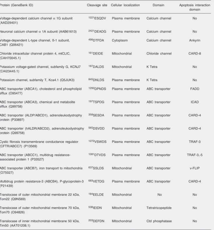
mu-Table 1. Identification, structural properties and localization of cation and anion channels, ATP-binding cassette (ABC) transporters and the translocases in the inner (Tim) and outer (Tom) mitochondrial membrane family containing potential cleavage sites for caspases and the apoptosis interaction domain.
Protein (GeneBank ID) Cleavage site Cellular localization Domain Apoptosis interaction
domain
Voltage-dependent calcium channel α 1G subunit 1221ESQDV Plasma membrane Calcium channel No (AAD29401)
Neuronal calcium channel α 1A subunit (AAB61613) 2421DEADG Plasma membrane Calcium channel No
Voltage-dependent L-type channel, ß-1 subunit, 498DTFDA Cytoplasm Calcium channel Ankyrin CAB1 (Q06421)
Chloride intracellular channel protein 4, mtCLIC, 161DEIDE Mitochondrial Chloride channel CARD-8 (CAH70045.1)
Potassium voltage-gated channel, subfamily G, KCNJ7 187DALDS Mitochondrial K Tetra No
(CAI23445.1)
Potassium channel, subfamily T, Kca4.1 (Q5JUK3) 845DNLDS Plasma membrane K Tetra No
ABC transporter (ABCA1), cholesterol and phospholipid 1292DPNDS Plasma membrane ABC transporter FADD efflux (O95477)
ABC transporter (ABCA3), chemical and metabolite 197TSPDG Plasma membrane ABC transporter ICAD efflux (Q99758)
ABC transporter (ALDP/ABCD1), adrenoleukodystrophy 358SESDA Plasma membrane ABC transporter CARD-4 protein (P33897)
ABC transporter (hALDR/ABCD2), adrenoleukodystrophy 565DSVDD Plasma membrane ABC transporter CARD-4 protein (Q99758)
Cystic fibrosis transmembrane conductance regulator 1272VSWDS Plasma membrane ABC transporter TRAF-3 (CFTR/ABCC7) (P13569)
ABC transporter (ABCC1), multidrug resistance- 1081DTVDS Plasma membrane ABC transporter TRAF-3,-5 associated protein 1 (P33527)
ABC transporter (ABCB7), iron transport to mitochondria 637SSLDS Mitochondrial ABC transporter v-FLIP (O75027)
Multidrug protein resistance-3 (ABCB4), P-glycoprotein-3 683VETDG Plasma membrane ABC transporter CARD-4 (P21439)
Translocase of outer mitochondrial membrane 22 kDa, 184EELDE Mitochondrial No No Tom22 (Q9NS69)
Translocase of outer mitochondrial membrane 70 kDa, 538IEIDN Mitochondrial Tetratricopeptide No Tom70 (O94826)
Translocase of inner mitochondrial membrane 50 kDa, 203DEFDN Mitochondrial Ctd phosphatase No Tim50 (AAT01208.1)
tant forms of human PMCA4 that lack the caspase cleavage site prevents the delayed rise of Ca2+ or delayed Ca2+ deregulation
(48). Bano and colleagues (49) have also shown that both calpains and caspases can cleave NCX during excitotoxicity in neu-ronal cell lines. The broad-spectrum caspase inhibitor z-VAD-fmk moderately reduced the caspase-mediated cleavage of NCX3 and delayed Ca2+ deregulation that causes
ne-crotic cell death (49). Therefore, caspases appear to be involved in the control of trans-membrane Ca2+ channels and pumps and
indirectly in the increase of intracellular Ca2+
pools that may precede the opening of mito-chondrial death decision pores.
The superfamily of ATP-binding cas-sette (ABC) transporter proteins consists of efflux pumps that have important roles in transporting a diverse group of toxicants including lipophilic cationic, anionic, and neutrally charged drugs, peroxidation prod-ucts and antigenic peptides (51,52). ABC transporters are localized in the plasma mem-brane, endoplasmic reticulum and in or-ganelles, including peroxisomes and mito-chondria. Five mammalian mitochondrial ABC transporters have been described based on the presence of mitochondrial targeting presequences (51,52). Members of this fam-ily are involved in the export of iron-sulfur-containing proteins such as apoproteins from the mitochondrial matrix to the cytosol (51). Deletions of mitochondrial ABC transport-ers disturb iron homeostasis and are lethal (53). At present, one study (54) has shown that the proteolytic cleavage of ABCA1, a transmembrane transporter involved in cy-tosolic cholesterol efflux by calpain, in-creases its activity. An uncleavable form of ABCA1 displayed a 4-fold increase in the efflux of cholesterol, which prevented mac-rophage cell death due to cholesterol over-load (54). We noted that calpain and cas-pases have cleavage sites within the same PEST sequence (Table 1). This raises the chance that both proteases could regulate the
pumping activity of ABC transporters. Mitochondrial chloride intracellular chan-nel protein 4 (mtCLIC) belongs to a CLIC family of soluble globular proteins that can form ion channels in organelles and plasma membranes similar to bacterial toxins, an-nexins and BCL-xL (55). It has been shown
that mtCLIC protein 4 is up-regulated dur-ing the cell death response to cytotoxic agents and DNA damage. Overexpression of the mtCLIC gene is sufficient to induce kerati-nocyte cell death, which is associated with the expression of p53, loss of mitochondrial membrane potential and cytochrome c re-lease (54). More importantly, cell death is inhibited by z-VAD-fmk, a pancaspase in-hibitor (55).
The translocase of the inner (Tim com-plex) and outer (Tom comcom-plex) mitochon-drial membranes is composed of large and small proteins that display an intrinsic ca-pacity to bind and transfer polypeptides into mitochondrial compartments (56). They con-tain water-filled pores that mediate translo-cation of proteins tagged with presequences that act as bipartite sorting signals. These N-terminal sorting signals form amphipathic helices with a hydrophobic and positively charged side. The sequences are first recog-nized by receptors and targeted to the pro-tein-conducting channel (Tom proteins). This translocation mechanism depends on both ATP and the mitochondrial membrane po-tential (56).
The role the Tim23 complex for translo-cation of AIF, Endo G, Smac/Diablo, and HtrA2/Omi into the intermembrane mito-chondrial space has been demonstrated (56). Furthermore, a study with yeast mitochon-dria has shown that perturbations of Tim23 conductance activity with synthetic peptides and specific antibodies cause matrix swell-ing and ultimately cytochrome c release (57).
dys-function and rapid apoptosis (58). Tom20 and Tom22 have a negatively charged N-terminal region exposed to the cytosol that contains a putative cleavage site for cas-pases. This region coordinates the recogni-tion and translocarecogni-tion of many nuclear pro-teins into mitochondria (58). Interestingly, deletion of the Tom22 gene is lethal (56). A complex of approximately 400 kDa contain-ing Tom20, Tom40 and Tom70 operates in the translocation of apocytochrome c inside the intermembrane space, where it incorpo-rates the heme co-factor and is released as holo-cytochrome c (56). The possible con-sequences of Tom22 cleavage by caspases are numerous. For example, it could inter-rupt cytochrome c import causing electron transport interruption. In conclusion, Tim/ Tom complexes appear to be interesting path-ways for apoptogenic factor release. Future studies are needed to challenge this hypo-thesis and to test how caspases can affect protein translocation across mitochondrial membranes.
Outer membrane rupture and mitochondrial remodeling
The occurrence of swelling is common in the mitochondria of the cells committed to die, as a result of inner membrane perme-ability transition (20,21). Since the surface of the inner membrane is greater than the surface of the outer membrane, the swelling forces the expansion of the inner membrane to the cytoplasm, causing the outer mem-brane to rupture (10). Korsmeyer’s group has also described the morphological fea-tures of degenerating mitochondria treated with tBID and crista ultrastructural remodel-ing usremodel-ing high-voltage electron microscopy and tomography (59). The authors proposed that tBID is necessary for mitochondrial re-modeling and further mobilization of cyto-chrome c (~85%) that is retained in
mito-chondrial crista stores. Based on their crista remodeling, the mitochondria were
classi-fied as class I (normal) or class II, III, and IV (59). Class II mitochondria with highly in-terconnected and condensed cristae occur after 2-5 min in cells treated with tBID, TNF and Fas and several intrinsic death stimuli including thapsigargin, tunicamycin and bre-feldin A (59). Class III mitochondria are swollen and their outer membrane are rup-tured. This is comparable to the type II mito-chondrial profile described by Sesso and colleagues (10).
Finally, fusion and fission (division) are two morphologically and physiologically opposite processes within mitochondrial dynamics. It has been demonstrated that per-turbations in these processes result in mito-chondrial outer membrane permeabilization and release of apoptogenic factors and apop-tosis (60). Cytosolic GTPases of the dynamin family, optic atrophy 1 and mitofusins 1 and 2, are required for mitochondrial fusion, while dynamin-related protein 1 and Fis1 are re-quired for mitochondrial fission (60).
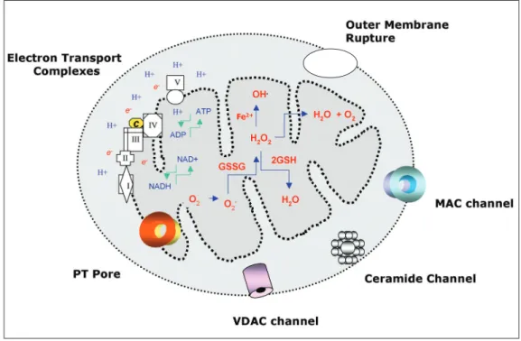
The present overview suggests that nu-merous biomolecules that are produced by biochemical and bioenergetic reactions in-side and outin-side the mitochondria, and regu-lators and effectors of apoptosis play a role in the coordination of events involved in mitochondrial membrane permeabilization and the release of cytochrome c, Smac/
Diablo, HtrA2/Omi and AIF. An integrated model is depicted in Figure 1. Permeabiliza-tion of the inner or outer membrane must be accompanied by exchange of cations, like Ca2+, K+, and Na+, and metabolites that have
ANT and pro-apoptotic proteins tBID, BAX and BAK, may participate in their assembly and operation. The MAC and a ceramide lipid pore may also be involved in the re-lease of the apoptogenic factors. Nonethe-less, the simple possibility that apoptogenic factor release occurs through a nonspecific mitochondrial outer membrane rupture also remains valid. Our understanding of the criti-cal role of caspases in mitochondrial dys-function is based on the experiments using recombinant caspases, their peptide inhibi-tors and mice deficient in both caspase-3 and caspase-7. Few components of the mito-chondrial machinery have been described as
caspase substrates. One study has shown that caspase-3 can enter the mitochondria and cleave NDUF1, a component of com-plex I of the respiratory chain. It remains unclear how a cytosolic protease can cross both mitochondrial membranes to cleave a matrix-exposed subunit embedded within complex I. However, biochemical analysis has demonstrated that pro-caspase and ac-tive caspase (-3, -7, -8, and -9) can partially co-localize in the outer membrane or are released into mitochondria via “death sig-naling vesicles” stimulated by TNF and CD95/Fas (45,46). In this scenario, it may be possible that caspases, especially the apical
Figure 1. Schematic representation of mitochondrial death decision pores with the biochemical ability to promote the inner and outer mitochondrial membrane permeabilization and the release of apoptogenic factors: cytochrome
References
1. Lemasters JJ. Dying a thousand deaths: redundant pathways from different organelles to apoptosis and necrosis. Gastroenterology
2005; 129: 351-360.
2. Chipuk JE, Bouchier-Hayes L, Green DR. Mitochondrial outer mem-brane permeabilization during apoptosis: the innocent bystander scenario. Cell Death Differ 2006; 13: 1396-1402.
3. Vander Heiden MG, Thompson CB. Bcl-2 proteins: regulators of apoptosis or of mitochondrial homeostasis? Nat Cell Biol 1999; 1: E209-E216.
4. Scorrano L, Korsmeyer SJ. Mechanisms of cytochrome c release by proapoptotic BCL-2 family members. Biochem Biophys Res Commun 2003; 304: 437-444.
5. Breckenridge DG, Xue D. Regulation of mitochondrial membrane permeabilization by BCL-2 family proteins and caspases. Curr Opin Cell Biol 2004; 16: 647-652.
6. Lucken-Ardjomande S, Martinou JC. Newcomers in the process of mitochondrial permeabilization. J Cell Sci 2005; 118: 473-483. 7. Zoratti M, Szabo I, De Marchi U. Mitochondrial permeability
transi-tions: how many doors to the house? Biochim Biophys Acta 2005; 1706: 40-52.
8. Dejean LM, Martinez-Caballero S, Kinnally KW. Is MAC the knife that cuts cytochrome c from mitochondria during apoptosis? Cell Death Differ 2006; 13: 1387-1395.
9. Siskind LJ. Mitochondrial ceramide and the induction of apoptosis. J Bioenerg Biomembr 2005; 37: 143-153.
10. Sesso A, Marques MM, Monteiro MM, Schumacher RI, Colquhoun A, Belizario J, et al. Morphology of mitochondrial permeability transi-tion: morphometric volumetry in apoptotic cells. Anat Rec A Discov Mol Cell Evol Biol 2004; 281: 1337-1351.
11. Magder S. Reactive oxygen species: toxic molecules or spark of life? Crit Care 2006; 10: 208.
12. Storz P. Reactive oxygen species-mediated mitochondria-to-nucleus signaling: a key to aging and radical-caused diseases. Sci STKE
2006; 2006: re3.
13. Shi Y. Caspase activation, inhibition, and reactivation: a mechanism view. Protein Sci 2004; 13: 1979-1987.
14. Fischer U, Janicke RU, Schulze-Osthoff K. Many cuts to ruin: a comprehensive update of caspase substrates. Cell Death Differ
2003; 10: 76-100.
15. Marzo I, Susin SA, Petit PX, Ravagnan L, Brenner C, Larochette N, et al. Caspases disrupt mitochondrial membrane barrier function.
FEBS Lett 1998; 427: 198-202.
16. Lakhani SA, Masud A, Kuida K, Porter GA Jr, Booth CJ, Mehal WZ, et al. Caspases 3 and 7: key mediators of mitochondrial events of apoptosis. Science 2006; 311: 847-851.
17. Orrenius S, Zhivotovsky B, Nicotera P. Regulation of cell death: the calcium-apoptosis link. Nat Rev Mol Cell Biol 2003; 4: 552-565. 18. Waring P. Redox active calcium ion channels and cell death. Arch
Biochem Biophys 2005; 434: 33-42.
19. Kowaltowski AJ, Castilho RF, Vercesi AE. Mitochondrial permeabil-ity transition and oxidative stress. FEBS Lett 2001; 495: 12-15. 20. Skulachev VP. Bioenergetic aspects of apoptosis, necrosis and
mitoptosis. Apoptosis 2006; 11: 473-485.
21. Bernardi P, Krauskopf A, Basso E, Petronilli V, Blachly-Dyson E, Di LF, et al. The mitochondrial permeability transition from in vitro
artifact to disease target. FEBS J 2006; 273: 2077-2099.
22. Launay S, Hermine O, Fontenay M, Kroemer G, Solary E, Garrido C. Vital functions for lethal caspases. Oncogene 2005; 24: 5137-5148. 23. Cain K, Bratton SB, Cohen GM. The Apaf-1 apoptosome: a large
caspase-activating complex. Biochimie 2002; 84: 203-214. 24. Vaux DL, Silke J. IAPs, RINGs and ubiquitylation. Nat Rev Mol Cell
Biol 2005; 6: 287-297.
25. Springs SL, Diavolitsis VM, Goodhouse J, McLendon GL. The kinet-ics of translocation of Smac/DIABLO from the mitochondria to the cytosol in HeLa cells. J Biol Chem 2002; 277: 45715-45718. 26. Rehm M, Dussmann H, Prehn JH. Real-time single cell analysis of
ones, gain access to mitochondria via altera-tion in endocytic traffic. Using a computa-tional program, we found that some mem-bers of cytoplasmic and mitochondrial ABC transporters and regulatory components of Tim and Tom complexes as well as some subunits of the plasma membrane and mito-chondrial membrane channels for Cl- and
K+ and Ca2+ are potential caspase substrates.
Their cleavage could cause changes in their structure and the switch to a nonspecific channel or pore with different conductance, selectivity or functional state. The validity of this mechanistic model needs to be exam-ined experimentally. Future studies for the characterization of the molecular compo-nents and of the mechanistic basis by which
the mitochondrial death decision pores op-erate will certainly help the design of new therapies to amplify or block the release of apoptogenic factors.
Acknowledgments
Smac/DIABLO release during apoptosis. J Cell Biol 2003; 162: 1031-1043.
27. Uren RT, Dewson G, Bonzon C, Lithgow T, Newmeyer DD, Kluck RM. Mitochondrial release of pro-apoptotic proteins: electrostatic interactions can hold cytochrome c but not Smac/DIABLO to mito-chondrial membranes. J Biol Chem 2005; 280: 2266-2274. 28. Arnoult D, Gaume B, Karbowski M, Sharpe JC, Cecconi F, Youle
RJ. Mitochondrial release of AIF and EndoG requires caspase acti-vation downstream of Bax/Bak-mediated permeabilization. EMBO J
2003; 22: 4385-4399.
29. Scorrano L, Oakes SA, Opferman JT, Cheng EH, Sorcinelli MD, Pozzan T, et al. BAX and BAK regulation of endoplasmic reticulum Ca2+: a control point for apoptosis. Science 2003; 300: 135-139. 30. Colombini M. VDAC: the channel at the interface between
mitochon-dria and the cytosol. Mol Cell Biochem 2004; 256-257: 107-115. 31. Lemasters JJ, Holmuhamedov E. Voltage-dependent anion channel
(VDAC) as mitochondrial governator - thinking outside the box.
Biochim Biophys Acta 2006; 1762: 181-190.
32. Rostovtseva TK, Antonsson B, Suzuki M, Youle RJ, Colombini M, Bezrukov SM. Bid, but not Bax, regulates VDAC channels. J Biol Chem 2004; 279: 13575-13583.
33. Shimizu S, Narita M, Tsujimoto Y. Bcl-2 family proteins regulate the release of apoptogenic cytochrome c by the mitochondrial channel VDAC. Nature 1999; 399: 483-487.
34. Crompton M, Barksby E, Johnson N, Capano M. Mitochondrial intermembrane junctional complexes and their involvement in cell death. Biochimie 2002; 84: 143-152.
35. Rostovtseva TK, Tan W, Colombini M. On the role of VDAC in apoptosis: fact and fiction. J Bioenerg Biomembr 2005; 37: 129-142. 36. Abu-Hamad S, Sivan S, Shoshan-Barmatz V. The expression level of the voltage-dependent anion channel controls life and death of the cell. Proc Natl Acad Sci U S A 2006; 103: 5787-5792.
37. Pavlov EV, Priault M, Pietkiewicz D, Cheng EH, Antonsson B, Manon S, et al. A novel, high conductance channel of mitochondria linked to apoptosis in mammalian cells and Bax expression in yeast.
J Cell Biol 2001; 155: 725-731.
38. Guo L, Pietkiewicz D, Pavlov EV, Grigoriev SM, Kasianowicz JJ, Dejean LM, et al. Effects of cytochrome c on the mitochondrial apoptosis-induced channel MAC. Am J Physiol Cell Physiol 2004; 286: C1109-C1117.
39. Guihard G, Bellot G, Moreau C, Pradal G, Ferry N, Thomy R, et al. The mitochondrial apoptosis-induced channel (MAC) corresponds to a late apoptotic event. J Biol Chem 2004; 279: 46542-46550. 40. Ricci JE, Waterhouse N, Green DR. Mitochondrial functions during
cell death, a complex (I-V) dilemma. Cell Death Differ 2003; 10: 488-492.
41. Lassus P, Opitz-Araya X, Lazebnik Y. Requirement for caspase-2 in stress-induced apoptosis before mitochondrial permeabilization.
Science 2002; 297: 1352-1354.
42. Enoksson M, Robertson JD, Gogvadze V, Bu P, Kropotov A, Zhivot-ovsky B, et al. Caspase-2 permeabilizes the outer mitochondrial membrane and disrupts the binding of cytochrome c to anionic phospholipids. J Biol Chem 2004; 279: 49575-49578.
43. Ricci JE, Munoz-Pinedo C, Fitzgerald P, Bailly-Maitre B, Perkins GA, Yadava N, et al. Disruption of mitochondrial function during apoptosis is mediated by caspase cleavage of the p75 subunit of
complex I of the electron transport chain. Cell 2004; 117: 773-786. 44. Waterhouse NJ, Sedelies KA, Sutton VR, Pinkoski MJ, Thia KY,
Johnstone R, et al. Functional dissociation of DeltaPsim and cyto-chrome c release defines the contribution of mitochondria upstream of caspase activation during granzyme B-induced apoptosis. Cell Death Differ 2006; 13: 607-618.
45. Lee KH, Feig C, Tchikov V, Schickel R, Hallas C, Schutze S, et al. The role of receptor internalization in CD95 signaling. EMBO J
2006; 25: 1009-1023.
46. Ouasti S, Matarrese P, Paddon R, Khosravi-Far R, Sorice M, Tinari A, et al. Death receptor ligation triggers membrane scrambling be-tween Golgi and mitochondria. Cell Death Differ 2006; 14: 456-461. 47. Garay-Malpartida HM, Occhiucci JM, Alves J, Belizario JE. CaSPre-dictor: a new computer-based tool for caspase substrate prediction.
Bioinformatics 2005; 21 (Suppl 1): i169-i176.
48. Schwab BL, Guerini D, Didszun C, Bano D, Ferrando-May E, Fava E, et al. Cleavage of plasma membrane calcium pumps by cas-pases: a link between apoptosis and necrosis. Cell Death Differ
2002; 9: 818-831.
49. Bano D, Young KW, Guerin CJ, Lefeuvre R, Rothwell NJ, Naldini L, et al. Cleavage of the plasma membrane Na+/Ca2+ exchanger in excitotoxicity. Cell 2005; 120: 275-285.
50. Bredesen DE, Mehlen P, Rabizadeh S. Receptors that mediate cellular dependence. Cell Death Differ 2005; 12: 1031-1043. 51. Lill R, Kispal G. Mitochondrial ABC transporters. Res Microbiol
2001; 152: 331-340.
52. Higgins CF, Linton KJ. The ATP switch model for ABC transporters.
Nat Struct Mol Biol 2004; 11: 918-926.
53. Senbongi H, Ling F, Shibata T. A mutation in a mitochondrial ABC transporter results in mitochondrial dysfunction through oxidative damage of mitochondrial DNA. Mol Gen Genet 1999; 262: 426-436. 54. Wang N, Chen W, Linsel-Nitschke P, Martinez LO, Agerholm-Larsen B, Silver DL, et al. A PEST sequence in ABCA1 regulates degrada-tion by calpain protease and stabilizadegrada-tion of ABCA1 by apoA-I. J Clin Invest 2003; 111: 99-107.
55. Fernandez-Salas E, Suh KS, Speransky VV, Bowers WL, Levy JM, Adams T, et al. mtCLIC/CLIC4, an organellular chloride channel protein, is increased by DNA damage and participates in the apopto-tic response to p53. Mol Cell Biol 2002; 22: 3610-3620.
56. Herrmann JM, Hell K. Chopped, trapped or tacked - protein translo-cation into the IMS of mitochondria. Trends Biochem Sci 2005; 30: 205-211.
57. Guo Y, Cheong N, Zhang Z, De Rose R, Deng Y, Farber SA, et al. Tim50, a component of the mitochondrial translocator, regulates mitochondrial integrity and cell death. J Biol Chem 2004; 279: 24813-24825.
58. Nargang FE, Rapaport D, Ritzel RG, Neupert W, Lill R. Role of the negative charges in the cytosolic domain of TOM22 in the import of precursor proteins into mitochondria. Mol Cell Biol 1998; 18: 3173-3181.
59. Scorrano L, Ashiya M, Buttle K, Weiler S, Oakes SA, Mannella CA, et al. A distinct pathway remodels mitochondrial cristae and mobi-lizes cytochrome c during apoptosis. Dev Cell 2002; 2: 55-67. 60. Martinou JC, Youle RJ. Which came first, the cytochrome c release