Effects of physical training and potassium supplementation
on blood pressure, glucose metabolism and albuminuria of
spontaneously hypertensive rats
Authors
Evelyn Manuella Martins Gomes Jodas1
Aline Francisco Voltera1 Milton Ginoza1
Osvaldo Kohlmann Junior1
Nelson Brancaccio dos Santos2
Mário Luís Ribeiro Cesaretti1,2
1 Federal University of São Paulo.
2 Pontifical Catholic University of São Paulo.
Submitted on: 08/15/2013. Approved on: 12/09/2013.
Correspondence to: Evelyn Manuella Martins Gomes Jodas.
Nephrology Program, UNIFESP/ EPM.
Rua Botucatu, nº 740. São Paulo, SP, Brasil. CEP: 04023-900. E-mail: ejodas@unifesp.br Tel: 5904-1699. Fax: 5904-1684. Coordination for the Improvement of Higher Education Personnel and the Oswaldo Ramos Foundation.
Introduction: It is still controversial whether there are synergistic effects among different non-pharmacological in-terventions used in the treatment of hy-pertension. Objectives: To evaluate the effect of aerobic exercise, oral supple-mentation of potassium and their com-bination on blood pressure, glucose me-tabolism, urinary albumin excretion and glomerular morphology in spontaneously hypertensive rats (SHR). Methods: SHR were divided into groups: Control Group (SHR; standard diet and sedentary, n = 10), Exercise Group (SHR + E; trained on a treadmill, standard diet, n = 10), Potas-sium Group (SHR + K; sedentary, potas-sium supplementation, n = 10) and Group Exercise + Potassium (SHR + E + K, exer-cise, potassium supplementation n = 10). Weekly, body weight (BW) and tail blood pressure (TAP) were measured. At the end of 16 weeks, a Oral Glucose Tolerance Test was performed. Albuminuria was de-termined in the baseline period, at 8th and
at 16th week. After sacrifice, the analysis
of glomerular sclerosis index and visceral fat weight was performed. Results: The TAP and BW did not change significantly. There was improvement in insulin sensi-tivity in SHR + E and SHR + K, but not in SHR + E + K. At week 16, albuminuria in all groups was significantly lower than the SHR control. The glomerular sclerosis in-dex and visceral fat content were also sig-nificantly lower in all groups compared to control. Conclusion: An oral supplemen-tation of potassium and exercise led to an improvement in glucose metabolism, in albuminuria and glomerular morphology, however, the overlap of the treatments did not show synergism.
A
BSTRACTKeywords: albuminuria; exercise; hyper-tension; kidney glomerulus; potassium chloride; rats, inbred SHR.
I
NTRODUCTIONSystemic hypertension is a multifac-torial clinical condition characterized by sustained high levels of blood pres-sure often associated with metabolic and hormonal disorders. It is frequen-tly associated with functional and/or structural alterations in target organs (heart, brain, kidneys, and blood ves-sels) and, consequently, with increased risk of fatal and non-fatal cardiovas-cular events.1
Insulin resistance is a genetic or acquired condition in which physio-logical concentrations of insulin elicit subnormal cell glucose uptake res-ponses, particularly in myocytes, he-patocytes, and adipocytes. Reduced glucose uptake levels lead to incre-ased production of insulin by the pancreas, which in turn increases the circulating levels of insulin and characterizes the coexistence of insu-lin resistance and hyperinsuinsu-linemia. However, the other roles played by insulin in the body remain unaltered.2
Insulin resistance and hyperinsuli-nemia have been described as com-mon elements in conditions such as hypertension, obesity and coronary disease.3,4
increased cell growth;5 and impaired ion
trans-port across cell membranes, causing increases in intracellular calcium and sodium levels and, con-sequently, in cell excitability.5
The non-pharmacological treatment of hypertension includes various health and dietary measures to lower blood pressure levels, such as reducing sodium intake, increasing potassium and magnesium intake, accompanied by aerobic exercises, and weight loss and stress management measures.6
Studies have shown that regular aerobic physical activity is an important item in hypertension therapy, as it may reduce blood pressure levels in hypertensive subjects. The literature has extensively described an inverse correlation between physical activity and the prevalence of hypertension.7
Evidence also indicates that potassium might have a mitigating role in hypertension, insulin resistance and related comorbidities.8
Increased potassium intake has been associa-ted with fewer deaths for stroke or heart di-sease.9 The literature further indicates that
in-creased potassium intake improves the binding of insulin to its receptors and decreases insulin resistance in human and experimental obesi-ty models, possibly leading to reduced blood pressure levels.10
Although evidence suggests that physical exercise and potassium-rich diets may be used in the non-pharmacological management of hypertension, a correlation is yet to be established between these two variables. This study aims to assess the effect of aerobic exercises, potassium overload, and the combined effect of both on blood pressure, urinary albumin excretion and glomerular morphology in spontaneously hypertensive rats.
M
ETHODSThis study used a strain of spontaneously hypertensive male rats (SHR) aged three months with blood pressure levels above 170 mmHg
Center for the Development of Experimental Models (CEDEME). The rats were kept in ideal vivarium conditions at the Department of Nephrology of the Federal University of São Paulo. The study protocol was reviewed and approved by the Ethics Committee of the Federal University of São Paulo (protocol number 0355/12).
EXPERIMENTALGROUPS
Four experimental groups were followed for 16 weeks:
• Control group (SHR, n = 10) - subjects were kept off physical exercises and on feed Nuvilab® throughout the study;
• Exercise group (SHR + EXE, n = 10) -
after baseline measurements, subjects were placed on a physical exercise protocol (see below) and kept on the standard diet;
• Potassium group (SHR + K, n = 10) - after baseline measurements, subjects were kept off exercises and on a diet with three times the amount of potassium contained in the standard diet (see below) throughout the study;
• SHR + Exercise + Potassium group (SHR + EXE + K, n = 10) - subjects were kept on the exercise protocol and on the potassium-rich diet.
Animal follow up time was 16 weeks.
POTASSIUM SUPPLEMENTATION
Potassium supplementation was calculated based on the 1.1 g of potassium per kilogram of feed reported by Nuvilab Nutrientes. The feed was ground and potassium chloride (Synth Diadema SP) added to bring the feed to a con-centration of 3.3 g/kg. The potassium-rich feed was moistened to form new pellets and hard-ened in an oven at 80 °C before it was fed to specific rat groups.
PHYSICAL TRAINING PROGRAM
minutes, for a period of 16 weeks. The rats were trained on a flat treadmill (Columbus Instruments, Treadmill Simplex II; height: 0.45 m, width: 0.70 m, length: 1.35 m) in individual 14-centimeter stalls. Physical training was started at a speed of 0.3 kph in the first session. Speed was gradually increased based on subject performance to a maximum of 1.1 kph. The chosen exercise intensity level corresponded to 70-80% of the subjects’ VO2 max and matched the anaerobic threshold of the maximal lactate steady state (MLSS) protocol.11
EXPERIMENTALPROTOCOL
The subjects had their tail blood pressure - using the oscillometric method - and bodyweight measured twice a week. The mean weekly blood pressure and bodyweight values were used as references for each week of the study.
At baseline and on weeks eight and sixteen into follow-up, the subjects were placed in metabolic cages (Nalgene, Rochester, NY) to have 24-hour urine samples collected. Urine output and urine albumin levels were verified by radial immunodiffusion.
After the end of the treatment/follow-up period, the subjects underwent oral glucose tolerance tests (OGTT). Twenty-four hours prior to OGTT, the rats were anesthetized with intraperitoneal ketamine (100 mg/kg) and xylazine (10 mg/kg) and placed in dorsal decubitus. An oblique inguinal incision was made to allow the implantation of a PE-10 polyethylene catheter (Intramedic, Clay Adams, NJ, USA) in the femoral artery. This catheter was connected to a 20-centimeter long PE50 catheter (CPL, São Paulo, Brazil). The catheters had been previously filled with heparinized saline (10 U/ml). The PE50 catheter was passed subcutaneously with the help of a trocar through the dorsum to the posterior neck, where it was exteriorized
and fixed. The rats were then anesthetized and placed in individual cages on solid food restriction for 12 hours prior to undergoing OGTT.
The first step in OGTT consisted of drawing blood from the subjects to find their fasting glucose and insulin levels. The arterial catheter was used to collect one drop of blood for glucose level testing and one milliliter of whole blood in an Eppendorf tube to test for insulin levels. After blood collection at baseline, the rats were force-fed 1.7 g/kg of anhydrous glucose diluted in distilled water. Additional blood samples were taken 15, 30, 60, 90 and 120 minutes after glucose administration.
A glucose meter (Optium - Abbott MediSense) and reagent strips (Optium Point-of-Care - Abbott MediSense) were used to assess blood glucose levels. In order to find plasma insulin levels, the collected blood was centrifuged in Eppendorf tubes (Eppendorf Centrifuge 5403) at 10,000 rpm for 10 minutes at 4 °C. Plasma was separated, put in new Eppendorf tubes and stored at -70 °C in a freezer (Revco Scientific Inc., Asheville, NC, USA). Coat-A-Acount reagent radioimmunoassay kits were used to measure insulin levels.
Blood glucose and plasma insulin levels found in OGTT were used to calculate the glucose and insulin areas under the curve (AUC) by the trapezoidal rule. Glucose and insulin AUC were used to calculate the insulin sensitivity index (ISI).
After the tests, the rats were euthanatized with anesthetics. Fat tissue from around the epididymis was removed and weighed to characterize visceral fat. Relative epididymal fat per 100 g of bodyweight was calculated.
The glomerular sclerosis index was calculated after staining with hematoxylin-eosin. A patholo-gist blinded to the rat groups assigned degrees of glomerular injury according to Chart 1 below:
Parametric and non-parametric tests were used to analyze the results, taking into consideration the nature of the studied variables and the variability of measurements. The following tests were used: a) repeated measures analysis of variance (parametric test) was used to compare temporal variations of blood pressure and bodyweight. Statistically significant differences were analyzed with the Bonferroni multiple comparison test; b) analysis of variance between the different groups (parametric test) was performed to compare between the epididymal fat relative weight values. Statistically significant differences were analyzed with the Bonferroni multiple comparison test; c) Kruskal-Wallis ANOVA by ranks (nonparametric test) was used to compare the OGTT values for glucose, insulin, and urine albumin AUC, ISI and glomerular sclerosis. Statistically significant differences were compared using Dunn’s multiple comparison test. A significance level of 5% (p < 0.05) was adopted to reject the null hypothesis in the tests.
R
ESULTSThe results were shown in the form of mean values and standard error of the mean.
BODYWEIGHTANDTAILBLOODPRESSURE
Our results showed that neither bodyweight nor tail blood pressure changed throughout the 16 weeks of the study (Table 1).
ORALGLUCOSETOLERANCETEST
Fasting blood glucose levels were similar (p = 0.08) for all groups, but after glucose overload the rats in groups SHR + Exe, SHR + K, and SHR + Exe + K had significantly lower blood glucose levels when compared to the subjects in group SHR in any OGTT stage. The lo-wer blood glucose levels of the rats in groups SHR + Exe, SHR + K, and SHR + Exe + K we-re we-reflected in their significantly lower glucose AUC values.
Fasting insulin levels were also similar between groups (p = 0.32). After glucose administration, lower insulin levels were observed in groups SHR + Exe and SHR + K. Thus, only these two groups had lower values for insulin AUC.
The calculation of the ISI revealed that groups SHR + Exe and SHR + K had a lower ISI than the rats included in group SHR. The subjects in the SHR + Exe + K group had lower insulin sensitivity indices, although without statistical significance (Table 1 and Figure 1).
URINEALBUMINANDGLOMERULARSCLEROSISINDEX
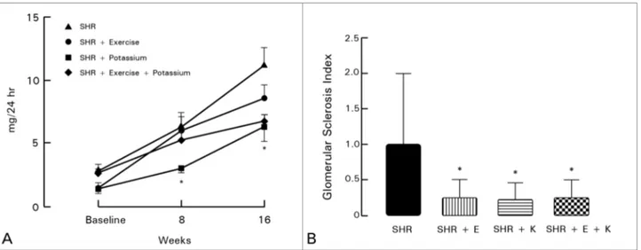
Baseline urine albumin levels were similar. On week eight, the rats in group SHR + Exe had significantly lower levels of urine albumin when compared to the subjects in group SHR. On week 16, groups SHR + Exe and SHR + K had significantly lower urine albumin levels than subjects on group SHR. The subjects in the SHR + Exe + K group also had significantly lower urine albumin levels, however not statistically significant.
CHART 1 GLOMERULARINJURYGRADESUSEDINTHECATEGORIZATIONOFTHEGLOMERULARSCLEROSISINDEX12
Grade 0: Vessels are unaltered or with minimal caliber reduction. Internal and external elastic laminae are well defined, showing a ratio between the lumen and the wall of the vessel greater than 1.5:1.0. Cell elements cannot be seen between the endothelium and the internal elastic lamina.
Grade I: Hyaline thickening of the vascular wall in varying degrees, while maintaining a lumen/wall ratio of 1.5:1.0. Slight increase in the number of elastic fibers in the vessel wall and vascular hypertrophy of the tunica media.
TABLE 1 MEAN ± STANDARDERROR - INITIALANDFINALBODYWEIGHT (INGRAMS), INITIALANDFINALTAILBLOODPRESSURE (IN
MILLIMETERSOFMERCURY), GLUCOSE (INMILLIGRAMSPERDECILITER) ANDINSULIN (INMILLIUNITS/DECILITER) AREAUNDER THECURVE, ANDINSULINSENSITIVITYINDEX (MG-1MU-1ML-1) FORGROUPS SHR, SHR + EXE, SHR + K AND SHR + EXE + K
SHR SHR + Exe SHR + K SHR + Exe + K
Initial bodyweight 283.4 ± 5.08 298.8 ± 6.56 280.2 ± 4.31 304.4 ± 7.57
Final bodyweight 354.0 ± 11.15 371.7 ± 6.34 337.0 ± 5.25 352.4 ± 9.20
Initial tail blood pressure 194.9 ± 2.10 193.5 ± 1.13 197.1 ± 1.16 194.8 ± 1.03
Final tail blood pressure 196.9 ± 2.47 198.7 ± 0.33 198.4 ± 0.37 196.4 ± 1.34 Glucose area under the curve 252.5 ± 10.2 144.1 ± 8.3* 156.2 ± 4.1* 165.1 ± 7.3*
Insulin area under the curve 25.80 ± 2.48 16.87 ± 1.65* 20.08 ± 2.97* 24.52 ± 1.56
Insulin sensitivity index 1.79 ± 0.20 4.43 ± 0.30* 4.89 ± 1.59* 2.72 ± 0.18 *p < 0.05 vs. SHR.
Figure 1. Insulin sensitivity index in mg-1 mU-1 mL-1, for groups SHR,
SHR + Exercise (SHR + EXE), SHR + Potassium (SHR + K) and SHR + Exercise + Potassium (SHR + EXE + K). * p < 0.05 vs. SHR.
The analysis of glomerular sclerosis by a pathologist indicated that the groups offered physical exercises, potassium-rich diet, or the combination of both had improved glomerular morphology (Figure 2 A-B).
VISCERALFATCONTENT
When compared to the SHR group, decreases in visceral fat were observed in all other groups (SHR = 3.49 ± 0.94; SHR + Exe = 2.48 ± 0.58*; SHR + K = 2.96 ± 0.57*; SHR + Exe + K = 3.09 ± 0.45* g/100 g, *p < 0.05 vs. SHR).
D
ISCUSSIONThis study aimed to assess glucose metabolism in spontaneously hypertensive rats submitted to non-pharmacological treatments for hypertension, namely: potassium supplementation, aerobic
exercises, and a protocol combining both. Although none of the approaches successfully reduced the subjects’ blood pressure levels, the three treatments resulted in improved glucose tolerance. However, only the rats fed a potassium-rich diet and the group offered physical exercises alone presented decreased insulin sensitivity indices. Significant decreases in urinary albumin excretion were observed in the last week of the study in these groups (SHR + K and SHR + Exe), a finding possibly correlated with the lower rates of glomerular sclerosis seen in treated rats. Treated subjects had lower levels of visceral fat.
Several factors have been associated with decreases in blood pressure due to physical training, such as reduced sympathetic vasomotor tone, decreased insulin resistance, decreased plasma volume and cardiac output,2 decreased
vascular reactivity, reduced peripheral resistance, decreased activity in the renin-angiotensin-aldosterone system, reduced oxidative stress, and increased levels of vasodilators in the endothelium.13
The reduction of blood pressure seen in our subjects differed from the literature. Amaral and Michelini13 reported 8-10% decreases in the BP of
male spontaneously hypertensive rats submitted to low-intensity exercises for three months. Recent studies have also described decreases in BP levels of spontaneously hypertensive rats trained on a treadmill.14 Some authors, however,
Figure 2 A-B. 24-hour urine albumin in milligrams (graph A) and glomerular sclerosis index (graph B) for groups SHR, SHR + Exercise (SHR + EXE), SHR + Potassium (SHR + K), and SHR + Exercise + Potassium (SHR + EXE + K). * p < 0.05 vs. SHR.
did not report BP reductions in spontaneously hypertensive rats submitted to physical exercises, particularly in rats started on physical training, as in this study, with previously established hypertension or rats submitted to longer training periods.12
Véras-Silva et al.15 found that 18 weeks of
high-intensity training on a treadmill were not enough to decrease the mean systolic and diastolic BP, or the heart rate of spontaneously hypertensive rats. Although no decreases were seen in the BP of trained spontaneously hypertensive rats, significant changes were observed in their cardiovascular parameters, thus eliciting the benefits of exercising. Physical exercises led to improved insulin action, especially in skeletal muscles,16 by increasing the expression
and translocation of glucose transporter type 4 (GLUT4) in the plasma membrane. The improvement in insulin sensitivity seen in group SHR + Exe can also be attributed to the lower sympathetic tone induced by physical training. According to the literature, physical training considerably reduces adrenergic tone.17
In an experimental study, potassium supplementation mitigated endothelial injury, reduced arterial wall thickening, decreased adhesion of macrophages to the vascular wall, and reduced mortality in stroke-prone spontaneously
potassium intake and prevalence of hypertension and cardiovascular disease.19
In this study, subject blood pressure levels were not altered by potassium overload. This finding goes against previous studies carried out in our laboratory20 and by other researchers.21
The non-reduction of blood pressure levels may be attributed to the sensitivity of the method used to measure tail BP levels. Other similar experimental studies in which physical exercises and potassium-rich diets were used failed to report changes in tail blood pressure.22
Pronounced BP reductions have not been described in every clinical study. Cohn et al.23
looked into the relationship between potassium intake and blood pressure levels in elderly individuals, and described that for each 1 g/ day of potassium above recommended levels a corresponding decrease of 0.9 mmHg in systolic BP and 0.8 mmHg in diastolic BP occurred.
The improvement in glucose metabolism in the rats given a potassium-rich diet (SHR + K group) was a significant finding in this study.
A previous study carried out in our laboratory reported significant reductions in BP and improvements in glucose metabolism in hypertensive rats fed a potassium-rich diet20
and rats with obesity induced by monosodium
4-aminopyridine, KATP, and glibenclamide and KCa++ channels with tetramethylammonium led to insulin resistance. The authors of this study further reported increases in glucose intake during hyperinsulinemic-euglycemic clamp when subjects were administered potassium channel activators such as nicorandil, pinacidil and chromakalin.
The pathophysiological mechanisms proposed for the reduction of blood pressure levels in animal models and patients given a potassium-rich diet involve endothelium dependent vasodilation through hyperpolarization of the endothelial cells, which diminishes the inflow of calcium,26
increases natriuresis, alters intracellular sodium content, modulates baroreceptors, reduces sensitivity to angiotensin and norepinephrine, increases serum and urinary levels of kallikrein, alters the balance of the sodium-potassium-ATPase pump, reduces levels of TGF-α, and alters DNA synthesis and smooth muscle cell proliferation.27
Microalbuminuria was reduced in the rats submitted to physical exercises in our study, a finding possibly reinforced by the lower degree of glomerular injury observed in the studied subjects. Physical training has been recognized as a way to increase antioxidant defenses and reduce oxidative stress,16 improve lipid profile,28 and
blood pressure levels,29 indicating the presence
of beneficial mechanisms that may reduce renal damage.
The greater vasodilation observed in the rats given potassium may be the cause of decreased microalbuminuria and improved glomerular morphology.30 The etiology of the
potassium-mediated improvement in glomerular function as seen in this study could be related to increased vasodilatory capacity (by nitric oxide or brady-kinin etc.),31 decreased synthesis of
vasoconstric-tors (angiotensin II and norepinephrine),32 and
decreased synthesis of cytokines.33 It is worth
noting that other studies with stroke-prone spon-taneously hypertensive rats failed to observe re-ductions in microalbuminuria when the subjects were fed a potassium-rich diet.34
The decrease in visceral fat seen in our experimental groups is important not only to improve the subjects’ glycemic profile, but also to improve the status of adipokines and decrease the production of non-esterified fatty acids which per se potentiate insulin resistance.35 In
addition to inducing increased concentrations of adiponectin,36 changes have also been also
observed in the activity of at least two cytokines involved in the pathophysiology of metabolic syndrome: TNF-α (tumor necrosis factor) and IL-6 (interleukin 6). Both the expression and plasma concentrations of IL-6 increase with physical exercises.37 When produced by muscle
cells, this cytokine, unlike when it is produced by macrophages, has anti-inflammatory properties as it inhibits inflammatory cytokines such as TNF-α, IL-1β, and IL-10.38 Exercise alone also
reduces TNF-α levels.39 The lessened deleterious
effects of cytokines reduce inflammation and insulin resistance.40
The fact that we have not observed a summation of effects in the administration of a potassium-rich diet along with aerobic exercises, although each separately produced similar impacts on the evaluated parameters, may suggest that common mechanisms mediate the effects of these therapeutic strategies upon metabolic parameters. This possibility seems plausible, as in many situations the summation of effects upon a given parameter has been observed when the mechanisms underlying such effects are different. A classic example lies in the combination of antihypertensive drugs when the maximum summation of effects occurs when antihypertensive drugs with different modes of action are combined.
R
EFERENCES1. Williams B. The year in hypertension. J Am Coll Car-diol 2009;55:65-73. PMID: 20117366 DOI: http://dx.doi. org/10.1016/j.jacc.2009.08.037
2. Kadowaki T. Insights into insulin resistance and type 2 diabetes from knockout mouse models. J Clin Invest 2000;106:459-65. PMID: 10953020 DOI: http://dx.doi.org/10.1172/JCI10830 3. Hall JE. Pathophysiology of obesity hypertension. Curr
Hyper-tens Rep 2000;2:139-47. DOI: http://dx.doi.org/10.1007/ s11906-000-0073-4
4. Kohlmann Jr O. Resistência à insulina e hipertensão arterial: relevância clínica. Hipertensão 1998;1:50-4.
5. Cartier EA, Conti LR, Vandenberg CA, Shyng SL. Defective trafficking and function of KATP channels caused by a sul-fonylurea receptor 1 mutation associated with persistent hype-rinsulinemic hypoglycemia of infancy. Proc Natl Acad Sci U S A 2001;98:2882-7. PMID: 11226335 DOI: http://dx.doi. org/10.1073/pnas.051499698
6. Malagris LEN, Brunini TMC, Moss MB, Silva PJA, Esposito BR, Ribeiro ACM. Evidências biológicas do treino de controle do stress em pacientes com hipertensão. Psicol Reflex Crit 2009;22:60-8. DOI: http://dx.doi.org/10.1590/S0102-79722009000100009
7. Medina FL, Lobo FS, Souza DR, Kanegusuku H, Forjaz CLM. Atividade física: impacto sobre a pressão arterial Physical activity: impact on blood pressure. Rev Bras Hipertens 2010;17:103-6.
8. He J, Gu D, Chen J, Jaquish CE, Rao DC, Hixson JE, et al.; GenSalt Collaborative Research Group. Gender difference in blood pressure responses to dietary sodium intervention in the GenSalt study. J Hypertens 2009;27:48-54.
9. Whelton PK, He J. Potassium in preventing and treating high blood pressure. Semin Nephrol 1999;19:494-9.
10. Ogihara T, Asano T, Ando K, Sakoda H, Anai M, Shojima N, et al. High-salt diet enhances insulin signaling and indu-ces insulin resistance in Dahl salt-sensitive rats. Hyperten-sion 2002;40:83-9. PMID: 12105143 DOI: http://dx.doi. org/10.1161/01.HYP.0000022880.45113.C9
11. Manchado-Gobatto FB, Protocolos invasivos e não invasivos para avaliação aeróbia e anaeróbia de ratos Wistar. 2007 [Tese de doutorado]. Rio Claro: Universidade Estadual Paulista, Ins-tituto de Biociências, 2007. 248f.
12. Schlüter KD, Schreckenberg R, da Costa Rebelo RM. Interac-tion between exercise and hypertension in spontaneously hyper-tensive rats: a meta-analysis of experimental studies. Hyper-tens Res 2010;33:1155-61. DOI: http://dx.doi.org/10.1038/ hr.2010.155
13. Amaral SL, Michelini LC. Effect of gender on training-induced vascular remodeling in SHR. Braz J Med Biol Res 2011;44:814-26. PMID: 21537612 DOI: http://dx.doi.org/10.1590/S0100--879X2011007500055
14. Ito D, Ito O, Cao P, Mori N, Suda C, Muroya Y. Effects of exercise training on nitric oxide synthase in the kidney of spontaneously hypertensive rats. Clin Exp Pharmacol Phy-siol 2013;40:74-82. DOI: http://dx.doi.org/10.1111/1440-1681.12040
15. Véras-Silva AS, Mattos KC, Gava NS, Brum PC, Negrão CE, Krieger EM. Low-intensity exercise training decreases cardiac output and hypertension in spontaneously hypertensive rats. Am J Physiol 1997;273:H2627-31. PMID: 9435596
16. De Angelis KL, Oliveira AR, Werner A, Bock P, Belló-Klein A, Fernandes TG, et al. Exercise training in aging: hemody-namic, metabolic, and oxidative stress evaluations. Hyper-tension 1997;30:767-71. DOI: http://dx.doi.org/10.1161/01. HYP.30.3.767
17. Bertagnolli M, Schenkel PC, Campos C, Mostarda CT,
Casa-18. Ishimitsu T, Tobian L. High potassium diets reduce endothelial permeability in stroke-prone spontaneously hypertensive rats. Clin Exp Pharmacol Physiol 1996;23:241-5. PMID: 8934615 DOI: http://dx.doi.org/10.1111/j.1440-1681.1996.tb02603.x 19. Stolarz-Skrzypek K, Bednarski A, Czarnecka D,
Kawecka--Jaszcz K, Staessen JA. Sodium and potassium and the patho-genesis of hypertension. Curr Hypertens Rep 2013;15:122-30. DOI: http://dx.doi.org/10.1007/s11906-013-0331-x
20. Shigehara N, Miyataka H, Kakegawa H, Nishiki M, Matsumo-to H, Isobe A, et al. InflammaMatsumo-tory action of 8-methoxypsora-len-spermine photoproduct (8-MOP-Spm-P(GFC)) and effects of various drugs on rat paw edema induced by 8-MOP-Spm--P(GFC). Biol Pharm Bull 1999;22:1202-6. PMID: 10598028 21. Zicha J, Dobešová Z, Behuliak M, Kuneš J, Vaněčková I.
Preventive dietary potassium supplementation in young salt--sensitive Dahl rats attenuates development of salt hyperten-sion by decreasing sympathetic vasoconstriction. Acta Physiol (Oxf) 2011;202:29-38. DOI: http://dx.doi.org/10.1111/j.1748-1716.2010.02248.x
22. Galdino GS, Lopes AM, Franca VM, Duarte ID, Perez AC. Eva-luation of exercise and potassium chloride supplementation on blood pressure and nociceptive threshold in hypertensive rats. Appl Physiol Nutr Metab 2010;35:184-7. DOI: http://dx.doi. org/10.1139/H09-138
23. Cohn JN, Kowey PR, Whelton PK, Prisant LM. New guidelines for potassium replacement in clinical practice: a contemporary review by the National Council on Potassium in Clinical Prac-tice. Arch Intern Med 2000;160:2429-36. PMID: 10979053 DOI: http://dx.doi.org/10.1001/archinte.160.16.2429
24. Kim YJ, CesarettI MLR, Ginoza M, Kohlmann NEB, Tavares A, Zanella MT, Ribeiro AB, Kohlmann O. Effects of dietary potassium overload (K+) on glucose metabolism of sodium monoglutamate-induced (SMG) obese rats. Am J Hyper-tens 2001;14:218A DOI: http://dx.doi.org/10.1016/S0895-7061(01)01867-2
25. Neves CRS, Ginoza M, Cesaretti MLR, Kohlmann NEB, Tava-res A, Zanella MT, et al. Bradykinin-induced (BK) improvement in insulin sensitivity: A role for potassium (K+) channel. Am J Hypertens 2001;14:218A. DOI: http://dx.doi.org/10.1016/ S0895-7061(01)01868-4
26. Haddy FJ, Vanhoutte PM, Feletou M. Role of potassium in regulating blood flow and blood pressure. Am J Physiol Regul Integr Comp Physiol 2006;290:R546-52. PMID: 16467502 27. Houston MC. The importance of potassium in managing
hypertension. Curr Hypertens Rep 2011;13:309-17. DOI: http://dx.doi.org/10.1007/s11906-011-0197-8
28. Lehmann R, Kaplan V, Bingisser R, Bloch KE, Spinas GA. Impact of physical activity on cardiovascular risk factors in IDDM. Diabetes Care 1997;20:1603-11. DOI: http://dx.doi. org/10.2337/diacare.20.10.1603
29. Chobanian AV, Bakris GL, Black HR, Cushman WC, Green LA, Izzo JL Jr, et al.; National Heart, Lung, and Blood Ins-titute Joint National Committee on Prevention, Detection, Evaluation, and Treatment of High Blood Pressure; National High Blood Pressure Education Program Coordinating Com-mittee. The Seventh Report of the Joint National Committee on Prevention, Detection, Evaluation, and Treatment of High Blood Pressure: the JNC 7 report. JAMA 2003;289:2560-72. PMID: 12748199 DOI: http://dx.doi.org/10.1161/01. HYP.0000107251.49515.c2
32. Jung JY, Kim S, Lee JW, Jung ES, Heo NJ, Son MJ, et al. Effects of potassium on expression of renal sodium trans-porters in salt-sensitive hypertensive rats induced by unine-phrectomy. Am J Physiol Renal Physiol 2011;300:F1422-30. PMID: 21389090 DOI: http://dx.doi.org/10.1152/ajpre-nal.00598.2010
33. Perticone F, Maio R, Tripepi G, Sciacqua A, Mallamaci F, Zoc-cali C. Microalbuminuria, endothelial dysfunction and inflam-mation in primary hypertension. J Nephrol 2007;20:S56-62. 34. Smeda JS, Lee RM, Forrest JB. Structural and reactivity
alte-rations of the renal vasculature of spontaneously hyperten-sive rats prior to and during established hypertension. Circ Res 1988;63:518-33. PMID: 3409484 DOI: http://dx.doi. org/10.1161/01.RES.63.3.518
35. Straznicky NE, Lambert EA, Nestel PJ, McGrane MT, Dawood T, Schlaich MP, et al. Sympathetic neural adaptation to hypoca-loric diet with or without exercise training in obese metabolic syndrome subjects. Diabetes 2010;59:71-9. PMID: 19833893 DOI: http://dx.doi.org/10.2337/db09-0934
36. McMurray RG, Andersen LB. The influence of exercise on meta-bolic syndrome in youth: a review. Am J Lifestyle Med 2010;4:176-86. DOI: http://dx.doi.org/10.1177/1559827609351234 37. Keller C, Steensberg A, Pilegaard H, Osada T, Saltin B, Pedersen
BK. et al. Transcriptional activation of the IL-6 gene in human contracting skeletal muscle: influence of muscle glycogen content. FASEB J 2001;15:2748-50.
38. Febbraio MA, Pedersen BK. Contraction-induced myokine production and release: is skeletal muscle an endocrine organ? Exerc Sport Sci Rev 2005;33:114-9. PMID: 16006818 DOI: http://dx.doi.org/10.1097/00003677-200507000-00003 39. van der Poll T, Coyle SM, Barbosa K, Braxton CC, Lowry SF.