57
SIGNALING VIA ITGB1/FAK AND MICROFILAMENT REARRANGEMENT MEDIATES THE
INTERNALIZATION OF LEPTOSPIRA INTERROGANS IN MOUSE J774A.1 MACROPHAGES
Xin Zhao1,2,3,4, Ai-Hua Sun5,*, Yu-Mei Ge1,2,3, Huan Wang1,2,3 and Jie Yan1,2,3
1 Collaborative Innovation Center for Diagnosis and Treatment of Infectious Diseases, Zhejiang University, Hangzhou, Zhejiang 310003, P. R. China
2 Division of Basic Medical Microbiology, State Key Laboratory for Diagnosis and Treatment of Infectious Diseases, the First Affiliated Hospital, Zhejiang University School of Medicine, Hangzhou, Zhejiang 310058, P. R. China 3 Department of Medical Microbiology and Parasitology, Zhejiang University School of Medicine, Hangzhou, Zhejiang
310058, P. R. China
4 Tianjin International Travel Health Care Center, Entry-Exit Inspection and Quarantine Bureau, Tianjin 300456, P. R. China.5Faculty of Basic Medicine, Zhejiang Medical College, Hangzhou, Zhejiang 310053, P. R. China
*Corresponding author: med_bp@zju.edu.cn
Abstract - Leptospirosis caused by pathogenic Leptospira species is a worldwide zoonotic infectious disease, but the mecha-nisms of leptospiral internalization remain poorly understood. Here, we report that mouse J774A.1 macrophages ex-pressed integrin-subfamily proteins (ITGB1, ITGB2 and ITGB3). Antibody blockage and siRNA-based knockdown of ITGB1 decreased the internalization of leptospires into mouse J774A.1 macrophage cells. The internalization required focal adhesion kinase (FAK) activation in J774A.1 cells rather than phosphoinositide-3-kinase (PI3K), and microfilament rather than microtubule aggregation during infection. The data indicated that the ITGB1/FAK/microfilament signaling pathway is responsible for leptospiral internalization in mouse macrophages.
Key words:Leptospira interrogans; mouse macrophage cell; internalization; signaling; cytoskeletal rearrangement
Received October 3, 2014; Revised November 7, 2014; Accepted November 14, 2014
INTRODUCTION
Infection with pathogenic Leptospira species causes leptospirosis, a global zoonotic infectious disease (Bharti et al., 2003). Southeast Asia and South Amer-ica are the long-term epidemic areas of the disease (Romero et al., 2003; Pappas et al., 2008; Zhang et al., 2012). However, in North America and Europe, leptospirosis is considered an emerging infectious disease due to the frequent case reports and several outbreaks in the last ten years (Meites et al., 2004; Ko et al., 2009; Hotez et al., 2011; Lo et al., 2011).
may cause septicemia, and then pass through small blood vessels to spread into internal organs such as the lungs, liver and kidneys within 3 to 5 days after infection (McBride et al., 2005; Levett et al., 2001). Clinical manifestations of human leptospi-rosis are characterized by high fever and myalgia in mild cases or hemorrhage, jaundice, renal impair-ment and septic shock in severe cases. However, the mechanism of internalization of leptospire is poorly understood.
Adherence to host cells is the initial step for microbial pathogens during interaction with host cells (Bhavsar et al., 2007). Several surface proteins such as LigA, LigB, Lsa21, Lsa24 and Lsa63 of Lep-tospira have been confirmed as the adhesins that bind to fibronectin (FN), laminin (LN), collagen I (COL1), COL3 and COL4 molecules in the extracel-lular matrix (ECM) of different host cells (Barbosa et al., 2006; Choy et al., 2007; Stevenson et al., 2007; Hauk et al., 2008; Atzingen et al., 2008; Vieira et al., 2010). After binding by microbial adhesins, allosteric changes in the ECM molecules enable them to bind to the N-terminus of integrins viatheir RGD motif. Many extracellular matrix (ECM) proteins contain the tripeptide arginine-glycine-aspartic acid (RGD) as the integrin recognition site; these proteins in-clude fibronectin, laminin and collagens (Secott et al., 2004; Hoffmann et al., 2011; Zhang et al., 2012; Ruoslahti et al., 1987). An integrin molecule is com-posed of two subunits (α and β), and all the integrins can be classified into β1, β2 and β3 subfamilies ac-cording to the identity of their β subunits (ITGB1, ITGB2 and ITGB3) (Takada et al., 2007). Binding of the ECM molecules induces the integrin polymeri-zation that triggers focal adhesion kinase (FAK)- or phosphoinositide-3-kinase (PI3K)-dependent intra-cellular signaling to mediate the internalization of microbial pathogens (Harburger et al., 2009). For ex-ample, group A streptococci bind to the ECM protein fibronectin, which in turn binds to integrins. These interactions then trigger the uptake of streptococci by encapsulating invaginations (Parton et al., 2009).
L. interrogans is the most prevalent pathogenic Leptospira species in the world (Ren et al., 2003;
Nas-cimento et al., 2004). In China, about 70% of lepto-spirosis cases are due to infection with L. interrogans serovar Lai (Zhang et al., 2012). In the present study, we characterized integrins expressed by mouse mac-rophages. The roles of different integrins and intra-cellular signaling pathways in the internalization of the spirochete were subsequently examined.
MATERIALS AND METHODS
Leptospiral strain and culture
L. interrogans serogroup Icterohaemorrhagiae ser-ovar Lai strain Lai was provided by the National In-stitute for Control of Pharmaceutical and Biological Products in Beijing, China. The strain was cultivated at 28°C in Ellinghausen-McCullough-Johnson-Har-ris (EMJH) liquid medium supplemented with 5% albumin bovine fraction V (Sigma, USA) and 0.05% Tween-80 (Difco, USA) (Hu et al., 2013).
Cell lines and culture
A mouse macrophage-like cell line (J774A.1) was provided by the Cell Bank of the Institute of Cytobi-ology, Chinese Academy of Science, Shanghai, Chi-na. The cells were maintained in RPMI-1640 liquid medium (Gibco, USA), supplemented with 10% fetal calf serum (FCS, Gibco), 100 U ml-1 penicillin (Sig-ma, USA) and 100 µg ml-1 streptomycin (Sigma) at 37°C in an atmosphere of 5% CO2.
Detection of intracellular leptospires by transmission electron microscopy
et al., 2012). The intracellular leptospires were ob-served under a transmission electron microscope (type TECNAI-10, Philips, Holland).
Detection of cellular integrins by flow cytometry
The ITGB1, ITGB2 or ITGB3 expressed by J774A.1 cells were detected as previously described (Gray et al., 2002). Briefly, J774A.1 cell (1×10⁶) were blocked with 2% donkey serum-PBS for 15 min at 4°C. Using goat anti-ITGB1, ITGB2 or ITGB3-IgG (Santa Cruz) as the primary antibody, and Alexa Fluor488-conjugated donkey anti-goat-IgG (In-vitrogen) as the secondary antibody, the ITGB1, ITGB2 or ITGB3 expressed by the cells was detect-ed using a flow cytometer (type FC500MCL, Beck-man, Germany). In the assay, the goat IgG of irrel-evant specificity (Santa Cruz) instead of the goat anti-ITGB-IgGs as the primary antibody was used as the isotype controls.
RNA interference
Knockdown of ITGB1, ITGB2 or ITGB3 genes in J774A.1 cells was achieved using a siRNA Transfec-tion Kit (Thermo Scientific) with 5-30 µM Stealth se-lect RNAi™ siRNAs from [Mus musculus] databases (Invitrogen) according to the manufacturer’s proto-cols.
Determination of the role of integrins in leptospiral internalization
Wild type J774A.1 cells or ITGB1, ITGB2, ITGB3-knockdown J774A.1 cells (1×10⁵ per well) were seeded in 12-well culture plates (Corning) for a 24-h incubation at 37°C. After washing with PBS, each of the wild-type cells was incubated with 30 μg goat anti-ITGB1, ITGB2 or ITGB3-IgG (Santa Cruz) (Wang et al., 2010),for 1 h at 37°C. After washing with PBS again, all the cells were infected with L. in-terrogans strain Lai at an MOI of 100 for 1 h at 37°C (Rejman et al., 2004), then treated with gentamicin and trypsin and centrifuged to detach the extracel-lular leptospires. The observation of intracelextracel-lular leptospires was by confocal microscopy. In the
de-tection, J774A.1 cells transfected with non-specific siRNAs (Invitrogen) as well as the goat IgG of irrel-evant specificity (Santa Cruz) were used as isotype controls.
Detection of AKT and PI3K phosphorylation in cells during infection
J774A.1 cells (1×10⁶ per well) were seeded in 6-well culture plates (Corning) for a 24-h incubation at 37°C. The cell monolayers were washed with PBS, and then infected with L. interrogans strain Lai at an MOI of 100 for 0.5 or 1 h at 37°C (Jin et al., 2009). After treatment with gentamicin and trypsin and centrifugation to detach the extracellular lep-tospires as above, the cells were lysed with RIPA ly-sis buffer (Millipore). The lysates were centrifuged at 400×g for 10 min (4°C) to remove cell debris, and the supernatants were harvested to measure protein concentration as above. The supernatants were collected to measure protein concentration using a BCA Protein Assay Kit (Thermo Scientif-ic); then 200 ng total proteins each sample were added. After SDS-PAGE and electro-transferring onto PVDF membrane (Millipore), the phospho-rylation of AKT or PI3K in the protein samples was detected by Western blot usingrabbit anti-to-tal FAK or phospho-FAK-IgG, anti-toanti-to-tal PI3K or phospho-PI3K-IgG (Cell Signaling Technology) as the primary antibody, and HRP-conjugated goat anti-rabbit-IgG (Cell Signaling Technology) as the secondary antibody. The immunoblotting signals reflecting phosphorylation levels of FAK and PI3K were quantified by densitometry (gray scale deter-mination) using an Image Analyzer (type Gel Doc XR-T2A, Bio-Rad) (Zhang et al., 2012). In the as-say, normal J774A.1 cells without infection served as controls.
Detection of cytoskeletal rearrangement in cells during infection
for 1 or 2 h (Jin et al., 2009). After treatment with gentamycin and trypsin, fixation with paraform-aldehyde, permeabilization with Triton X-100 and centrifugation as above, the precipitated cells were incubated with phalloidin-FITC (Sigma) for 40 min to stain cellular microfilaments or with rat anti-tu-bulin-IgG (Abcam) as the primary antibody and Alexa Fluor488-conjugated donkey anti-rat-IgG (In-vitrogen) as the secondaryantibody to stain cellular microtubules for 1 h at room temperature. The cells were then incubated with rabbit anti-L. interrogans strain Lai-IgG prepared by our laboratory, followed by incubation with Alexa Fluor594-conjugated don-key anti-rabbit-IgG (Invitrogen) for 1 h to stain in-tracellular leptospires. Finally, the cells were smeared on glass slides and observed under a laser confocal microscope (type FV1000, Olympus). In this assay, the normal J774A.1 cells without infection were used as the controls.
Determination of the roles of AKT and PI3K signaling and cytoskeletal rearrangement in
leptospiral internalization
J774A.1 cells (1×10⁵ per well) were seeded in 12-well plates (Corning) for a 24-h incubation at 37°C. After washing with PBS, the cell monolayers were treated with 5 µM FAK inhibitor-I (Calbiochem, Germany) (34), 20 µM PI3K inhibitor LY294002 (Calbiochem) (35), 2 µM microfilament inhibitor cytochalasin D (Sigma) or 10 µM microtubule inhibitor colchicine (Sigma) at 37°C for 1 h (36), and then infected with L. interrogans strain Lai at an MOI of 100 for 1 h (Jin et al., 2009). The subsequent steps as well as ob-servation of cytoskeletal rearrangement and intra-cellular leptospires by confocal microscopy were the same as described above. In this assay, the normal J774A.1 cells without treatment of any inhibitors were used as the controls.
Statistical analysis
Data from a minimum of three independent experi-ments were averaged and presented as mean ± stand-ard deviation (SD). One-way analysis of variance fol-lowed by Dunnett’s multiple comparisons test were
used to determine significant differences. Statistical significance was defined as p <0.05.
RESULTS
Internalization of L. interrogans through endocytosis
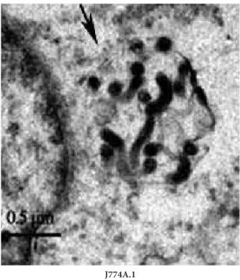
When L. interrogans strain Lai was incubated with J774A cells for 1 h, leptospires within phagocytic vesicles could be found in the cytosol of J774A.1 cells under the transmission electron microscope (Fig. 1).
Expression of integrins on J774A.1 cells
The flow cytometric examination demonstrated that J774A.1 cells expressed ITGB1, ITGB2 and ITGB3 (Fig. 2A).
Fig. 1. Internalization of L. interrogans into different host cells. Leptospires in J774A.1 cells under transmission electron micro-scope after infection with L. interrogans strain Lai for 1 h. The arrows indicate the leptospires within phagocytic vesicles of the host cells.
Role of ITGB1 in internalization of L. interrogans
The ITGB1-, ITGB2- or ITGB3-knockdown J774A.1 cells obtained by RNA interference had growth ki-netics similar to the wild-type cells (Fig S1. A). The qRT-PCR and Western blot confirmed the significant decrease of both mRNA and protein ITGB1, ITGB2 and ITGB3 in the target gene-knockdown cells (Fig S1. B and C).
When ITGB1 in J774A.1 cells was blocked with ITGB1-IgG or the expression of ITGB1 gene was in-hibited with RNA interference, the number of
lepto-spires in the cells during infection with L. interrogans strain Lai were significantly decreased compared to the wild-type cells (Fig. 2B and C). However, neither blockage of ITGB2 and ITGB3 protein or knock-down of ITGB2 and ITGB3 genes affected the inter-nalization of leptospires. Moreover, the transfection with non-specific negative siRNAs and blockage with goat IgG of irrelevant specificity did not affect the internalization of the leptospires into J774A.1 cells (Fig S2. A and B). The data suggest that ITGB1 mediated the internalization of L. interrogans in the J774A.1 cells.
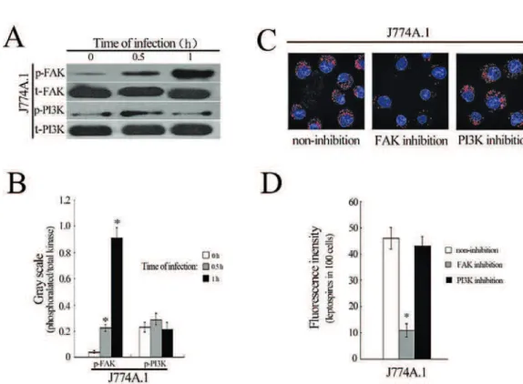
Activation of FAK or PI3K in different cells during infection
FAK and PI3K signaling pathways play crucial roles in cell membrane function such as phagocytosis and pathogen internalization through regulation of cytoskeletal rearrangement (Doherty et al., 2009). However, our Western blot revealed that the phos-phorylation levels of FAK, but not PI3K in J774A.1 cells were increased during infection with L. interro-gans strain Lai (Fig. 3A and B). When J774A.1 cells were pretreated with FAK inhibitor-I, the internali-zation of the spirochete was significantly decreased (Fig 3C and D). However, the inhibition of PI3K in
J774A.1 cells had no influence on the internalization of leptospires.
Microfilament rearrangement in cells during leptospiral internalization
that rearrangement of microfilaments rather than microtubules in J774A.1 is responsible for internal-ization of L. interrogans.
DISCUSSION
Leptospirosis is a typical systemic infectious disease that depends on the invasive ability of pathogenic Leptospira species (McBride et al., 2005; Levett et al., 2001). Macrophages play a crucial role in the innate immune response against infection by pathogenic Leptospira species. Adherence is a prerequisite for bacterial invasion into host cells (Pizarro-Cerda et al., 2006). Fibronectin (FN), laminin (14), collagen
ternalization of L. interrogans into mouse J774A.1 macrophages,
During bacterial internalization, the binding of integrin dimers to RGD-motif-containing ECM molecules has been shown to activate FAK and PI3K signaling pathways (Owen et al., 2007). Both FAK and PI3K pathways can mediate internalization of bacteria into different host cells through microfila-ment- and/or microtubule-dependent cytoskeletal rearrangements (Liu et al., 2007;, Wang et al., 2012). However, in this study, only FAK activation and mi-crofilament aggregation in the leptospire-infected J774A.1 cells could be found. More importantly, in-hibition of FAK and blockage of microfilament ag-gregation resulted in noticeable attenuation of lepto-spiral internalization.
Taken together, our findings indicated that ITGB1/FAK/microfilament-signaling mediate the internalization of L. interrogans in mouse J774A.1 macrophages, documenting one potential host-de-pendent factor of the virulence of L. interrogans in mice.
Acknowledgments - This work was supported by grants (81171534 and 81261160321) from the National Natural Sci-ence Foundation of China, and a grant ([2012]241) from the Zhejiang Provincial Program for the Cultivation of High-lev-el Innovative Health Talents, China. We also thank Dr. I.C. Bruce (Zhejiang University School of Medicine) for reading the manuscript.
Authors’ contribution
XZ and AS contributed equally to this work.
REFERENCES
Adler, B. and A. de la Pena Moctezuma (2010). Leptospira and leptospirosis. Vet. Microbiol. 140, 287-296.
Atzingen, M.V., Barbosa, A.S., De Brito, T., Vasconcellos, S.A., de Morais, Z.M., Lima, D.M., Abreu, P.A. and A.L. Nasci-mento (2008). Lsa21, a novel leptospiral protein binding adhesive matrix molecules and present during human in-fection. BMC Microbiol.8, 70.
Barbosa, A.S., Abreu, P.A., Neves, F.O., Atzingen, M.V., Watanabe, M.M., Vieira, M.L., Morais, Z.M., Vasconcellos, S.A. and A.L. Nascimento (2006). A newly identified leptospiral ad-hesin mediates attachment to laminin. Infect. Immun. 74, 6356-6364.
Bharti, A.R., Nally, J.E., Ricaldi, J.N., Matthias, M.A., Diaz, M.M., Lovett, M.A., Levett, P.N., Gilman, R.H., Willig, M.R., Go-tuzzo, E. and J.M. Vinetz (2003). Leptospirosis: a zoonotic disease of global importance. The Lancet infectious dis-eases 3, 757-771.
Bhavsar, A.P., Guttman, J.A. and B.B. Finlay (2007). Manipula-tion of host-cell pathways by bacterial pathogens. Nature
449, 827-834.
Choy, H.A., Kelley, M.M., Chen, T.L., Moller, A.K., Matsunaga, and, D.A. Haake(2007). Physiological osmotic induction of Leptospira interrogans adhesion: LigA and LigB bind extracellular matrix proteins and fibrinogen. Infect. Im-mun.75, 2441-2450.
Doherty, G.J. and H.T. McMahon (2009). Mechanisms of endocy-tosis. Ann. Rev. Biochem. 78, 857-902.
Faine, S., Adler, B. and C. Bolin(1999). Leptospira and Leptospi-rosis. MediSci, 274-287 Melbourne. Australia
Gray, D.H., Chidgey, A.P. and R.L. Boyd (2002). Analysis of thy-mic stromal cell populations using flow cytometry. J. Im-munol. Meth.260, 15-28.
Harburger, D.S., and D.A.Calderwood 2009. Integrin signalling at a glance. J. Cell Sci.122, 159-163.
Hauk, P., Macedo, F., Romero, E.C., Vasconcellos, S.A., de Morais, Z.M., Barbosa, A.S. and P.L. Ho (2008). In LipL32, the ma-jor leptospiral lipoprotein, the C terminus is the primary immunogenic domain and mediates interaction with col-lagen IV and plasma fibronectin. Infect. Immun. 76, 2642-2650.
Hoffmann, C., Ohlsen, K. and C.R. Hauck (2011). Integrin-me-diated uptake of fibronectin-binding bacteria. Eur. J. Cell Biol.90, 891-896.
Hotez, P.J. and M. Gurwith (2011). Europe’s neglected infections of poverty. Int. J. Infect. Dis.15, e611-619.
Hu, W., Ge, Y., Ojcius, D.M., Sun, D., Dong, H., Yang, X.F. and J.Yan (2013). p53 signalling controls cell cycle arrest and caspase-independent apoptosis in macrophages infected with pathogenic Leptospira species. Cell. Microbiol.15, 1642-1659
Ko, A.I., Goarant, C. and M. Picardeau (2009). Leptospira: the dawn of the molecular genetics era for an emerging zoo-notic pathogen. Nature Reviews. Microbiology7, 736-747.
Levett, P.N. (2001). Leptospirosis. Clin. Microbiol. Rev.14, 296-326.
Liu, Y., Zheng, W., Li, L., Mao, Y. and J. Yan (2007). Pathogenesis of leptospirosis: interaction of Leptospira interrogans with in vitro cultured mammalian cells. Med. Microbiol. Immu-nol.196, 233-239.
Lo, Y.C., Kintziger, K.W., Carson, H.J., Patrick, S.L., Turabelidze, G., Stanek, D., Blackmore, C., Lingamfelter, D., Dudley, M.H., Shadomy, S.V., Shieh, W.J., Drew, C.P., Batten, B.C. and S.R. Zaki (2011). Severe leptospirosis similar to pan-demic (H1N1) 2009, Florida and Missouri, USA. Emerg-ing Infect. Dis.17, 1145-1146.
McBride, A.J., Athanazio, D.A., Reis, M.G. and A.I. Ko (2005). Leptospirosis. Curr. Opin. Infect. Dis.18, 376-386.
Meites, E., Jay, M.T., Deresinski, S., Shieh, W.J., Zaki, S.R., Tomp-kins, L. and D.S. Smith (2004). Reemerging leptospirosis, California. Emerg. Infect. Dis.10, 406-412.
Mostowy, S. and P. Cossart (2009). Cytoskeleton rearrangements during Listeria infection: clathrin and septins as new play-ers in the game. Cell Mot. Cytoskel.66, 816-823.
Nascimento, A.L., Ko, A.I., Martins, E.A., Monteiro-Vitorello, C.B., Ho, P.L., Haake, D.A., Verjovski-Almeida, S., Harts-keerl, R.A., Marques, M.V., Oliveira, M.C., Menck, C.F., Leite, L.C., Carrer, H., Coutinho, L.L., Degrave, W.M., Dellagostin, O.A., El-Dorry, H., Ferro, E.S., Ferro, M.I., Furlan, L.R., Gamberini, M., Giglioti, E.A., Goes-Neto, A., Goldman, G.H., Goldman, M.H., Harakava, R., Jeronimo, S.M., Junqueira-de-Azevedo, I.L., Kimura, E.T., Kuramae, E.E., Lemos, E.G., Lemos, M.V., Marino, C.L., Nunes, L.R., de Oliveira, R.C., Pereira, G.G., Reis, M.S., Schriefer, A., Siqueira, W.J., Sommer, P., Tsai, S.M., Simpson, A.J., Ferro, J.A., Camargo, L.E., Kitajima, J.P., Setubal, J.C. and M.A. Van Sluys (2004). Comparative genomics of two Leptospi-ra interrogans serovars reveals novel insights into physiol-ogy and pathogenesis. J. Bacteriol. 186, 2164-2172.
Oelschlaeger, T.A., Guerry, P. and Kopecko, D.J. (1993). Unusual microtubule-dependent endocytosis mechanisms trig-gered by Campylobacter jejuni and Citrobacter freundii. Proc. Natl. Acad. Sci. U. S. A.90, 6884-6888.
Owen, K.A., Thomas, K.S. and A.H. Bouton (2007). The differ-ential expression of Yersinia pseudotuberculosis adhesins determines the requirement for FAK and/or Pyk2 during bacterial phagocytosis by macrophages. Cell. Microbiol.9, 596-609.
Pappas, G., Papadimitriou, P., Siozopoulou, V., Christou, L. and N. Akritidis (2008). The globalization of leptospirosis: worldwide incidence trends. Int. J. Infect. Dis.12, 351-357.
Parton, R.G. and K. Simons (2007). The multiple faces of caveo-lae. Nature reviews. Molec. Cell. Biol. 8, 185-194.
Patti, J.M., Bremell, T., Krajewska-Pietrasik, D., Abdelnour, A., Tarkowski, A., Ryden, C. and M. Hook (1994). The Staphy-lococcus aureus collagen adhesin is a virulence determi-nant in experimental septic arthritis. Infect. Immun. 62, 152-161.
Pizarro-Cerda, J. and P. Cossart (2006). Bacterial adhesion and entry into host cells. Cell124, 715-727.
Ren, S.X., Fu, G., Jiang, X.G., Zeng, R., Miao, Y.G., Xu, H., Zhang, Y.X., Xiong, H., Lu, G., Lu, L.F., Jiang, H.Q., Jia, J., Tu, Y.F., Jiang, J.X., Gu, W.Y., Zhang, Y.Q., Cai, Z., Sheng, H.H., Yin, H.F., Zhang, Y., Zhu, G.F., Wan, M., Huang, H.L., Qian, Z., Wang, S.Y., Ma, W., Yao, Z.J., Shen, Y., Qiang, B.Q., Xia, Q.C., Guo, X.K., Danchin, A., Saint Girons, I., Somerville, R.L., Wen, Y.M., Shi, M.H., Chen, Z., Xu, J.G. and G.P Zhao. (2003). Unique physiological and pathogenic fea-tures of Leptospira interrogans revealed by whole-genome sequencing. Nature 422, 888-893.
Romero, E.C., Bernardo, C.C. and P.H. Yasuda (2003). Human leptospirosis: a twenty-nine-year serological study in Sao Paulo, Brazil. Revista do Instituto de Medicina Tropical de Sao Paulo45, 245-248.
Ruoslahti, E. and M.D. Pierschbacher (1987). New perspectives in cell adhesion: RGD and integrins. Science238, 491-497.
Scibelli, A., Roperto, S., Manna, L., Pavone, L.M., Tafuri, S., Della Morte, R. and N. Staiano (2007). Engagement of integrins as a cellular route of invasion by bacterial pathogens. Vet. J.173, 482-491.\
Secott, T.E., Lin, T.L. and C.C. Wu (2004). Mycobacterium avium subsp. paratuberculosis fibronectin attachment protein facilitates M-cell targeting and invasion through a fibro-nectin bridge with host integrins. Infect. Immun.72, 3724-3732.
Stevenson, B., Choy, H.A., Pinne, M., Rotondi, M.L., Miller, M.C., Demoll, E., Kraiczy, P., Cooley, A.E., Creamer, T.P., Suchard, M.A., Brissette, C.A., Verma, A. and D.A. Haake (2007). Leptospira interrogans endostatin-like outer membrane proteins bind host fibronectin, laminin and regulators of complement. PloS ONE 2, e1188.
Takada, Y., Ye, X. and S. Simon (2007). The integrins. Genome biology8, 215.
Tang, C.L., Zhao, H.B., Li, M.Q., Du, M.R., Meng, Y.H. and D.J. Li (2012). Focal adhesion kinase signaling is necessary for the Cyclosporin A-enhanced migration and invasion of human trophoblast cells. Placenta33, 704-711.
Mitochondri-al complex III ROS regulate adipocyte differentiation. Cell Metabol.14, 537-544.
Vieira, M.L., de Morais, Z.M., Goncales, A.P., Romero, E.C., Vas-concellos, S.A. and A.L. Nascimento (2010). Lsa63, a newly identified surface protein of Leptospira interrogans binds laminin and collagen IV. J. Infect.60, 52-64.
Wang, H., Leavitt, L., Ramaswamy, R. and A.C. Rapraeger (2010). Interaction of syndecan and alpha6beta4 integrin cyto-plasmic domains: regulation of ErbB2-mediated integrin activation. J. Biol. Chem. 285, 13569-13579.
Zhang, C., Wang, H. and J. Yan (2012). Leptospirosis prevalence in Chinese populations in the last two decades. Microbes and infection / Institut Pasteur 14, 317-323.
Zhang, L., Zhang, C., Ojcius, D.M., Sun, D., Zhao, J., Lin, X., Li, L., Li, L. and J. Yan (2012). The mammalian cell entry (Mce) protein of pathogenic Leptospira species is responsible for RGD motif-dependent infection of cells and animals. Mol. Microbiol.83, 1006-1023.
SUPPLEMENTARY MATERIAL
RESULTS
Characterization of the target gene-knockdown cells
The J774A.1 cells whose ITGB1, ITGB2 or ITGB3 gene had knocked down with RNA interference showed growth kinetics similar to the wild-type cells (Fig S1. A). The qRT-PCR and Western Blot assay confirmed the significant decrease of both mRNA and protein expressed by the ITGB1, ITGB2 or ITGB3 in the target gene knockdown J774A.1 cells (Fig S1. B and C).
Internalization of L. interrogans into non-specific siR-NAs-transfected or goat IgG-blocked host cells
Fig. S1. Characterization of target gene knockdown cells. (A) Growth kinetics of the target gene knockdown cells determined by cell enumeration. The wild-type and gene knockdown cells were seeded in cell plate at 1×10⁴ separately, cell number were counted at 24h,
48h and 72h. The data show the means ± SD of three independent experiments. (B) Significant decrease of ITGB1-, ITGB2- or ITGB3-mRNA levels in the target gene-knockdown cells determined by qRT-PCR. Bars show the means ± SD of three independent experi-ments. The ITGB1-, ITGB2- or ITGB3 mRNA levels in the wild-type J774A.1 was set as 100%. *: p < 0.05vs.the ITGB1-, ITGB2- or ITGB3-mRNA levels in the wild-type J774A.1 cells. (C) Significant decrease of ITGB1, ITGB2 or ITGB3 protein expressedby the target gene-knockdown cells determined by Western blot assay.