Factors Affecting Cerebral Oxygenation in
Hemodialysis Patients: Cerebral Oxygenation
Associates with pH, Hemodialysis Duration,
Serum Albumin Concentration, and Diabetes
Mellitus
Kiyonori Ito1, Susumu Ookawara1,2*, Yuichiro Ueda1, Sawako Goto1, Haruhisa Miyazawa1, Hodaka Yamada3, Taisuke Kitano1, Mitsunobu Shindo1, Yoshio Kaku1, Keiji Hirai1,
Masashi Yoshida3, Taro Hoshino1, Aoi Nabata1, Honami Mori1, Izumi Yoshida1, Masafumi Kakei3, Kaoru Tabei1
1Division of Nephrology, Department of Integrated Medicine, Saitama Medical Center, Jichi Medical University, Saitama, Japan,2Department of Internal Medicine, Nishikawa Town Hospital, Yamagata, Japan,
3Division of Endocrinology and Metabolism, Department of Integrated Medicine Saitama Medical Center, Jichi Medical University, Saitama, Japan
*su-ooka@hb.tp1.jp
Abstract
Background
Patients undergoing hemodialysis (HD) often develop cerebral disease complications. Fur-thermore, cerebral regional saturation of oxygen (rSO2) was previously reported to be
signif-icantly lower in HD patients than in healthy subjects. We aimed to identify the factors affecting the cerebral rSO2in HD patients.
Methods
Fifty-four HD patients (38 men and 16 women; mean age, 67.7±1.2 years, HD duration, 6.5±1.9 years) were recruited. Cerebral rSO2was monitored at the forehead before HD
using an INVOS 5100C (Covidien Japan, Tokyo, Japan).
Results
The rSO2levels were significantly lower in HD patients compared with healthy controls
(49.5±1.7% vs. 68.9±1.6%, p<0.001). Multiple regression analysis showed that cerebral rSO2 independently associated with pH (standardized coefficient: -0.35), HD duration (stan-dardized coefficient: -0.33), and serum albumin concentration (stan(stan-dardized coefficient: 0.28). Furthermore, the rSO2was significantly lower in HD patients with diabetes mellitus
(DM), compared with patients without DM (46.8±1.7% vs. 52.1±1.8%, p<0.05).
OPEN ACCESS
Citation:Ito K, Ookawara S, Ueda Y, Goto S, Miyazawa H, Yamada H, et al. (2015) Factors Affecting Cerebral Oxygenation in Hemodialysis Patients: Cerebral Oxygenation Associates with pH, Hemodialysis Duration, Serum Albumin Concentration, and Diabetes Mellitus. PLoS ONE 10(2): e0117474. doi:10.1371/journal.pone.0117474
Academic Editor:Kandiah Jeyaseelan, National University of Singapore, SINGAPORE
Received:August 26, 2014
Accepted:December 24, 2014
Published:February 23, 2015
Copyright:© 2015 Ito et al. This is an open access article distributed under the terms of theCreative Commons Attribution License, which permits unrestricted use, distribution, and reproduction in any medium, provided the original author and source are credited.
Data Availability Statement:All relevant data are within the paper.
Funding:These authors have no support or funding to report.
Conclusions
In HD patients, cerebral rSO2was affected by multiple factors, including pH, HD duration,
and serum albumin concentration. Furthermore, this is the first report describing significant-ly lower levels of rSO2in HD patients with DM than in those without DM.
Introduction
Central nervous system (CNS) dysfunction, such as uremic encephalopathy, cognitive im-pairment, and dementia, is a frequent complication of patients undergoing hemodialysis (HD). [1] Cerebrovascular accident (CVA) was described as the fourth leading cause of death in HD patients according to the annual report of the Japanese Society for Dialysis Therapy in 2011. [2] Magnetic resonance imaging (MRI) is a useful tool for detecting morphological changes in the brain and therefore evaluating CVA; in addition, silent cerebral infarction detected by MRI has been found to associate with the severity of cognitive impairment in HD patients. [3] How-ever, imaging methods like MRI and computed tomography can only provide information about organic lesions in the brain, and cannot evaluate the functional status such as cerebral blood flow and cerebral oxygenation. Recently, near-infrared spectroscopy (NIRS) has been used as a tool to measure the regional saturation of oxygen (rSO2), a marker of tissue
oxygen-ation, at the frontal cerebral cortex in a variety of clinical situations, and has shown the change of critical balance between arterial oxygen delivery and cerebral oxygen consumption. [4–7] Cerebral rSO2was reported to be significantly lower in HD patients than in healthy controls.
[1,8] Few reports, however, have examined the relationship between cerebral oxygenation in HD patients and clinical parameters. Therefore, in this study, we aimed to elucidate the clinical factors influencing cerebral rSO2in HD patients.
Methods
Monitoring of cerebral oxygenation and clinical laboratory measurement
Cerebral rSO2was monitored at the forehead with an INVOS 5100C saturation monitor(Covi-dien Japan, Tokyo, Japan), which utilizes NIRS technology. This instrument uses a light-emit-ting diode, which transmits near-infrared light at 2 wavelengths (735 and 810 nm), and 2 silicon photodiodes which act as light detectors; results are read as a single numerical value that represents the rSO2[9,10]. All data obtained by this instrument were immediately and
automatically stored in sequence. Interobserver variance for this instrument, that is, reproduc-ibility of the rSO2measurement, is acceptable as previously reported. Therefore, rSO2is
consid-ered reliable when estimating the actual cerebral oxygenation. [11]
Prior to HD, the recruited patients rested in the supine position for at least 10 min in order to reduce the influence of postural change. An rSO2measurement sensor was attached to the
patient’s forehead for measurement in the resting state. Thereafter, rSO2was measured for
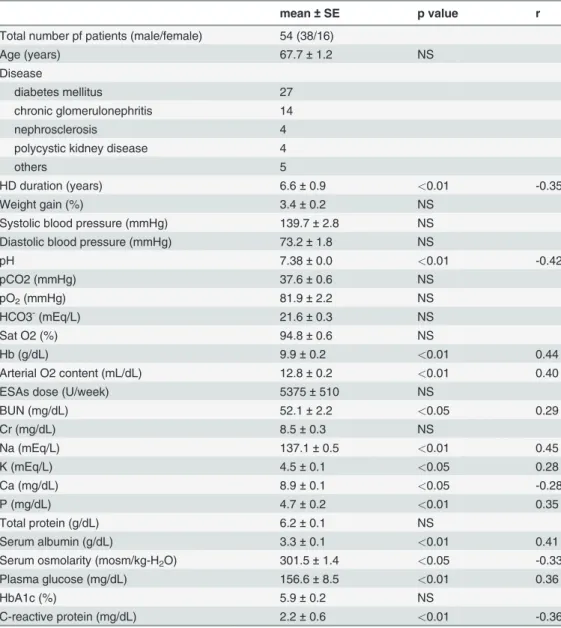
Table 1. Patients Characteristics and the correlation between cerebral rSO2and clinical parameters.
mean±SE p value r
Total number pf patients (male/female) 54 (38/16)
Age (years) 67.7±1.2 NS
Disease
diabetes mellitus 27
chronic glomerulonephritis 14
nephrosclerosis 4
polycystic kidney disease 4
others 5
HD duration (years) 6.6±0.9 <0.01 -0.35
Weight gain (%) 3.4±0.2 NS
Systolic blood pressure (mmHg) 139.7±2.8 NS
Diastolic blood pressure (mmHg) 73.2±1.8 NS
pH 7.38±0.0 <0.01 -0.42
pCO2 (mmHg) 37.6±0.6 NS
pO2(mmHg) 81.9±2.2 NS
HCO3-(mEq/L) 21.6±0.3 NS
Sat O2 (%) 94.8±0.6 NS
Hb (g/dL) 9.9±0.2 <0.01 0.44
Arterial O2 content (mL/dL) 12.8±0.2 <0.01 0.40
ESAs dose (U/week) 5375±510 NS
BUN (mg/dL) 52.1±2.2 <0.05 0.29
Cr (mg/dL) 8.5±0.3 NS
Na (mEq/L) 137.1±0.5 <0.01 0.45
K (mEq/L) 4.5±0.1 <0.05 0.28
Ca (mg/dL) 8.9±0.1 <0.05 -0.28
P (mg/dL) 4.7±0.2 <0.01 0.35
Total protein (g/dL) 6.2±0.1 NS
Serum albumin (g/dL) 3.3±0.1 <0.01 0.41
Serum osmolarity (mosm/kg-H2O) 301.5±1.4 <0.05 -0.33
Plasma glucose (mg/dL) 156.6±8.5 <0.01 0.36
HbA1c (%) 5.9±0.2 NS
C-reactive protein (mg/dL) 2.2±0.6 <0.01 -0.36
5 min before HD, and we evaluated the mean rSO2for 5 min, as a marker of cerebral
oxygen-ation, in each patient. Blood samples were obtained from each patient under room air. It was previously reported that samples obtained from the radial artery or those from an arterial line at the arteriovenous fistula presented similar values when evaluating the parameters of oxygen status, including pH, oxygen pressure (pO2: mmHg), and oxygen saturation (SpO2: %). [12]
Therefore, prior to HD we obtained all blood samples, including blood gas analysis, from the arterial site of arteriovenous fistulae in each patient.
Arterial O2 content (CaO2) and serum osmolality (sOsm) were calculated using the
follow-ing equations:
CaO2ðmL=dLÞ ¼1:34 Hb SpO2 100 þ ð0:0031 pO2Þ
sOsmðmosm=kg H2OÞ ¼ ð2 NaÞþPG 18 þ BUN 2:8
where Hb represents the hemoglobin concentration (g/dL). Na represents the serum sodium concentration (mEq/L), PG represents the plasma glucose level (mg/dL), and BUN represents the blood urea nitrogen concentration (mg/dL).
Erythropoiesis-stimulating agents (ESAs) were administered for the treatment of renal ane-mia, and calculations of the optimum ESA dose (U/week) were based on a method reported previously, [15] where a ratio of 1:200 was used to convert the dose for long-acting ESAs, in-cluding darbepoetin-αand continuous erythropoietin receptor activator, into a short-acting
re-combinant human erythropoietin equivalent dose for each patient. [15] The rSO2in healthy
controls was measured for at least 5 min in the supine position in a manner similar to that in HD patients.
Analysis
Data were expressed as mean ± standard error (SE). The Student’s t-test for non-paired values was used for comparing 2 groups, and Mann–Whitney U test was used for comparison of non-parametric variables between 2 groups. Correlations between 2 groups were evaluated by Pear-son’s correlation coefficient and linear regression analysis. Multiple regression analysis was performed using parameters that showed a significant correlation with cerebral rSO2. A
differ-ence of p<0.05 was considered significant.
Results
Cerebral rSO2at rest in HD patients was compared with that in healthy controls, and there was
a significant difference between the 2 groups (HD patients: 49.5 ± 1.7%, healthy controls: 68.9 ± 1.6%, p<0.001) (Fig. 1). Recently, cerebral rSO2was reported to be significantly lower in
HD patients than in healthy controls [1,8], and our results are consistent with these reports.
Table 1shows patients’characteristics, and correlations between the cerebral rSO2and
clini-cal parameters. Cerebral rSO2showed significant positive correlations with CaO2, hemoglobin
(Hb) level, serum sodium concentration, serum potassium concentration, serum inorganic phosphate concentration, serum albumin concentration, and plasma glucose level. A simple linear regression analysis revealed that cerebral rSO2was negatively correlated with pH, serum
calcium concentration, sOsm, HD duration, and C-reactive protein.
We performed a multivariate linear regression analysis using variables that showed a signifi-cant correlation with the cerebral rSO2in a simple linear regression analysis (Table 2). The
multivariate regression analysis found that the cerebral rSO2was independently associated
with pH (standardized coefficient: -0.35), HD duration (standardized coefficient: -0.33), and serum albumin concentration (standardized coefficient: 0.28). On the other hand, the cerebral
[13]
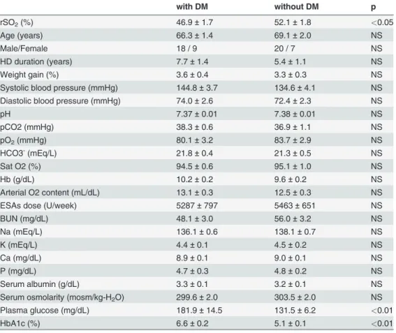
rSO2was independent of Hb and CaO2. We also evaluated the influence of DM on the cerebral
rSO2values (Table 3). The cerebral rSO2was significantly lower in patients with DM than in
those without DM (46.8 ± 1.7% vs. 52.1 ± 1.8, p<0.05) (Fig. 2). In addition to the difference in cerebral rSO2, there were significant differences in serum sodium concentration, plasma
glu-cose level, and HbA1c levels between the 2 groups.
Fig 1. Comparison between cerebral rSO2in hemodialysis patients and healthy controls.
Discussion
Regional saturation of oxygen (rSO2) is widely used for monitoring cerebral function during
cerebral surgery, as rSO2measured using NIRS can provide accurate yet non-invasive
informa-tion on cerebral oxygen saturainforma-tion, and can be easily performed in the clinical setting. [1,4–11] Recently, cerebral rSO2was reported to be significantly lower in HD patients than in healthy
controls. [1,8] The reasons for this however, remain uncertain, and the factors affecting the de-terioration of cerebral rSO2in HD patients have not been determined. In this study, we
identi-fied modifiable factors, including pH, HD duration, and serum albumin concentration, as being independently associated with cerebral rSO2; we also demonstrated that cerebral rSO2
was significantly lower in HD patients with DM than in those without DM.
Among the modifiable factors identified as being independently associated with cerebral rSO2, pH was the factor most strongly affecting cerebral rSO2in HD patients. A decrease of
Table 2. Multivariate linear regression analysis: Independent factors of cerebral rSO2in hemodialysis patients.
Variables Coefficient Standardized coefficient p
pH -62.5 -0.35 0.012
HD duration -0.47 -0.33 0.006
Serum albumin 4.12 0.28 0.041
doi:10.1371/journal.pone.0117474.t002
Table 3. Different clinical parameters for hemodialysis patients with and without diabetes mellitus.
with DM without DM p
rSO2(%) 46.9±1.7 52.1±1.8 <0.05
Age (years) 66.3±1.4 69.1±2.0 NS
Male/Female 18 / 9 20 / 7 NS
HD duration (years) 7.7±1.4 5.4±1.1 NS
Weight gain (%) 3.6±0.4 3.3±0.3 NS
Systolic blood pressure (mmHg) 144.8±3.7 134.6±4.1 NS
Diastolic blood pressure (mmHg) 74.0±2.6 72.4±2.3 NS
pH 7.37±0.01 7.38±0.01 NS
pCO2 (mmHg) 38.3±0.6 36.9±1.1 NS
pO2(mmHg) 80.1±3.2 83.7±2.9 NS
HCO3-(mEq/L) 21.8±0.4 21.3±0.5 NS
Sat O2 (%) 94.5±0.6 95.1±1.0 NS
Hb (g/dL) 10.2±0.2 9.6±0.2 NS
Arterial O2 content (mL/dL) 13.1±0.3 12.5±0.3 NS
ESAs dose (U/week) 5287±797 5463±651 NS
BUN (mg/dL) 48.1±3.0 56.0±3.2 NS
Na (mEq/L) 136.1±0.6 138.1±0.7 NS
K (mEq/L) 4.4±0.1 4.5±0.2 NS
Ca (mg/dL) 8.9±0.1 9.0±0.1 NS
P (mg/dL) 4.7±0.3 4.8±0.2 NS
Serum albumin (g/dL) 3.3±0.1 3.2±0.1 NS
Serum osmolarity (mosm/kg-H2O) 299.6±2.0 303.5±2.0 NS
Plasma glucose (mg/dL) 181.9±14.5 131.5±6.2 <0.01
HbA1c (%) 6.6±0.2 5.1±0.1 <0.01
extracellular pH, even without partial pressure of carbon dioxide (pCO2) increase, was previ-ously reported to induce a dilation of the cerebral artery, [16] and therefore, regional cerebral blood flow (rCBF) would increase in response to the decrease in pH. Thus, in the brain, it is possible that cerebral rSO2increases via the increase of arterial oxygen delivery accompanying
rCBF increase induced by pH decrease. This mechanism could explain why cerebral rSO2
shows an inverse relationship with pH change. Furthermore, changes in cerebral rSO2were
re-cently shown to be independently and negatively associated with changes in pH in patients un-dergoing liver transplantation, [17] and our results, which indicated an inverse relationship between cerebral rSO2and pH, were consistent with this report. Thus far, however, the change
of rSO2affected by pH remains unclear, so further examination would be required regarding
the association between cerebral rSO2change and pH.
Serum albumin concentration also showed a significant positive correlation with cerebral rSO2. Serum albumin concentration was previously reported to be a prognostic marker of
sur-vival in HD patients, similar to nutritional status. [18,19] The decrease in its concentration has often been observed in patients with protein-energy malnutrition, leading to prognostic
Fig 2. Comparison of cerebral rSO2in hemodialysis patients (HD) with and without diabetes mellitus (DM).*<0.05 at HD patients with DM vs. those
without DM.
aggravation in HD patients. In general, serum albumin concentration contributes to the forma-tion of colloid osmotic pressure in vessels and associates with body-fluid movement, mainly between the vessels and the interstitium. In addition, serum albumin concentration was recent-ly shown to positiverecent-ly correlate with the regional cerebral blood flow in patients with liver cir-rhosis. [20] Based on our results, we propose that an increase in serum albumin concentration might lead to increased rCBF and improved cerebral oxygenation, which can then be measured as cerebral rSO2. Therefore, serum albumin concentration would appear to associate not only
with nutritional status and prognosis, but also with cerebral oxygenation in HD patients, al-though its precise mechanism remains uncertain.
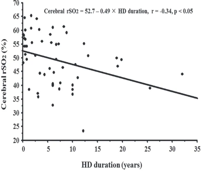
Furthermore, in this study, the cerebral rSO2was negatively affected by HD duration, and
the annual rSO2decline in HD patients was predicted to be -0.49%/year by simple linear
re-gression analysis (Fig. 3). It was previously reported that, in HD patients, rCBF to the frontal cortex decreased with an increase in HD duration, resulting in white matter lesions. [21] As the cerebral rSO2mainly indicates the condition of rCBF, the negative impact of HD duration on
cerebral rSO2might be due to a decrease in the rCBF.
On the other hand, although CaO2and Hb levels significantly correlated with cerebral rSO2
in a simple linear regression analysis, these associations disappeared upon multivariate linear regression analysis. Hemoglobin is an important factor in oxygen supply to the peripheral tis-sues and organs, including the brain; CaO2is a marker for oxygen supply. Thus, cerebral rSO2
might be expected to show a strong correlation with Hb and CaO2levels; however, no
signifi-cant correlations were observed in the present study. Positron emission tomography analysis has revealed an association between the rCBF and Hb levels in HD patients. [22] In this report, regional cerebral blood flow decreased significantly, and oxygen metabolism was disrupted de-spite the increase in Hb levels. Decreased blood cell deformability and increased plasma viscos-ity resulted in decreased erythrocyte velocviscos-ity in the cerebral capillaries, leading to an increase in Hb levels. Furthermore, in HD patients, CaO2was reported to be the most important
deter-minant of inter-individual middle cerebral artery (MCA) blood flow velocity variance and vari-ation; in addition, an increase in CaO2could induce the decrease of MCA blood flow velocity
via vasoconstrictions in small intracerebral vessels to maintain oxygen delivery to the brain. [23,24] Indeed, in an experimental study, cerebral O2 transport (CBF × CaO2) was regulated at
constant levels, independently of alterations in Hb levels and CaO2values. [25] Therefore, it is
unlikely that oxygen metabolism including rSO2could directly associate with Hb and CaO2
levels. Furthermore, ESA administration was reported to reduce the development of brain edema, and preserve the local brain oxygen saturation and brain tissue oxygenation after trau-matic brain injury. [26,27] However, there was no association between cerebral rSO2values
and ESA doses in HD patients.
This study included the results obtained from 1 patient with DM, which showed an ex-tremely low cerebral rSO2value. Thus, we analyzed the cerebral rSO2values using the
Grubbs-Smirnov rejection test to clarify whether the extremely low rSO2value should be excluded. The
test results were not significant; therefore, based on the data obtained in this study, a compari-son of cerebral rSO2values was performed for cohorts of HD patients with, and without, DM.
The results, which showed significant differences in cerebral rSO2in HD patients with DM
hyperglycemia-induced endothelial dysfunction. Furthermore, dynamic cerebral autoregula-tion was reported to be impaired even during an early phase in type 2 DM patients. [32] In this study, plasma glucose and HbA1c levels were significantly higher in patients with DM than in those without DM; therefore, the significant decrease in cerebral rSO2with DM might be
in-duced by the dysregulation of regional cerebral perfusion due to hyperglycemia-inin-duced endo-thelial dysfunction. In the present study, there was a difference of ~5% in the cerebral rSO2
between HD patients with and without DM, and this value corresponds to cerebral rSO2
de-cline for nearly 10 years, at an annual rate of dede-cline of -0.49%/year for rSO2in HD patients;
calculations for the rate of decline were based on the negative correlation between cerebral rSO2and HD duration. Therefore, these results might explain the mechanisms underlying the
frequent occurrence of cerebral complications such as dementia and cognitive impairment in HD patients with DM.
This study faced the limitation of a relatively small sample size; therefore, further study is re-quired to fully elucidate the correlation of cerebral rSO2with various clinical parameters.
Fig 3. Correlation between hemodialysis duration and rSO2.
In conclusion, cerebral rSO2was affected by multiple factors in HD patients, including pH,
HD duration, and serum albumin concentration. Furthermore, rSO2was significantly lower in
HD patients with DM than in those without DM.
Acknowledgments
We thank the study participants and the clinical engineers, Katsunobu Ando, Takayuki Uchida, Masaya Kofuji, and Tsukasa Higuchi, for their day-to-day clinical care about hemodialysis.
Author Contributions
Conceived and designed the experiments: KI SO. Analyzed the data: KI SO YU SG H. Miya-zawa TK MS YK KH TH AN H. Mori IY. Wrote the paper: KI SO KT. Supervised data collec-tion and manuscript preparacollec-tion: HY MY MK KT.
References
1. Prohovnik I, Post J, Uribarri J, Lee H, Sandu O, et al. (2007) Cerebrovascular effects of hemodialysis in chronic kidney disease. J Cereb Blood Flow Metab 27: 1861–1869. PMID:17406658
2. Nakai S, Watanabe Y, Masakane I, Wada A, Shoji T, et al.(2013) Overview of regular dialysis treatment in Japan (as of 31 December 2011). Ther Apher Dial 17: 567–611. doi:10.1111/1744-9987.12147 PMID:24330555
3. Naganuma T, Uchida J, Tsuchida K, Takemoto Y, Tatsumi S, et al. (2005) Silent cerebral infarction pre-dicts vascular events in hemodialysis patients. Kidney Int 67: 2434–2439. PMID:15882289
4. Parnia S, Nasir A, Ahn A, Malik H, Yang J, et al. (2014) A feasibility study of cerebral oximetry during in-hospital mechanical and manual cardiopulmonary resuscitation. Crit Care Med 42: 930–933. doi:10.
1097/CCM.0000000000000047PMID:24247475
5. Ono M, Arnaoutakis GJ, Fine DM, Brady K, Easley RB, et al. (2013) Blood pressure excursions below the cerebral autoregulation threshold during cardiac surgery are associated with acute kidney injury. Crit Care Med 41: 464–471. doi:10.1097/CCM.0b013e31826ab3a1PMID:23263580
6. McCusker K, Chalafant A, de Foe G, Gunaydin S, Vijay V (2006) Influence of hematocrit and pump prime on cerebral oxygen saturation in on-pump coronary revascularization. Perfusion 21: 149–155. PMID:16817287
7. Calderon-Arnulphi M, Alaraj A, Amin-Hanjani S, Mantulin WW, Polzonetti CM, et al. (2007) Detection of cerebral ischemia in neurovascular surgery using quantitative frequency-domain near-infrared spec-troscopy. J Neurosurg 106: 283–290. PMID:17410713
8. Hoshino T, Ookawara S, Miyazawa H, Ito K, Ueda Y, et al. (2014) Evaluation of cerebral oxygenation in patients undergoing long-term hemodialysis. Nephron Clin Pract 126: 57–61. doi:10.1159/000358432 PMID:24526002
9. Tobias JD (2006) Cerebral oxygenation monitoring: near-infrared spectroscopy. Expert Rev Med De-vices 3:235–243. PMID:16515389
10. Ferrari M, Mottola L, Quaresima V (2004) Principles, techniques, and limitations of near infrared spec-troscopy. Can J Appl Physiol 29: 463–487. PMID:15328595
11. Lemmers PMA, Toet MC, van Bel F (2008) Impact of patent ductus arteriosus and subsequent therapy with indomethacin on cerebral oxygenation in preterm infants. Pediatrics 121: 142–147. doi:10.1542/
peds.2007-0925PMID:18166568
12. Nielsen AL, Thunedborg P, Brinkrnfeldt H, Hegbrant J, Jensen HA, et al. (1999) Assessment of pH and oxygen status during hemodialysis using the arterial blood line in patients with an arteriovenous fistula. Blood Purif 17: 206–212. PMID:10494023
13. Roach RC, Koskolou MD, Calbert JAL, Saltin B (1999) Arterial O2 content and tension in regulation of cardiac output and leg blood flow during exercise in humans. Am J Physiol Heart Circ Physiol 276: H438–H445.
14. Gennari FJ (1984) Serum osmolality. Uses and limitation. N Engl J Med 310: 102–105. PMID:6361557 15. Portoles JM, de Francisco ALM, Gorriz JL, Martines-Castelao A, Lopes-Gomez JM, et al. (2008)
16. Kontos HA, Raper AJ, Patterson JL (1977) Analysis of vasoactivity of local pH, PCO2 and bicarbonate on pial vessels. Stroke 8: 358–360. PMID:16363
17. Jun IG, Shin WJ, Park YS, Song JG, Kim YK, et al. (2012) Factors affecting intraoperative changes in regional cerebral oxygen saturation in patients undergoing liver transplantation. Transplant Proc 45: 245–50.
18. Lowrie EG, Lew NL (1990) Death risk in hemodialysis patients: the predictive value of commonly mea-sured variables and an evaluation of death rate differences between facilities. Am J Kidney Dis 15: 458–482. PMID:2333868
19. Stenvinkel P, Heimbürger O, Lindholm B, Kaysen GA, Bergström J (2000) Are there two types of mal-nutrition in chronic renal failure? Evidence for relationships between malmal-nutrition, inflammation and ath-erosclerosis (MIA syndrome). Nephrol Dial Transplant 15: 953–960. PMID:10862630
20. Tanaka H, Maeshima S, Ueda H, Shigekawa Y, Fukuchi H, et al. (2007) Reduction of regional cerebral blood flow of patients with liver cirrhosis and its correlation with serum albumin. International Medical Journal 14: 35–39.
21. Kanai H, Hirakata H, Nakane H, Fujii K, Hirakata E, et al. (2001) Depressed cerebral oxygen metabo-lism in patients with chronic renal failure: a positron emission tomography study. Am J Kidney Dis 38: S129–33. PMID:11576938
22. Metry G, Wikström B, Valind S, Sandhagen B, Linde T, et al. (1999) Effect of normalization of hemato-crit on brain circulation and metabolism in hemodialysis patients. J Am Soc Nephrol 10: 854–863. PMID:10203371
23. Stefanidis I, Bach R, Mertens PR, Liakopoulos V, Liapi G, et al. (2005) Influence of hemodialysis on the mean blood flow velocity in the middle cerebral artery. Clin Nephrol 64: 129–137. PMID:16114789 24. Macko RF, Ameriso SF, Akmal M, Paganini-Hill A, Mohler JG, et al. (1993) Arterial oxygen content and
age are determinants of middle cerebral artery blood flow velocity. Stroke 24: 1025–1028. PMID:
8322377
25. Ulatowski JA, Bucci E, Razynska A, Traystman RJ, Koehler RC (1998) Cerebral blood flow during hyp-oxic hypoxia with plasma-based hemoglobin at reduced hematocrit. Am J Physiol 274: H1933–1942. PMID:9841479
26. Verdonck O, Lahrech H, Francony G, Carle R, Farion R, et al. (2007) Erythropoietin protects from post-traumatic edema in the rat brain. J Cereb Blood Flow Metab 27: 1369–1376. PMID:17264861 27. Bouzat P, Millet A, Boue Y, Pernet-Gallary K, Trouve-Buisson T, et al. (2013) Changes in brain tissue
oxygenation after treatment of diffuse traumatic brain injury by erythropoietin. Crit Care Med 41: 1316– 1324. doi:10.1097/CCM.0b013e31827ca64ePMID:23591210
28. Ott A, Stolk RP, van Harskamp F, Pols HA, Hofman A, et al. (1999) Diabetes mellitus and the risk of de-mentia: The Rotterdam Study. Neurology 53: 1937–1942. PMID:10599761
29. Murray AM, Tupper DE, Knopman DS, Gilbertson DT, Pederson SL, et al. (2006) Cognitive impairment in hemodialysis patients is common. Neurology 67: 216–23. PMID:16864811
30. Ohara T, Doi Y, Ninomiya T, Hirakawa Y, Hata J, et al. (2011) Glucose tolerance status and risk of de-mentia in the community: the Hisayama study. Neurology 77: 1126–1134. doi:10.1212/WNL.
0b013e31822f0435PMID:21931106
31. Cosentino F, Battista R, Scuteri A, De Sensi F, De Siati L, et al. (2009) Impact of fasting glycemia and regional cerebral perfusion in diabetic subjects: a study with technetium-99m-ethyl cysteinate dimer single photon emission computed tomography. Stroke 40: 306–308. doi:10.1161/STROKEAHA.108.
520627PMID:18845804