Cop
yright
© ABE&M t
odos os dir
eit
os r
eser
vados
.
Correspondence to: Amélio F. Godoy-Matos Instituto Estadual de Diabetes e Endocrinologia
Rua Moncorvo Filho, 90 20211-340 − Rio de Janeiro, RJ, Brazil
Received on Jan/5/2011 Accepted on Feb/11/2011
1 Instituto Estadual de Diabetes e
Endocrinologia do Rio de Janeiro (IEDE), Rio de Janeiro, RJ Brazil
2 Pontifícia Universidade Católica
do Rio de Janeiro (PUC-RJ), Rio de Janeiro, RJ, Brazil
3 Sérgio Franco Medicina
Diagnóstica, Rio de Janeiro, RJ, Brazil
Serum retinol binding protein 4 is not
decreased in congenital generalized
lipodystrophy: a case series
Os níveis plasmáticos da retinol binding protein 4 (rbP4) não estão reduzidos na lipodistroia congênita generalizada: uma série de casos
Amélio F. Godoy-Matos1,2, Rodrigo O. Moreira1,2, Renata MacDowell1,2, Izidro Bendet3
ABSTRACT
Introduction: Previous studies have suggested that Retinol Binding Protein 4 (RPB4), a protein produced by the adipose tissue, is associated with insulin resistance (IR). Congenital Generalized Lipodystrophy (CGL) is a rare disease characterized by IR and paucity of adipose tissue. Our objec-tive was to determine RBP4 levels in patients with CGL. Subjects and methods: Six (6) patients with CGL and a healthy control group were selected to participate in the study. Anthropometric and biochemical variables were compared between groups. Results: No difference was observed in RBP4 levels between the two groups (CGL 42.5 [12.5 – 127] vs. control 57.4 [15.9 – 165]; p = 0.78). On
the other hand, leptin levels were significantly lower in CGL patients (CGL 0.65 [0.2 – 0.7] vs. control
10.9 [0.9 – 38.6]; p = 0.015). No correlation was found between RBP-4 and waist circunference (r = 0.18, p = 0.57), or BMI (r = 0.24, p = 0.45). Conclusion: RBP4 is not decreased in CGL. These results suggest that adipose tissue may not be the main source of RBP4. Arq Bras Endocrinol Metab. 2011;55(4):279-83
Keywords
Retinol binding protein 4; lipodystrophy; obesity
RESUMO
Introdução: Estudos prévios sugeriram que os níveis plasmáticos da retinol binding protein (RBP4), uma proteína do tecido adiposo, estão associados com a resistência à insulina (RI). A lipodistrofia congênita generalizada (LCG) é uma doença rara caracterizada por ausência de tecido adiposo e RI. O objetivo é determinar os níveis de RBP4 em pacientes com LCG. Sujeitos e métodos: Seis (6) pacientes com LCG e um grupo controle saudável foram selecionados para participar no estudo. As variáveis antropométricas e bioquímicas foram comparadas quando comparados os grupos. Resultados: Nenhuma diferença foi observada entre os níveis de RBP4 log entre os grupos (LCG 42,5 [12,5 – 127] vs. controle 57,4 [15,9 – 165]; p = 0,78). Por outro lado,
os níveis de leptina foram menores em pacientes com LCG (LCG 0,65 [0,2 – 0,7] vs. controle 10.9
[0,9 – 38,6]; p = 0,015). Nenhuma correlação foi encontrada entre RBP4 e cintura (r = 0,18, p = 0,57) ou IMC (r = 0,24, p = 0,45). Conclusão: RBP4 não está diminuída na LCG. Esses resultados suge-rem que o tecido adiposo pode não ser a principal fonte de RBP4. Arq Bras Endocrinol Metab. 2011;55(4):279-83
Descritores
Retinol binding protein 4; lipodistroia congênita generalizada; obesidade
INTRODUCTION
A
dipose tissue was traditionally considered only an energy storage organ. Over the past decades, how-ever, it has also emerged as an endocrine organ. It is now recognized that adipose tissue secretes multipleIn-Cop
yright
© ABE&M t
odos os dir
eit
os r
eser
vados
.
deed, one of the most important regulators of IR in obese patients is glucose-transporter 4 (Glut-4).
In 2001, Abel and cols. (3) developed a mouse mo-del selectively knocked-out for Glut-4 gene in the adi-pose tissue (adipose GLUT4 -/- mice). Those animals developed IR in muscle and liver, suggesting that impai-ring glucose uptake by adipose tissue would interrupt a “dialogue” between fat and muscle or liver. Lately, the same group analyzed DNA arrays of adipose-tissue samples from these mice, and showed a substantial in-crease in mRNA expression of retinol binding protein 4 (RBP4). They also demonstrated that subjects with IR also presented higher RBP4 plasma levels (4). Moreo-ver, injection of RBP4 in rodents or its overexpression impairs glucose uptake by skeletal muscle and increases expression of the gluconeogenic enzyme phosphoenol-pyruvate carboxykinase (PEPCK) in the liver, deining a link between RBP4 with insulin and glucose metabo-lism (4-6).
Previously, RBP4 was known as the speciic trans-port protein for retinol (vitamin A) in the circulation. RBP4 is a 21-KDa protein that binds to transthyretin to form a complex that prevents its renal clearance (4,6). Since those initial cornerstone reports, several authors have shown that RBP4 correlates to anthropo-metric indicators of overweight and/or fat distribution (i.e. body mass index and waist-to-hip ratio) (5,6), and associated metabolic abnormalities such as triglyceride levels, systolic blood pressure and low levels of high--density lipoprotein cholesterol (6,7). Further, RBP4 is increased in type 2 diabetes (4). Studies have not con-clusively demonstrated if RBP4 plays a causal role in IR and, therefore, will be a future goal for therapy, or if it acts only as a biomarker for type 2 diabetes and insulin resistance (5,7).
One way to investigate the association between adipokines and metabolic diseases would be states of absence (or signiicant reduction) of fat tissue. One im-portant disease that presents such feature is congenital generalized lipodystrophy (CGF) (8). For instance, it has been shown that leptin is signiicantly decreased in total lipodystrophy and anorexia nervosa (9-12). To the best of our knowledge, RBP4 has not been stu-died in such situation. Therefore, we aimed at studying serum RBP4 levels in patients with CGL due to their complete absence of adipose tissue. Considering leptin as a recognized biomarker of body fat, we established this adipokine as an adjustment factor, and obtained a ratio between RBP4 and leptin.
STUDY DESIGN AND METHODS
Subjects
Six (6) patients with congenital generalized lipodystro-phy were invited to participate in the study. All partici-pants were carefully examined by an experienced endo-crinologist, and provided a detailed medical history at baseline evaluation. The protocol was approved by the Ethics Committee of the institution. A written infor-med consent was obtained from each patient or a legal tutor, after the procedures involved in the study were thoroughly explained.
The group of 6 patients with generalized lipodys-trophy presented typical phenotype for generalized lipodystrophy type 1, characterizing the Berardinelli--Seip Syndrome. A control group was also carefully and sequentially selected in order to match each lipodystro-phic patient by sex, BMI and age.
Anthropometrical examination
All participants had the following anthropometric data recorded: body weight (kg), height (m), body mass in-dex (BMI), waist circumference (WC), waist-to-hip ra-tio (WHR) and blood pressure. BMI was calculated as weight in kilograms divided by the square of height in meters (kg/m2). Waist circumference was determined at the midpoint between the lowest rib and the iliac crest. WHR was deined as the ratio of waist girth to the largestcircumference of the hips, measured at the trochanter major.
Laboratory evaluation
Cop
yright
© ABE&M t
odos os dir
eit
os r
eser
vados
.
Statistical analysis
Statistical analysis was performed with GraphPad InStat 3.00 for Windows 95 (GraphPad Software, San Diego, California, USA). Unpaired t test was used for parametric variables, and Mann-Whitney for non-parametric varia-bles. The strength of the linear relationship between two continuous variables was evaluated by means of Pearson’s correlation coeficient or Spearman’s correlation coefi-cient. The level of statistical signiicance was 5%.
RESULTS
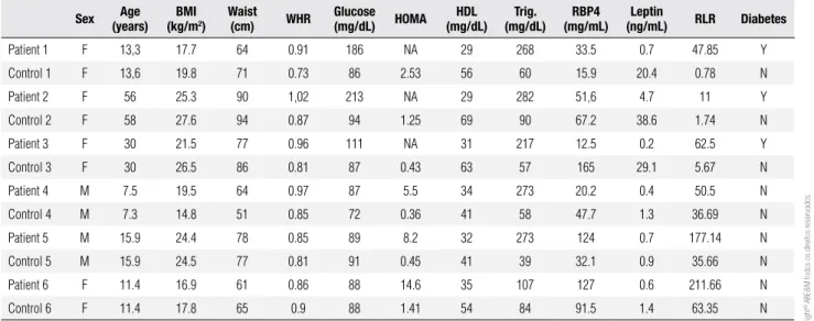
Table 1 presents the individual characteristics of pa-tients and controls. No patient or control individual had blood ties. Three patients in the CGL group (pa-tients 1, 2 and 3) had diabetes (lipoatrophic diabetes) and were under treatment with insulin. Patients 4, 5 and 6 have extreme insulin resistance, as measured by HOMA-IR. All patients in the CGL group presented high triglyceride and low HDL-cholesterol levels. Five out of 6 patients in CGL group had liver ultrasound results indicative of steatosis (data not shown).
Table 2 displays the statistical comparisons betwe-en CGL group and control patibetwe-ents. RBP-4 levels were not different in CGL patients as a group, when com-pared to their matched controls (p = 0.78). However, RLR tended to be increased in CGL (p = 0.064). Sig-niicant differences were observed in waist-to-hip ratio (p = 0.02), systolic blood pressure (p = 0.019), diastolic blood pressure (p = 0.052), alanine transferase (ALT; p = 0.002); HDL cholesterol (p = 0.002) and
triglyce-ride levels (p = 0.002). Glucose and insulin levels were not included for comparison in Table 2 because 3 pa-tients in lipodystrophic group had already been diagno-sed with lipoatrophic diabetes.
The relationship between anthropometric indicators of overweight, leptin and RBP-4 was also evaluated in the entire sample. No correlation was found between RBP-4 and waist (r = 0.18, p = 0.57), waist-to-hip ratio (r = -0.23, p = 0.46) and BMI (r = 0.24, p = 0.45). No correlation was found between RBP-4 and Leptin, either (r = 0.34, p = 0.27).
Table 1. Individual characteristics of patients with congenital generalized lipodystrophy and in the control group
Sex Age (years)
BMI (kg/m2)
Waist (cm) WHR
Glucose (mg/dL) HOMA
HDL (mg/dL)
Trig. (mg/dL)
RBP4 (mg/mL)
Leptin
(ng/mL) RLR Diabetes
Patient 1 F 13,3 17.7 64 0.91 186 NA 29 268 33.5 0.7 47.85 Y
Control 1 F 13,6 19.8 71 0.73 86 2.53 56 60 15.9 20.4 0.78 N
Patient 2 F 56 25.3 90 1,02 213 NA 29 282 51,6 4.7 11 Y
Control 2 F 58 27.6 94 0.87 94 1.25 69 90 67.2 38.6 1.74 N
Patient 3 F 30 21.5 77 0.96 111 NA 31 217 12.5 0.2 62.5 Y
Control 3 F 30 26.5 86 0.81 87 0.43 63 57 165 29.1 5.67 N
Patient 4 M 7.5 19.5 64 0.97 87 5.5 34 273 20.2 0.4 50.5 N
Control 4 M 7.3 14.8 51 0.85 72 0.36 41 58 47.7 1.3 36.69 N
Patient 5 M 15.9 24.4 78 0.85 89 8.2 32 273 124 0.7 177.14 N
Control 5 M 15.9 24.5 77 0.81 91 0.45 41 39 32.1 0.9 35.66 N
Patient 6 F 11.4 16.9 61 0.86 88 14.6 35 107 127 0.6 211.66 N
Control 6 F 11.4 17.8 65 0.9 88 1.41 54 84 91.5 1.4 63.35 N
BMI: body mass index; WHR: waist-to-hip ratio; HOMA: homeostasis model assessment; RBP4: retinol binding protein 4; RLR: RBP4/Leptin Ratio; Trig: triglycerides.
Table 2. Comparison between control group and patients with congenital generalized lipodystrophy (CGL)
Control (n = 6)
Congenital generalized Lipodystrophy
(n = 6)
p
Age (years) 22.7 ± 18.9 22.3 ± 18.2 0.97
BMI (kg/m2) 21.8 ± 5.1 20.8 ± 3.4 0.71
Waist (cm) 74.0 ± 15.3 72.3 ± 11.2 0.83
Waist-to-hip ratio 0.82 ± 0.06 0.92 ± 0.06 0.02
SBP (mmHg) 85.0 ± 13.7 120.0 ± 27.5 0.019
DBP (mmHg) 56.6 ± 10.3 75.0 ± 17.6 0.052
HDL Chol. (mg/dL) 54.0 ± 11.3 31.6 ± 2.5 0.002
Tryglicerides (mg/dL) 64.4 ± 53.5 236.6 ± 67.6 0.002
ALT (mg/dL) 10.1 ± 3.9 33.6 ± 13.3 0.002
RBP4 (mg/ml)* 57.4 (15.9 – 165) 42.5 (12.5 – 127) 0.78
Leptin (ng/ml)* 10.9 (0.9 – 38.6) 0.65 (0.2 – 0.7) 0.015
RLR 23.9 ± 25.3 93.4 ± 80.8 0.064
Cop
yright
© ABE&M t
odos os dir
eit
os r
eser
vados
.
DISCUSSION
Congenital generalized lipodystrophy is a rare disease characterized by a severe lack of adipose tissue. There-fore, it is an excellent model to study hormones and/ or substances produced by adipocytes. Indeed, patients with CGL present very low levels of some adipokines, including leptin (11).
Over the last few years, RBP-4 appeared as one more adipokine. Several studies have demonstrated that RBP-4 levels correlated directly with anthropo-metric measurements of adiposity, including BMI and waist circumference (4-6). It was then hypothesized that RBP-4 levels would indirectly indicate the amount of adipose tissue, and that the absence of adipose tis-sue would be accompanied by reduced levels of RBP-4. This was not the case: RBP4 levels in patients with CGL did not differ from a well-matched healthy con-trol group, despite the difference demonstrated in lep-tin levels (transformed in log). A ratio between RBP4 and leptin (RLR) was also calculated, and an increasing tendency was observed (p = 0.06). If RBP4 levels were to be decreased in CGL, one would expect RLR to be decreased, or close to one. Therefore, this inding un-derscores that RBP4 is really not altered in states of severe paucity of adipose tissue.
Detection of normal levels of RBP4 in patients with CGL indicates that the adipocyte is not the main sour-ce for RBP-4; there may be some other sour-cells and/or organs that also participate in the regulation of RBP4 production. Some studies have already indicated that the liver may be the most important regulator of RBP-4 levels (13,1RBP-4). Recently, Stefan and cols. (15) found no association between RBP-4 levels and total, subcu-taneous, visceral and intramyocellular fat. On the other hand, RBP-4 was positively and signiicantly correlated to liver fat.
These indings were also demonstrated in diabetic patients with nonalcoholic fatty liver disease (NAFLD). Patients with NAFLD presented signiicant higher le-vels of serum RBP-4 in comparison with patients that did not it ultrasound criteria for NAFLD (13). Fi-nally, Yagmur and cols. (14) have also demonstrated that RBP-4 levels correlated directly with the severity of chronic liver disease. Indeed, in patients with liver cirrhosis, RBP4 levels correlated negatively with Child--Pugh score and laboratorial indicators of liver func-tion, including albumin levels and bilirrubins (14).
Some indings in our study support the importance of the liver in regulating RBP-4 levels. First, in our CGL
group, 5 out of 6 patients presented hepatomegaly, in-creased levels of ALT and ultrasound evidence of NA-FLD. Remarkably, the only patient exhibiting very low levels of serum RBP4 [Table 1 (patient 3; RBP-4 = 12.5 mcg/mL)], has developed moderate hepatic failure wi-thin few weeks of evaluation. Taken together, these data suggest that the liver, and not adipose tissue, may act as the main determinant of RBP4 concentrations.
This study has some limitations. First, the use of a small sample may impact our results. However, CGL is a rare and well-deined disease and the identiication of normal levels of RBP4 in almost all CGL patients is of great value. It may signiicantly change some pre-viously deined concepts for RBP4. Second, we did not perform any genetic evaluation of our patients. Althou-gh this would be important to conirm the diagnosis of CGL, we believe that phenotypic and biochemical cha-racteristics of our patients (presented in tables 1 and 2), specially leptin levels, are suficient to establish the diag-nosis. Third, a reference range for RBP-4 levels, accor-ding to gender and age, is still needed. Unfortunately, our population showed a huge variation in age (from 7 to 58 years old), and this may have affected our statisti-cal analysis. Finally, most of the ELISA assays for RBP-4 determination currently available in the market show se-veral discrepancies, and variability may compromises the comparison of results (16). However, this is the most commonly used method in the majority of studies so far.
In conclusion, this study demonstrated that RBP-4 levels are not decreased in patients with CGL and su-ggests that adipose tissue is not its main source. It also indirectly reinforces the capital role of the liver in rela-tion to circulating RBP4.
Conlict of interest disclosure: Amélio F. Godoy-Matos, Rodrigo O. Moreira and Renata MacDowell declare that there is no con-lict of interest Izidro Bendet is a scientiic consultant in immu-nology at Sérgio Franco Medicina Diagnóstica.
REFERENCES
1. Kershaw EE, Flier JS. Adipose tissue as an endocrine organ. J Clin Endocrinol Metab. 2004;89:2548-56.
2. Ronti T, Lupattelli G, Mannarino E. The endocrine function of adi-pose tissue: an update. Clin Endocrinol. 2005;64:355-65. 3. Abel ED, Peroni O, Kim JK, Kim YB, Boss O, Hadro E, et al.
Adipo-se-selective targeting of the GLUT4 gene impairs insulin action in muscle and liver. Nature. 2001;409:729-33.
Cop
yright
© ABE&M t
odos os dir
eit
os r
eser
vados
.
5. Polonsky KS. Retinol-binding protein 4, insulin resistance, and type 2 diabetes. N Eng J Med. 2006;354(24):2596-8.
6. Graham TE, Yang Q, Blüher M, Hammarstedt A, Ciaraldi TP, Henry RR, et al. Retinol-binding protein 4 and insulin resistance in lean, obese, and diabetic subjects. N Engl J Med. 2006;354:2552-63. 7. Craig RL, Chu WS, Elbein SC. Retinol binding protein 4 as a
candi-date gene for type 2 dibetes and prediabetic intermediate traits. Mol Genet Metabol. 2007;90:338-44.
8. Janke J, Engeli S, Boschmann M, Adams F, Böhnke J, Luft FC, et al. Re-tinol-binding protein 4 in human obesity. Diabetes. 2006;55:2805-10. 9. Garg A. Acquired and inherited lipodystrophies. N Engl J Med.
2004;350:1220-34.
10. Garg A. Gender differences in the prevalence of the metabolic complications in familial partial lipodystrophies. J Clin Endocri-nol Metab. 2000;85:1776-82.
11. Haque WA, Shimomura I, Matsuzawa Y, Garg A. Serum adiponec-tin and lepadiponec-tin levels in patients with lipodystrophies. J Clin Endo-crinol Metab. 2002;87:2395-8.
12. Mantzoros C, Flier JS, Lesem MD, Brewerton TD, Jimerson DC. Cerebrospinal fluid leptin in anorexia nervosa: correlation with nutritional status and potential role in resistance to weight gain. J Clin Endocrinol Metab. 1997;82:1845-51.
13. Wu H, Jia W, Bao Y, Lu J, Zhu J, Wang R, et al. Serum retinol bin-ding protein 4 and nonalcoholic fatty liver disease in patients with type 2 diabetes mellitus. Diabetes Res Clin Pract. 2008;79:185-90. 14. Yagmur E, Weiskirchen R, Gressner AM, Trautwein C, Tacke F. Insu-lin resistance in liver cirrhosis is not associated with circulating retinol-binding protein 4. Diabetes Care. 2007;30:1168-72. 15. Stefan N, Hennige AM, Staiger H, Machann J, Schick F, Schleicher