I
ntroductIonCerebral venous thrombosis (CVT), which is, the thrombosis affecting cerebral veins and sinus is a rare condition, responding for less than 1% of strokes. An epidemiological study on stroke
in 164 young patients (15-49 years), in Brazil, identiied CVT
in only seven cases.1 Incidence in adults is higher in the third decade of life with a ratio between male/female sex of 1.5-5.2 The involvement of young women is important, which can be attributed to the use of oral contraceptives, main risk factor asso-ciated.3 The use of oral contraceptives, as well as the prothrombin gene mutation (G20210A) are signiicant risk factors for CVT and
should be routinely investigated.4,5
In 15% of cases, the cause might not be identiied.6 The diagnosis may be late or neglected due to the great clinical spectrum of symptoms, various forms of initial presentation and
unspeciic signs of neuroimaging.
This study intended to analyze 15 CVT patients seen in a neurology service, comparing them with data found in literature.
M
ethodsEpidemiological features, clinical picture, risk factors, and prognosis were assessed in all the 15 patients with cerebral venous sinus thrombosis seen consecutively in the Neurology service of Santa Casa de Belo Horizonte, in the period of April 2007 to December 2008, and results were compared with data found in literature.
r
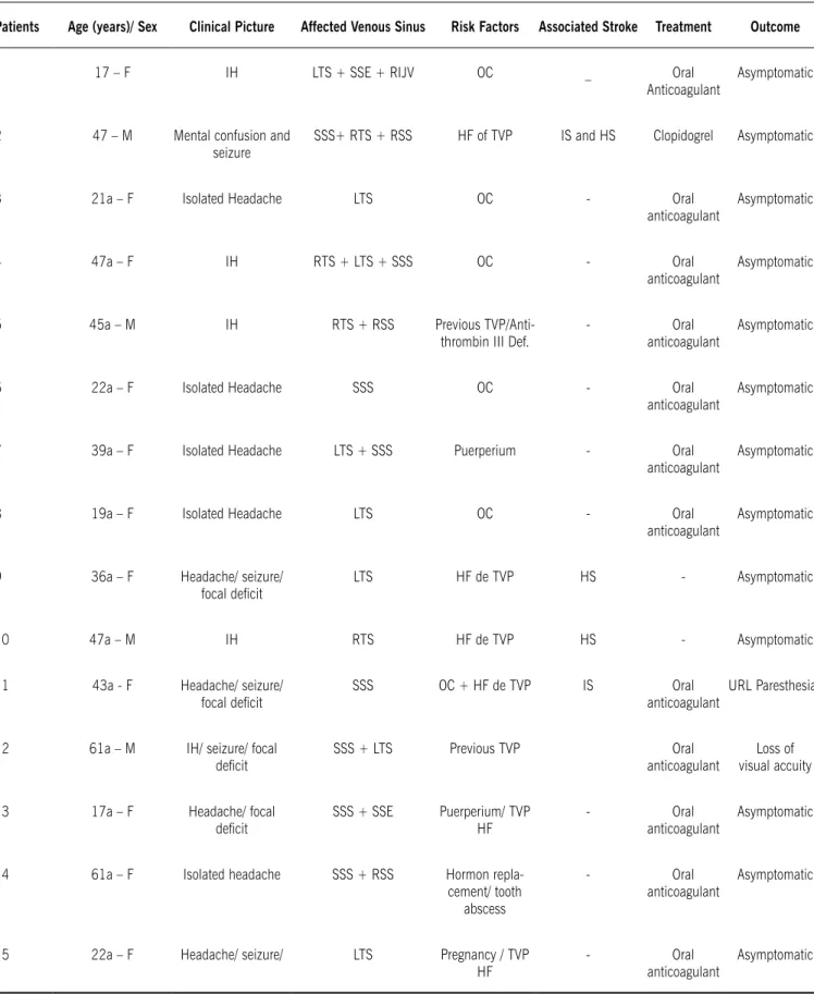
esultsOf the 15 patients assessed (Table 1), 73% were women. Average and median of age were respectively 36.3 and 39 years. The most prevalent symptom was headache, found in all the patients, and in 5 of them (33.3%) it was the only one presented. The most affected sinus were superior sagittal and transverse. Four patients presented stroke, two of them had hemorrhage, one had ischemia, and another one had both (hemorrhage and ischemia). The most important risk factor found was the use of OAC (40%). Thrombophilia was found in only two patients (13%) in this series, particularly the anti-thrombin III deficiency.
*Correspondence: Av. Professor Alfredo Balena, 189, Sala 1708-
Santa Eigênia
Belo Horizonte- MG, Brazil CEP: 30130-100
AbstrACt
objectIve. To analyze a series of 15 patients with cerebral venous thrombosis (CVT) and analyze the results were compared with data in literature.
Methods. In a retrospective, cross-sectional and descriptive study, epidemiological features, clinical pictures, risk factors and prognosis were assessed in 15 patients with CVT admitted in the Neurology division of Santa Casa de Belo Horizonte during the period of April 2007 to December 2008. results Diagnoses were reached through magnetic resonance imaging of the brain in 14 cases and through an angiography in one. The main risk factors identiied were use of birth control pills (40%)
and history of family member with deep venous thrombosis. Thrombophilia was found in two patients (13%). The veins more affected were the transverse sinus (73%) followed by the upper sagittal sinus
(53%). Four patients had strokes and ive had only isolated headache. Twelve patients were treated
with heparin and oral anticoagulant.
conclusIon Treatment with heparin in the acute phase followed by an oral anticoagulant was shown as safe and eficient to prevent worsening of the disease, recurrence and for quick improvement of
neurological symptoms of all treated patients. CVT is one of the possible diagnoses of secondary headache even in patients with no signs and symptoms.
Keywords:Intracranial thrombosis. Venous thrombosis. Heparin. Thrombophilia. Signs and symptoms.
cerebral
venous
sInus
throMbosIs
:
study
of
fIfteen
cases
and
lIterature
revIew
Paulo PereIra chrIsto1*, Gustavo MartInsde carvalho2, antonIo PereIra GoMes neto3
Research conducted in the Neurology division at Santa Casa de Belo Horizonte, Belo Horizonte, MG, Brazil
1. Doutor em Neurologia - Assistente da clínica neurológica da Santa Casa de Belo Horizonte e Hospital das Clínicas da Universidade Federal de Minas Gerais – UFMG, Belo Horizonte, MG
2. Clínica Médica - Residente de neurologia pela Santa Casa de Misericórdia de Belo Horizonte, Belo Horizonte, MG
table 1 - Epidemiological and clinical features of 15 patients assessed with CVt
Patients Age (years)/sex Clinical Picture Affected Venous sinus risk Factors Associated stroke treatment Outcome
1 17 – F IH LTS + SSE + RIJV OC _ Oral
Anticoagulant
Asymptomatic
2 47 – M Mental confusion and
seizure
SSS+ RTS + RSS HF of TVP IS and HS Clopidogrel Asymptomatic
3 21a – F Isolated Headache LTS OC - Oral
anticoagulant
Asymptomatic
4 47a – F IH RTS + LTS + SSS OC - Oral
anticoagulant
Asymptomatic
5 45a – M IH RTS + RSS Previous
TVP/Anti-thrombin III Def.
- Oral
anticoagulant
Asymptomatic
6 22a – F Isolated Headache SSS OC - Oral
anticoagulant
Asymptomatic
7 39a – F Isolated Headache LTS + SSS Puerperium - Oral
anticoagulant
Asymptomatic
8 19a – F Isolated Headache LTS OC - Oral
anticoagulant
Asymptomatic
9 36a – F Headache/ seizure/
focal deicit LTS HF de TVP HS - Asymptomatic
10 47a – M IH RTS HF de TVP HS - Asymptomatic
11 43a - F Headache/ seizure/
focal deicit SSS OC + HF de TVP IS anticoagulantOral
URL Paresthesias
12 61a – M IH/ seizure/ focal
deicit SSS + LTS Previous TVP anticoagulantOral
Loss of visual accuity
13 17a – F Headache/ focal
deicit SSS + SSE Puerperium/ TVP HF
- Oral
anticoagulant
Asymptomatic
14 61a – F Isolated headache SSS + RSS Hormon
repla-cement/ tooth abscess
- Oral
anticoagulant
Asymptomatic
15 22a – F Headache/ seizure/ LTS Pregnancy / TVP
HF
- Oral
anticoagulant
Asymptomatic
The CVT diagnosis was conirmed by a magnetic nuclear
resonance (MNR) of encephalon in 14 cases, by means of a demonstration of the thrombosed sinus by the alteration of the signal in the images pondered in T1 and T2 by cerebral angio-graphy in one case.
The follow up time of patients varied from 2 to 20 months and none of the cases presented new thrombotic events. Thirteen patients (87%) presented a good evolution with total recovery of signs and symptoms in the early phase of the treatment (attack
phase). Only two remained with neurological deicits in the
follow up: in one patient there was permanent visual loss due to intracranial hypertension, and in another one, paresthesia
in upper right member, consequence of an ischemic cerebral
infarction associated to CVT.
d
IscussIonIn this series of cases, we have identiied two main risk
factors: the use of oral contraceptive in six patients and the pres-ence of deep venous thrombosis in lower limbs preceding the
picture in two patients or a history of the disease in irst degree
relatives in other six. Azin et al.3observed that the oral contra-ceptive was the main risk factor associated in a study with 61 patients. Other important risk factors associated are: pregnancy and puerperium, primary antiphospholipid syndrome,
heredi-tary thrombophilias (C and S protein deiciency, antithrombin III deiciency, Leiden V factor, prothrombin gene mutation) and
pre-meningeal infections.7
Indeed various disturbances may cause or predispose CVT patients as all the gyneco-obstetric, surgical causes that lead to thrombosis in lower limbs, cancer, hematologic diseases, vascu-lites, and cranioencephalic trauma.8 However, CVT is typically multifactorial, which means that the identiication of a risk factor
or cause should not interrupt the propaedeutics. Sometimes idiopathic cases are elucidated months later.
Headache was reported by all the patients of the series, datum
veriied in the literature,2 and it appeared in each ive of them as the only symptom. Five patients presented with intracranial hypertension syndrome. There was a cerebral infarction in four
cases and convulsive crises in ive. Seizure is more frequent in
CVT, comparing arterial strokes, possibly present in up to 40% of the cases.2
CVT might manifest itself with an ample spectrum of signs and symptoms, possibly simulating various other neurological diseases; nevertheless, four patterns develop more usually: isolated intracra-nial hypertension, focal syndrome, cavernous sinus syndrome, and sub-acute encephalopathy.8,9 The possibility of headache as single symptom has been described recently and made, therefore, the
suspicion and diagnosis in such patients dificult.10
Initial exam for assessing patients with CVT may be both a
cranial tomography (CT) with technical reinement (other
cutting-planes, bony window, and study with emphasis on venous circu-lation) and, when available, encephalic MNR that may show a higher number of alterations resulting from venous congestion and perform the diagnosis. Around 30% of CVT patients present normal cranial CT in the beginning of the clinical picture.11 Angioresonance has the advantage of being a non-invasive exam
capable of conirming suspect or inconclusive cases, indicated
by MNR images.12 (Figure 1)
Most patients with CVT present a seric increase in the D dimer. Its elevation intensity is related to the time of installation and extension of cerebral disease. Normal levels make the diagnosis little probable, but not impossible, so it should not replace, yet, the clinical suspicion and imaging exams at diagnosis.13 Dimer D may in the future become part of CVT propaedeutics, helping in the exclusion of this diagnosis
The most affected sinus was the transverse, in 73% of cases, followed by the upper sagittal, in 53%. In more than half the cases, thrombosis was found in two or more sinus. Wysokinska et al.,6 in a study with 163 patients, showed that transverse sinus was affected in 79%, sigmoid in 50%, upper sagittal in 49%, and in 66% of cases two or more sinus were involved.
Twelve patients received heparin in the acute phase and warfarin for maintenance. In three patients anticoagulant therapy was discouraged by the assistant doctor due to the presence of associated cerebral hemorrhage. Nevertheless, despite the little evidence based on randomized tests, it is a current consensus that CVT patients receive anticoagulant treatment with low molecular weight heparin or unfractionated heparin; and the presence of spontaneous cerebral hemorrhage does not contraindicate its use.2 In the 19th Century, CVT was commonly diagnosed by autopsy and generally showed hemorrhagic lesions, which,
by analogy with arterial stroke, lead physicians to think about contraindicating the use of heparin.8
According to the guide of the European Federation of Neurology and CVT Treatment, in cases secondary to transitory risk factors, the use of oral anticoagulant must be kept for 3 months. In these idiopathic with less severe thrombophilia, as
C and S protein deiciency, heterozygosis for V Leiden factor or
prothrombin gene mutation for up to 12 months. In the presence of relapsing disease and severe thrombophilic factors, such as
antithrombin III deiciency, homozygosis of the mutant V Leiden
factor or two or more associated factors, therapy should be kept
indeinitely.2
The objectives of antithrombotic treatment in CVT are recana-lization of the sinus or occluded vein, prevention of the propaga-tion of the thrombus and treatment of the underlying prothrom-botic state, preventing venous thrombosis in other part of the body, such as pulmonary embolia, and the recurrence of CVT.
In the series presented, the three patients with associated cerebral hemorrhage did not use anticoagulant and had a good neurological evolution. This result might be explained partly by the small number of patients in the series.
As predicting factors for death or dependence in the cerebral sinus thrombotic disease are cited: age over 37, altered mental state, coma, cerebral hemorrhage at admission, deep veins thrombosis, among others.14,15
In a study with 624 adult patients, Ferro JM et al.14 reported 13% of mortality and permanent dependence. Factors related to a poor prognosis were coma, hemorrhage, and malignities.
In our small series, the four patients who presented stroke at admission had a benign course of the disease. Even the three
patients who did not use speciied therapeutics presented a
favorable outcome.
In a series of 24 patients treated with anticoagulation, they did not present new bleedings or worsening of previous hemor-rhages. Patients with parenchymatous lesions or thrombophilias
had an increased risk of neurological sequelae. In another series
of 50 CVT cases, a worse outcome of the disease was observed in African-descendant patients in comparison to Caucasians.16
There is no consensus yet on the eficacy or security of
chemical thrombolysis and thrombectomy in the treatment of the disease. Tsai FY et al.,17 treating 25 patients, suggested the use of chemical or mechanical thrombolytic therapy in cases experiencing a clinical worsening, despite the use of heparin, or in those that present with evidence of hemorrhage or edema (venous congestion). Stam et al.,18 in a prospective study with 20 patients, concluded that endovascular treatment may be
beneicial in patients with severe disease, but it might increase
the risk of cerebral hemorrhage.
The ive patients who presented seizures, three of them
associated with cerebral infarctions, remained under antiepi-leptic therapy, based on literature data that suggest its use, for a minimal period of one year, in the cases associated with focal
neurological deicits, edema, and cerebral infarctions.2 Supraten-torial lesions and convulsive crises at presentation are predicting factors for crises in a short period of time.19
Various studies have demonstrated the increase of cere-bral venous thrombosis risk in patients using oral contra-ceptives and thrombophilia, particularly in the presence of
hyper-homocysteinemia, mutation of the V Leiden factor and prothrombin gene mutation.20 The suspension of oral contracep-tives should be recommended, so, for the patients that presented CVT, highlighting alternative contraceptive methods. In the pres-ence of thrombophilia suspension is mandatory.20 Due to the rarity of thrombophilic factors, a screening for women who want contraception might be based on the previous history of extra-cerebral venous thrombosis or familial history of the disease.20 Patients with venous thrombosis associated to Antiphospholipid
Antibody Syndrome should be indeinitely anticoagulated, and
the RNI kept between 2 and 3 and between 3 and 4 in recur-rent cases.21
Thrombophilia was found in 2 patients (13%) of this series, an approximate value to the one found by Wysokinska et al.,6 who have identiied the presence of thrombophilia in 10% in a
cohort of 163 patients.
c
onclusIonCVT, due to the broad spectrum of clinical presentation,
might be confused with other pathologies and, so, frequently
neglected. In this series of 15 cases, the clinical picture varied from a headache refractory to analgesic treatment to severe forms
as intracranial hypertension syndrome, focal deicits and coma.
CVT, therefore, is within the differential diagnosis for secondary headaches even in the absence of other signs and symptoms.
The main risk factors identiied were the use of oral contracep -tive and a previous or familial history of deep venous thrombosis.
Treatment with heparin in the acute phase followed by oral anticoagulant has shown to be safe and effective in the preven-tion of disease progression, its relapse and rapid recuperapreven-tion of neurological picture in the big majority of patients.
No conlict of interest declared concerning the publication of
this article.
r
eferences1. Zétola VH, Nóvak EM, Camargo CH, Carraro H Jr, Coral P, Muzzio JÁ, et al.
Acidente vascular cerebral em pacientes jovens: análise de 164 casos. Arq. Neuropsiquiatr. 2001;59(3B):740-5.
2. Einhäupl K, Bousser MG, Bruijn SF, Ferro JM, Martinelli I, Masuhr F, et al. EFNS guideline on the treatment of cerebral venous and sinus thrombosis. Eur J Neurol. 2006;13(6):553-9.
3. Azin H, Ashjazadeh N. Cerebral venous sinus thrombosis - clinical features, predisposing and prognostic factors. Acta Neurol Taiwan. 2008;17(2):82-7. 4. Rodrigues CA, Rocha LK, Morelli VM, Franco RF, Lourenço DM. Prothrombin G20210A mutation, and not factor V Leiden mutation, is a risk factor for cerebral venous thrombosis in Brazilian patients. J Thromb Haemost. 2004;2(7):1211-2.
5. Gadelha T, André C, Jucá AA, Nucci M. Prothrombin 20210A and oral contra-ceptive use as risk factors for cerebral venous thrombosis. Cerebrovasc Dis. 2005;19(1):49-52.
6. Wysokinska EM, Wysokinski WE, Brown RD, Karnicki K, Gosk-Beirska I, Grill D. Thrombophilia differences in cerebral venous sinus and lower extremity deep venous thrombosis. Neurology. 2008;70:627-633.
7. Camargo ECS, Bacheschi LA. Trombose venosa cerebral: como identiicá-la?.
Rev Assoc Med Bras. 2001;47(4):278.
8. Bousser MG, Ferro JM. Cerebral venous thrombosis: an update. Lancet Neurol. 2007;6(2):162-70.
9. Stam J. Thrombosis of the cerebral veins and sinuses. N Engl J Med. 2005;352(17):1791-8.
10. Cumurciuc R, Crassard I, Sarov M, Valade D, Bousser MG. Headache as the only neurological sign of cerebral venous thrombosis: a series of 17 cases. J Neurol Neurosurg Psychiatry. 2005;76(8):1084-7
12. Ferreira CS, Pellini M, BoasquevisqueE, Souza, LAM. Alterações parenquima -tosas na trombose venosa cerebral: aspectos da ressonância magnética e da angiorressonância. Radiol Bras. 2006;39(5):315-21.
13. Haapaniemi E, Tatlisumak T. Is D-dimer helpful in evaluating stroke patients?
A systematic review. Acta Neurol Scand. 2009;119(3):141-50.
14. Ferro JM, Canhão P, Stam J, Bousser MG, Barinagarrementeria F. Prognosis of cerebral vein and dural sinus thrombosis: results of the International Study on Cerebral Vein and Dural Sinus Thrombosis (ISCVT). Stroke. 2004;35(3):664-70.
15. Canhão P, Ferro JM, Lindgren AG, Bousser MG, Stam J, Barinagarrementeria F. Causes and predictors of death in cerebral venous thrombosis. Stroke. 2006;37(2):331-2.
16. Appenzeller S, Zeller CB, Annichino-Bizzachi JM, Costallat LT, Deus-Silva L,
Voetsch B, et al. Cerebral venous thrombosis: inluence of risk factors and imaging indings on prognosis. Clin Neurol Neurosurg. 2005;107(5):371-8.
17. Tsai FY, Kostanian V, Rivera M, Lee KW, Chen CC, Nguyen TH. Cerebral venous congestion as indication for thrombolytic treatment. Cardiovasc Intervent Radiol. 2007;30(4):675-87.
18. Stam J, Majoie CBLM, Delden OMV, Lieden KPV, Reekers JA. Endovascular thrombectomy and thrombolysis for severe cerebral sinus thrombosis. A prospective study. Stroke. 2008;39(5):1487-90.
19. Ferro JM, Canhão P, Bousser MG, Stam J, Barinagarrementeria F. Early seizures in cerebral vein and dural sinus thrombosis: risk factors and role of antiepi-leptics. Stroke. 2008;39(4):1152-8.
20. Saadatnia M, Tajmirriahi M. Hormonal contraceptives as a risk factor for cerebral venous and sinus thrombosis. Acta Neurol Scand. 2007;115(5):295-300. 21. Tuthill JI, Khamashta MA. Management of antiphospholipid syndrome. J
Autoimmun. 2009;33(2):92-8.