174
EPIDEMIOLOGICAL INVESTIGATIONS OF A PESTE DES PETITS RUMINANTS (PPR) OUTBREAK IN AFGHAN SHEEP IN PAKISTAN
A. B. ZAHUR, A. ULLAH, H. IRSHAD, M. S. FAROOQ, M. HUSSAIN AND M. JAHANGIR
Animal Health Laboratories, Animal Sciences Institute, National Agricultural Research Centre, Park Road, Islamabad, Pakistan
ABSTRACT
Epidemiological and virological investigations were carried out during an outbreak of Peste des petits
ruminants (PPR) in Afghan (Bulkhi) sheep in Pakistan. The overall morbidity, mortality and case fatality
rates were 41.0, 1.2 and 3.0%, respectively. The epidemic curve was plotted and the values for basic reproductive number (R0) and herd immunity threshold (HIT) for the affected flock were estimated to be 6.85 and 85.4%, respectively. The morbid material analysis by immuno-capture ELISA (Ic-ELISA) and haemagglutination assay (HA) revealed the presence of PPR virus. The PPR virus was isolated and identified through cytopathic effects, Ic-ELISA and transmission electron microscopy (TEM).
Key words: PPR, sheep, basic reproduction number, herd immunity threshold, epidemic curve. INTRODUCTION
Peste des Petits Ruminants (PPR) is a disease of
major economic importance and imposes a significant constraint upon sheep and goat production owing to its high mortality rate (Asim et al., 2008; 2009). It is an acute, highly contagious and frequently fatal disease of sheep and goats caused by PPR virus (PPRV), a member of genus morbillivirus of family Paramy- xoviridae. Therefore, it poses serious threat to the development of small ruminants production in many countries where it is endemic and is a source of great financial loss to the farmers and livestock owners.
The disease was first reported in Pakistan during 1994 when the confirmatory diagnosis was made by polymerase chain reaction (Amjad et al., 1994). This was further validated when the PPR virus (PPRV) antigen was detected during outbreaks at different areas using Ic-ELISA (Hussain et al., 2002).
An outbreak of PPR occurred in the Bulkhi sheep of Small Ruminant’s Research Programme, Livestock Research Station, National Agricultural Research Centre, Islamabad, Pakistan during June, 2006. The epidemiological data, clinical/pathological features of this outbreak and recovery and identification of PPR virus are reported in this paper.
MATERIALS AND METHODS Epidemiological observations
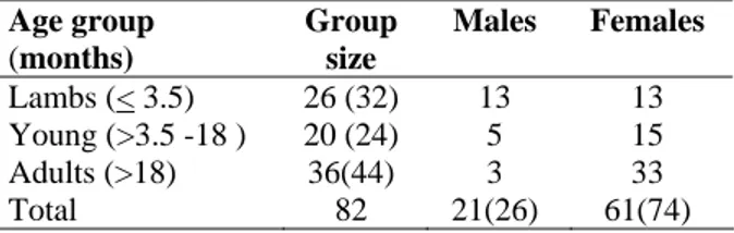
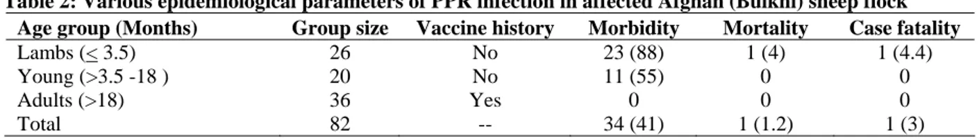
Data regarding flock size, age, sex, vaccination history and possible source of virus transmission were recorded. The flock comprised a total of 82 sheep. Of these 26 were lambs (<3.5 months old), 20 young stock (>3.5 months to 18 months old) and 36 adults (>18 months old). There were 21 males and 61 females.
Adults were vaccinated against PPR vaccine (Jovac, Jordan), while the other two groups (lambs and young stock) were not vaccinated (Table 1).
Table 1: Parameters of affected Afghan (Bulkhi) sheep flock
Age group (months)
Group size
Males Females
Lambs (< 3.5) 26 (32) 13 13
Young (>3.5 -18 ) 20 (24) 5 15 Adults (>18) 36(44) 3 33
Total 82 21(26) 61(74)
Numbers in parenthesis show percentages.
The epidemiological curve was drawn to see the magnitude and progression/ time course of the outbreak. The basic reproduction number (R0) and herd immunity threshold (HIT) values were estimated following Anderson and May (1992):
R0 = 1/ proportion susceptible HIT = 1-1/R0
Clinical and post-mortem examination
Clinical examination of the affected flock and necropsy of dead animal was carried out and ocular (n = 6) and nasal (n = 6) swabs from all suspected live animals were collected. A piece of lung, spleen, kidney, liver and mesenteric lymph nodes (MLNs) from one dead animal were collected for laboratory confirmation.
Laboratory analysis
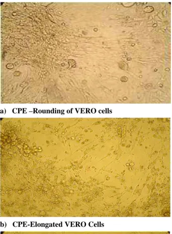
was identified by cytopathic effects (CPE), Ic-ELISA for PPR virus and transmission electron microscopy (TEM), following standard operating procedures (SOPs).
RESULTS Epidemiological observations
The flock consisted of 82 sheep of different age groups (Table 2). The vaccinated animals (36) did not experience any morbidity or mortality. Only the non vaccinated animals (46) experienced the disease. Of 26 lambs, one died resulting in a group case fatality of 4.4%. However, lambs experienced the highest morbidity of 88% (23/26), showing overt disease. There were 55% (11/20) cases among young stock. The overall morbidity, mortality and case fatality were 41, 1.2 and 3%, respectively (Table 2).
The epidemiological curve was plotted to see the pattern of outbreak (Fig. 1). It resembles a typical propagated outbreak with peaks of primary, secondary and tertiary generations of cases spaced by incubation period. The most probable period of exposure of the susceptible animals to PPRV was calculated from 3 to 5 days before the index case was identified (Fig. 1). The R0 and HIT values for affected flock were estimated to be 6.85 and 85.4%, respectively.
Clinical and post-mortem findings
The clinical examination of the affected animals revealed high fever (106-107°F), mild conjunctivitis, congestion of the third eye lids and mild ocular and nasal discharges (Fig. 2a). Erosive lesions were present on the inner side of the upper lip (Fig. 2b). All animals exhibited diarrhea. On the external examination, the carcass was dehydrated (sunken eyes) along with the soiling of hind quarters. While on internal examination red raw area was observed on the dental pad beneath the incisors. Both lungs were pneumonic particularly the cardiac lobe (Fig. 2c). Haemorrhages were seen on liver (Fig. 2d), kidneys, mucosal surface of abomasum (Fig. 2e) and on the mucosa of large intestine (Fig. 2g)
Fig. 1: Epidemic curve of PPR outbreak in Afghan sheep: a) Most probable time of exposure to PPRV (day 3-5); b) Primary cases which acquired infection from index case on day 9. All expressed clinical signs after a 4-6 days incubation period; c) Secondary cases which acquired infection from primary cases. Most probably, these were infected between day 11-13; d) Tertiary cases. Intervention my have resulted in reduction in the number of cases evident by lower frequency of cases here. and the intestinal contents were watery. Lymph nodes (LN) particularly the mesenteric lymph nodes (MLN) were found reactive (inflammed).
Laboratory analysis
All the ocular and nasal swabs and lungs, spleen and MLNs were tested positive for PPRV antigen by Ic-ELISA. The spleen and MLNs homogenates exhibited agglutination of chicken erythrocytes at the concentration of 0.5% ranging from1:8 to 1:16. However, the ocular and nasal swabs from live animals did not demonstrate HA activity.
a) Mild conjunctivitis and nasal discharge
b) Erosions on the inner side of upper lip
c) Congestion of cardiac lobe of lungs
d) Haemorrhages on liver
e) Haemorrhages in abomasum
g) Haemorrhages in large intestine Fig. 2: Clinical signs and necropsy findings of PPR outbreak in Bulkhi sheep
DISCUSSION
PPR has been reported in sheep in a number of countries in the region including Afghanistan, Iran and India (Shaila et al., 1989; Majok, 2001; Abdollahpour
et al., 2006). Although the clinical and postmortem
a) CPE –Rounding of VERO cells
b) CPE-Elongated VERO Cells
c) CPE-Clumping of VERO Cells
d) Transmission Electron micrograph (TEM) of PPR virus
Fig. 3: Cytopathic effects (CPE) on VERO cells due to PPRV and TEM of PPR virion isolated from Bulki sheep.
to those produced by rinderpest virus on VERO cells (Gopilo, 2005). Similar observations were made by Ozkul et al. (2002), who reported initial rounding of VERO cells and later on development of syncitia.
The Ic-ELISA proved suitable for both diagnosis from field samples and identification of PPRV in cell culture supernatant. The TEM of PPRV displayed a typical structure for family paramyxoviridae (Losos, 1989).
During the outbreak, the epidemic curve revealed that cases occurred over more than a single incubation periods. The shape and other features of an epidemic curve can suggest hypotheses about the time and source of exposure, the mode of transmission, the causative agent, incubation period and the efficacy of control measures (Dicker and Gathany, 1992). The outbreak had a successive series of peaks reflecting increasing or decreasing numbers of cases in each generation. The epidemic waned after three generations, probably because either the number of susceptible animals fell below some critical level, or the zoo-sanitary measures taken to control the disease became effective. The most probable period of exposure of the susceptible animals to PPRV ranges between 3 and 5 days when the index case must have been infected, perhaps during grazing with the other small ruminants. The R0 value indicates the transmissibility of a pathogen. The estimated R0 value (6.85) for PPRV seems to be very close to those for other morbillivirus. Anderson (1995) documented that the value of R0 varies with the virulence of the pathogen and susceptibility of the host population. It was further reported that rinderpest virus can establish infection in relatively small populations of susceptible species but large populations are necessary for the infection to sustain. In the present outbreak, the susceptible population was very small and the interventions were instituted on the 4th day of the outbreak. This however, is not applicable for the field conditions where the chances of contact transmission are great (extensive production system) and farmers usually do not recognize the disease in its early stages.
animal husbandry, breed, age and other factors. In Pakistan, goats react severely to the exposure of PPRV like other parts of the world where the disease is endemic. It has been reported that during an outbreak of PPR in Pakistan, no clinical signs were observed in sheep kept with the sick goats in the same premises under one roof but they got sero-converted only (Hussain et al., 2002). Diallo (1997) reported that the reasons for these outbreaks in different epidemiological condition, like those where flocks consist of only sheep or goats or both, are unknown and was further reported that partial N and H gene sequences of the virus isolate involved in sheep or goat outbreaks have not revealed a clue to explain this situation. It highlights the need for concerted efforts to study the role of Bulkhi sheep in the epidemiology of the disease.
In the present study, lambs and young stock were affected among the susceptible population, while those which were vaccinated 18 months earlier remained un-affected. These observations are in agreement with Awa
et al. (2002), who reported that maternal immunity
decayed after 12 weeks in lambs from experimentally vaccinated ewes. It was also elucidated that attenuated PPR vaccine is capable of providing protection for at least 1.5 years and possibly for the whole economic life of small ruminants (about two years). Further studies may be needed to investigate this issue. The HIT is the proportion of susceptible population needed to be immune for a disease to become stable. The HIT for the flock indicated that we need to achieve more than 85.4% vaccination coverage for control of PPR infection in sheep population. For rinderpest, HIT has been estimated as 75-80% (Rossiter and James, 1989).
It is therefore recommended that before opting for vaccination as a national policy for controlling PPR, R0 value and HIT for both sheep and goat population may be estimated in endemic areas.
REFERENCES
Abdollahpour, G., A. Roofi, J. Najafi, F. Sasani and E. Sakhaie, 2006. Clinical and para-clinical findings of a recent outbreak of Peste des petits ruminants in Iran. J. Vet. Med. B, 53: 14-16.
Amjad, H., Q. U. Islam, M. Forsyth, T. Barret and P. B. Rossitter, 1994. Peste des petits ruminants in goats in Pakistan. Vet. Rec., 139(5): 118-119.
Anderson, E. C., 1995. Morbillivirus infections in wildlife (in relation to their population biology and disease control in domestic animals). Vet. Microbiol., 44: 319-332
Anderson, R. M. and R. M. May, 1992. Infectious Diseases of Humans: dynamics and control. Oxford Science Publications, Oxford, UK.
Anonymous, 2002. Peste des petits ruminants ELISA kit, Ic-ELISA for detection of antigen to rinderpest virus and PPR virus. Bench protocol, version-ICE 2.1, January 2002. Joint FAO/IAEA Programme,
Animal Production and Health, Pirbright, United Kingdom and CIRAD-EMV, Montplier, France. Asim, M., A. Rashid and A. H. Chaudhary, 2008.
Effect of various stabilizers on titre of lyophilized live-attenuated Peste des petits ruminants (PPR) vaccine. Pakistan Vet. J., 28(4): 203-204.
Asim, M., A. Rashid, A. H. Chaudhary and M. S. Noor, 2009. Production of homologous live attenuated cell culture vaccine for the control of Peste des
petits ruminants in small ruminants. Pakistan Vet.
J., 29(2): 72-74.
Awa, D. N., A. Ngagnou, E. Tefiang, D. Yaya and A. Njoya, 2002. Post vaccination and colostral Peste des petits ruminants antibody dynamics in research flocks of Kirdi goats and Foulbe sheep of north Cameroon. Prev. Vet. Med., 55: 265-271.
Diallo, A., 1997. Peste des petits ruminants: An overview. FAO-EMPRES FMS Contingency Planning Workshop. Hanoi.
Diallo, A., 2006. Control of Peste des petits ruminants and poverty alleviation. J. Vet. Med., 53: 11-13 Dicker, R. and N. C. Gathany, 1992. Principles of
Epidemiology. 2nd Ed., U. S. Department of Health and Human Services, Public Health Service, Centers for Disease Control and Prevention (CDC), Atlanta, USA.
Gopilo, A., 2005. Epidemiology of Peste des petits ruminants virus in Ethiopia and molecular studies on virulence. PhD Thesis, Le titre de docteur de l’institut national poly technique de Toulouse, France.
Hussain, M., R. Muneer, M. Jahangir, A. H. Awan, M. A. Khokhar, A. B. Zahoor, M. Zulfiqar and A. Hussain, 2002. Chromatographic strip technology: A pen side test for the diagnosis of Peste des petits ruminants in sheep and goats. On–line J. Biol. Sci., 3(1): 1-7.
Losos, G. J., 1989. Infectious tropical diseases of domestic animals. Longman Scientific Technical, Canada, 12: 549-556.
Majok, A. A., 2001. Animal Health Component. AFG/00/015, Annual Report. Islamabad, Pakistan. Ozkul, A., Y. Akca, F. Alkan, T. Barrett, T. Karaoglu,
S. B. Dagalp, J. Anderson, K. Yesilbag, C. Cokcaliskan, A. Gencay and I. Burgu, 2002. Prevalence, distribution and hosts range of Peste des petits ruminants virus in Turkey. Emerging Infec. Dis., 8(7): 708-712.
Rossiter, P. B. and A. D. James, 1989. An epidemiological model of rinderpest: II. Simulations of the behavior of rinderpest virus in populations. Trop. Anim. Hlth. Prod., 21: 69-84. Shaila, M. S., V. Purushothaman, D. Bhavasar, K.
Venugopal and R. A. Venkatesan, 1989. Peste des petits ruminants of sheep in India. Vet. Rec., 125: 602.