Vojnosanit Pregl 2016; 73(4): 393–396. VOJNOSANITETSKI PREGLED Page 393
Correspondence to: Ksenija Božić, Clinic of Rheumatology, Military Medical Academy, Crnotravska 17, 11 000 Belgrade, Serbia. E-mail: [email protected]
C A S E R E P O R T UDC: 612.017:616.72-002.77]:[617.764.1+616.316
DOI: 10.2298/VSP150118120B
Case report of Mikulicz`s disease – a modern concept of an old entity
Prikaz bolesnika sa Mikuli
č
evom bolesti – savremeni koncept starog entiteta
Ksenija Božić*, Branislava Glišić*†, Olga Radić-Tasić‡, Bojana Knežević*
*Clinic of Rheumatology, ‡Institute of Pathology, Military Medical Academy, Belgrade, Serbia; †Faculty of Medicine of the Military Medical Academy, University of Defence,
Belgrade, Serbia
Abstract
Introduction. Modern knowlegde defines Mikulicz´s dis-ease as a part of immunoglobulin G4-related disdis-ease. The main feature is the presence of lymphoplasmacytic infil-trates, immunoglobulin G4 plasma cells positivity, distinc-tive storiform fibrosis and moderate eosinophilia. Case re-port. A 59-years old male presented with a mild keratocon-juctivitis sicca and enlarged lacrimal and salivary glands dur-ing the last two years. Althought clinical presentation of the patient was typical, earlier testing did not pinpoint Miku-licz´s disease. By typical clinical presentation, elevated se-rum immunoglobulin G4 level and histopathological finding of lacrimal glands tissue we diagnosed Mikulicz´s disease successfully treated with corticosteroid therapy. Conclu-sion. We reported the first case of IgG4-related Mikulicz´s disease in Serbia. Our report highlights IgG4-related Miku-licz`s disease as an important differential diagnosis with Sjögren`s syndrome and lymphoproliferative disease in rheumatological practice.
Key words:
mikulicz' disease; diagnosis, differential; diagnosis; lacrimal apparatus; salivary glands; immunoglobulin G; histological techniques; glucocorticoids; treatment outcome.
Apstrakt
Uvod. Savremena saznanja svrstala su Mikuličevu bolest u
grupu bolesti posredovanih imunoglobulinom G4, čija je glavna odlika histološki nalaz limfoplazmocitnih infiltrata, imunoglobulin G4 pozitivnih plazma ćelija, uz storiformnu fibrozu i umerenu eozinofiliju. Prikaz bolesnika. Prikazan je 59-godišnji bolesnik koji je dve godine imao umeren suvi keratokonjuktivitis i uvećane pljuvačne i suzne žlezde. Iako je klinička slika bila karakteristična, ranijim ispitivanjem nije se došlo do dijagnoze Mikuličeve bolesti. Na osnovu tipič-nog kliničkog nalaza, visoke serumske koncentracije imu-noglobulina G4, uz patohistološki nalaz biopsije tkiva suznih žlezda, dijagnostikovali smo Mikuličevu bolest. Pri-menjena terapija kortikosteroidima bila je efikasna.
Zaklju-čak. Prikazali smo prvog bolesnika u Srbiji sa Mikuličevom
bolesti posredovanim imunoglobulinom G4. Našim prika-zom istakli smo značaj poznavanja Mikuličeve bolesti pos-redovane IgG4, kao i diferencijalnu dijagnozu sa Sjögreno-vim sindromom i limfoproliferativnim bolestima u reuma-tološkoj praksi.
Ključne reči:
mikuličeva bolest; dijagnoza, diferencijalna; dijagnoza; suzni aparat; pljuvačne žlezde; IGG; histološke tehnike; glukokortikoidi; lečenje, ishod.
Introduction
Immunoglobulin G4 (IgG4)-related diseases (IgG4-RD) are new clinical entity of fibro-inflammatory conditions, charac-terized by the tendency to form tumorous lesions, dense lymphoplasmacytic infiltration abudant of IgG4-positive plasma cells and storiform fibrosis in relevant organs and often, but not always, elevated serum IgG4 levels 1. The concept is based on the discovery of increased serum IgG4 levels in patients with sclerosing pancreatitis 2. IgG4-RD can affect various organs, pancreas more often than the others as well as hepatobiliary tract, salivary and lacrimal glands, orbits and lymph nodes.
Very little data exists on the incidence and prevalence of IgG4-RD. Most epidemiological studies come from Japan and they are focused on autoimmune pancreatitis. It was es-timated that the incidence of new cases with IgG4-RD is 2.63–10.2/million, with newly diagnosed 336–1,300 patients per year 3.
pati-Page 394 VOJNOSANITETSKI PREGLED Vol. 73, No. 4
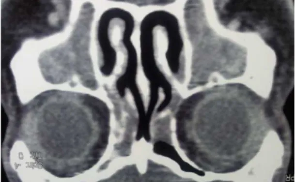
Božić K, et al. Vojnosanit Pregl 2016; 73(4): 393–396. Fig. 1 – Computed tomography (CT) images reveal enlargement of the lacrimal glands.
A B
Fig. 2 – A) Dacryoadenitis manifests as bilateral swelling of upper eyelieds, and B) Sialadenitis manifests as bilateral swelling of parotid regions in the same patient.
ents with MD and defined histopathological findings in glands tissue, presenting MD as part of IgG4-RD 4.
Today, the focus of interest is how to differentiate the diagnosis of MD and SS. MD is usually observed in patients older than 50 years, both males and females. In spite of the persistent swelling of salivary and lacrimal glands, their fun-ction is not significantly reduced. The number of positive an-tinuclear antibody (ANA) is small, while anti-Ro and anti-La antibodies are negative. Patients with SS are mostly females around 50 years old. Although gland swellings in SS are in-termittent, keratoconjunctivitis sicca is present. Serum tests in most patients show positive ANA, 70% of them have anti-Ro and 30% anti-La antibodies. High serum level of IgG4 is specific for MD, but is usually not seen in SS. The basics for MD diagnosing is a histopathological feature. Although the lymphocytic infiltrates are typical for MD and SS, their in-fluence is different. In MD lymphoid follicules are placed around the duct protecting it while in SS they create lymphoepithelial lesions and destroy the duct. This explains less frequent keratoconjunctivitis sicca in MD in spite of si-gnificant gland swellings. Infiltration of IgG4 positive plas-ma cells is the plas-main difference between MD and SS. The ra-tio of IgG4 positive cells to IgG positive cells is higher than 40%. Steroid therapy is efficient in MD, but partially in SS. In spite of certain clinical similarities, MD and SS are two different diseases 5, 6.
We presented the first patient with proven IgG4-related MD in our country.
Case report
A 59-year-old man was admitted to our hospital due to chronic dacryoadenitis and sialadenitis, with suspicion of the
existence of lymphoproliferative disease. The patient had dry mouth and painless swelling in the area of parotid salivary glands during two years. A year before hospitalization he no-ticed swollen eyelids. His other problem was nasal obstructi-on. The diagnosis of keratoconjunctivitis sicca was made in other hospital a year before. There was no evidence for the existence of SS (Schirmer`s test was 7 and 8 mm; scintigraphy showed mildly reduced accumulative and excretory function of salivary glands; ANA, anti-Ro and an-ti-La antibodies were negative; ultrasound showed enlarged parotid and submandibular salivary glands; biopsy of the mi-nor labial salivary gland was negative). The computed tomography (CT) scan showed enlarged lacrimal glands mo-re on the left side (Figumo-re 1). Biopsy of the both glands was performed. The results showed chronic dacryoadenitis followed by significant polyclonal proliferation of the plas-mocites. Lymphoproliferative disease was suspected.
On admission the patient was obese, body mass index (BMI) of 34 kg/m2 (normal range 18.5–25 kg/m2), with en-larged, painless parotid, submandibular and lacrimal glands (Figure 2).
le-Vol. 73, No. 4 VOJNOSANITETSKI PREGLED Page 395
Božić K, et al. Vojnosanit Pregl 2016; 73(4): 393–396.
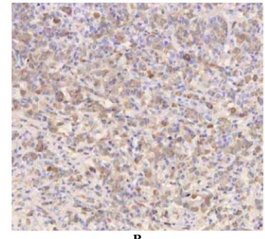
ast 40% (Figure 3). There was no histological criteria for a lymphoproliferative neoplasm.
Additional tests were done including myelogram and analysis of clonality in bone marrow lymphocytes. Using polymerase chain reaction method, no lymphoproliferative disease was found.
On the seventh hospital day, the patient showed signs of compressive, peripheral paresis of the left facial nerve. It was the reason for urgent corticosteroid therapy with prednisone 0.5 mg/kg/day. Two weeks later, swelling disappeared completely and the facial nerve recovered its function. The
corticosteroid dosage was tapering gradually 2.5 mg per week. After a 3-mount follow-up, the patient was still without any complaints. He continued to receive prednisolo-ne 10 mg per day as maintenance therapy.
Discussion
By the end of the 19th century, Johan von Mikulicz-Radecki described a patient with symmetrical swellings of the lacrimal, submandibular and parotid glands with massive infiltration of the glands by mononuclear cells 7. Later, these clinical features were observed in patients with tuberculosis, sarcoidosis and lymphomas 8. Schafer and Jacobsen 9 formed a group of patients with typical clinical features known as Mikulicz`s syndrome. In 1933, Henrik Sjögren 10 described histopathological lymphocyte infiltrates in salivary gland of patients with keratoconjunctivitis sicca and swollen main salivary glands. But his findings were forgotten until the mid 20th century, when Morgan and Castelman observed that salivary glands tissues in MD and SS are similar 11. Since then, MD is considered as a subtype of SS chronical form of dacryoadenitis and sialadenitis of an autoimmune etiology 11.
The research of Japanese authors in the 21st century shows elevated serum IgG4 levels and histopathologically abundant infiltration of IgG4 positive plasma cells in lacri-mal and salivary glands of patients with MD. This is the cre-ation of a modern clinical concept clearly differentiating MD from SS, putting MD to the group of IgG4-RD 12. The Japa-nese Society for Sjögren`s Syndrome has published
diagnos-tic criteria for related MD. According to them, IgG4-related MD defines with persistent (longer than 3 months) symmetrical swellings of at least 2 pairs of lacrimal, parotid or submandibular glands, elevated serum IgG4 levels (> 135 mg/dL) or histopathologically marked infiltration of IgG4 positive plasma cells with a ratio of IgG4 /IgG > 40%, with typical tissue fibrosis or sclerosis 13. So, patients with swelling of salivary and lacrimal glands, elevated serum IgG4 levels and significant infiltration of IgG4 positive pla-sma cells in lacrimal gland tissue fulfil all the criteria for IgG4-related MD.
The clonality of lymphocytes is necessary to be tested due to histopathological similarities between IgG4-related MD and lymphoma. In the early diagnosing of B-cell lymphomas, they are predominantly represented by B-cell in-filtrates unlike primary T-cell inin-filtrates in IgG4- RD 14.
Allergic rhinitis and bronchial asthma are more frequent in MD than in SS. A high occurrence of allergic conditions is explained by domination of type 2 helper T (Th2) cells immu-ne response, which raises the concentration of IgG4 and IgE 15.
Corticosteroids are the standard first-line treatment for IgG4-related MD. They rapidly reduce swellings of lacrimal and salivary glands, recover their functions and reduce IgG4 concentration in serum 16.
A number of patients with IgG4-related MD was desc-ribed in Japan with just a few individual cases in the western world. In spite of low prevalence of IgG4-related MD, it is necessary to have it in mind when dealing with patients pre-senting with swollen lacrimal and salivary glands.
Conclusion
In suspicion for Mikulicz´s disease serum levels of munoglobulin G4 as well as biopsies from glands with im-munohistochemical evaluation should be assessed. It is nessesery to distinguish immunoglobulin G4-related Miku-licz´s disease from other distinct disorders, including Sjö-gren´s syndrome and lymphoproliferative disease. Therapy with corticosteroids is efficient and recommended for a lon-ger period of time.
A B
Page 396 VOJNOSANITETSKI PREGLED Vol. 73, No. 4
Božić K, et al. Vojnosanit Pregl 2016; 73(4): 393–396. R E F E R E N C E S
1. Khosroshahi A, Stone JH. A clinical overview of IgG4-related systemic disease. Curr Opin Rheumatol 2011; 23(1): 57−66. 2. Hamano H, Kawa S, Horiuchi A, Unno H, Furuya N, Akamatsu T,
et al. High serum IgG4 concentrations in patients with scleros-ing pancreatitis. N Engl J Med 2001; 344(10): 732−8. 3. Hamano H, Arakura N, Muraki T, Ozaki Y, Kiyosawa K, Kawa S.
Prevalence and distribution of extrapancreatic lesions complicating autoimmune pancreatitis. J Gastroenterol 2006; 41(12): 1197−205. 4. Yamamoto M, Takahashi H, Sugai S, Imai K. Clinical and patho-logical characteristics of Mikulicz's disease (IgG4-related plas-macytic exocrinopathy). Autoimmun Rev 2005; 4(4): 195−200. 5. Guma M, Firestein GS. Best Practice. Res Clin Rheumatol 2012;
26(4): 425−38.
6. Yamamoto M, Takahashi H, Ohara M, Suzuki C, Naishiro Y, Ya-mamoto H, et al. A new conceptualization for Mikulicz's disease as an IgG4-related plasmacytic disease. Mod Rheumatol 2006; 16(6): 335−40.
7. Mikulicz JH. Uber eine eigenartige symmetrische Erkrankung der Tranen- und Mundspeicheldrusen. Stuttgart: Beitr Chir Fortschr; 1892.
8. Napp O. Über die Beziehungen der Mikuliczschen Erkrankung Tuberkulose. Stschr Augenheilk 1907; 17: 513.
9. Schaffer AJ, Jacobsen AW. Mikulicz's syndrome: a report of ten cases. Am J Dis Child 1927; 34(3): 327−46.
10.Sjögren HS. Zur kenntnis der keratoconjunctvitis sicca (Kerati-tis folliformis bei hypofunktion der tranendrusen). Acta Op-thalmol (Copenh) 1933; 2: 1−151.
11.Morgan WS. The probable systemic nature of Mikulicz's disease and its relation to Sjögren's syndrome. N Engl J Med 1954; 251(1): 5−10.
12.Umehara H, Okazaki K, Masaki Y, Kawano M, Yamamoto M, Saeki T, et al. A novel clinical entity, IgG4-related disease (IgG4RD): general concept and details. Mod Rheumatol 2012; 22(1): 1−14.
13.Umehara H, Okazaki K, Masaki Y, Kawano M, Yamamoto M, Saeki T, et al. Comprehensive diagnostic criteria for IgG4-related disease (IgG4-RD), 2011. Mod Rheumatol 2012; 22(1): 21−30.
14.Stone JH, Zen Y, Deshpande V. IgG4-related disease. N Engl J Med 2012; 366(6): 539−51.
15.Masaki Y, Dong L, Kurose N, Kitagawa K, Morikawa Y, Yama-moto M, et al. Proposal for a new clinical entity, IgG4-positive multiorgan lymphoproliferative syndrome: analysis of 64 cases of IgG4-related disorders. Ann Rheum Dis 2009; 68(8): 1310−5.
16.Khosroshahi A, Stone JH. Treatment approaches to IgG4-related systemic disease. Curr Opin Rheumatol 2011; 23(1): 67−71.