J of Evolution of Med and Dent Sci/ eISSN- 2278-4802, pISSN- 2278-4748/ Vol. 3/ Issue 10/Mar 10, 2014 Page 2408
TUBERCULAR MASTITIS
–
AN UNCOMMON ENTITY OF THE COMMON
DISEASE: A CASE REPORT
C. P. Madhu1, Sudhir S2, Madhu Srivasarangan3, Preetham Dev4, Nithyananda H. A5, Mittal N. Patel6
HOW TO CITE THIS ARTICLE:
C. P. Madhu, Sudhir S, Madhu Srivasarangan, Preetham Dev, Nithyananda H. A, Mittal N. Patel. Tubercular Mastitis – an Uncommon Entity of the Common Disease: A Case Report . Journal of Evolution of Medical and Dental Sciences 2014; Vol. 3, Issue 10, March 10; Page: 2408-2412, DOI: 10.14260/jemds/2014/2152
ABSTRACT: Tuberculosis (TB) is a very common infection affecting the Indian population. Breast TB is a very rare disease and constitutes only 0.025% - 1.040% of breast diseases. 25 year old married female was diagnosed as breast abscess and treated in the peripheral hospital with antibiotics and anti-inflammatory for period of one year and later referred to our institution. Even after starting with antitubercular treatment (ATT), size of lump did not reduce, she had to undergo simple mastectomy. Medical therapy is the mainstay of treatment with ATT, surgical intervention may be needed in upto 14% of the patients. Thus high index of suspicion is necessary in diagnosing the disease. Greater awareness of this uncommon entity of a common disease along with early institution of medical line of management with ATT may avoid surgery.
KEYWORDS: Mastitis, Mammary Tuberculosis, Primary Tuberculosis
INTRODUCTION: Tuberculosis (TB) is a very common infection affecting the Indian population. The infection can involve any organ and can mimic any other illness, hence know as great mimicker 1.
Amongst the extrapulmonary sites, breast is an infrequent site of TB. Breast TB is a very rare disease and constitutes only 0.025% - 1.040% of breast diseases. 2
Sir Astley Cooper in first described breast TB as Scrofulous swelling at the bosom of
young women suffering from enlargement of cervical glands . It has been classified into three categories namely nodular, disseminated and abscess varieties 2. The diagnosis of mammary TB can
be confirmed with a combination of strong clinical suspicion and cytological findings. The demonstration of acid fast bacilli (AFB) on ZN staining or culture remains the gold standard for diagnosis. Less than 100 cases of breast tuberculosis were reported from India till 19873. Few
scattered case reports and case series have been reported.
CASE REPORT: 25 year old female, mother of a 3 year old child was referred from a peripheral hospital for the complaint of lump in the left breast for 1 year. Lump was painless to start with, but noticed pain over the lump for last 6 months. Initially she was treated in the peripheral hospital with analgesics and other anti-inflammatory drugs; she also received several courses of antibiotics. On examination: 6*7 cm hard lump in the inner quadrant of left breast with ulceration of overlying skin noted, lump was not fixed to chest wall, nipple retraction and distortion of nipple areola complex were seen. Multiple sinuses opening were noted in lower inner and lower outer quadrants. No axillary lymphadenopathy. No evidence of tuberculosis elsewhere in the body and no past history of tuberculosis or antitubercular treatment (ATT).
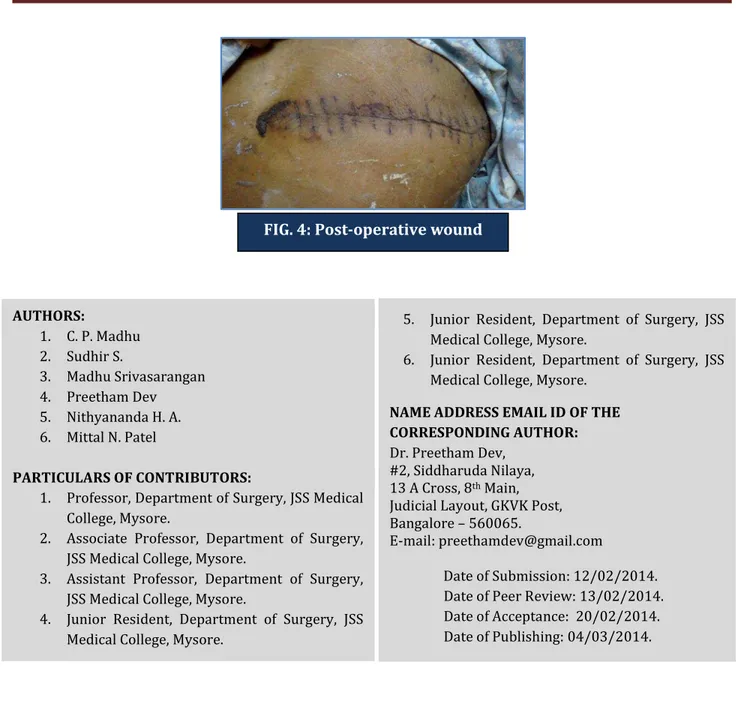
J of Evolution of Med and Dent Sci/ eISSN- 2278-4802, pISSN- 2278-4748/ Vol. 3/ Issue 10/Mar 10, 2014 Page 2409 of lump with persistence of non-healing ulcer with discharge. Simple mastectomy with axillary lymph node biopsy was done and the specimen was sent for histopathological examination (HPE). HPE showed features suggestive of granulomatous mastitis probably tubercular. Postoperative period was uneventful and patient was discharged on post op day 10 after removing the sutures. Wound healed well. Patient received complete course of ATT. Patient was followed upto 6 months with no evidence of disease.
DISCUSSION: TB of the mammary gland is a rare disorder; it is often mistaken for other benign, but most commonly to malignant lesions of the breast 4. TB breast constitutes approximately 0.25- 1% of
all surgically treated breast disease and 0.1 % to 0.5% of all tubercular cases 5, 6.
TB mastitis is more commonly seen in females of reproductive age group, especially during the lactation period when they are more susceptible, as the lactating breast is more vascular and predisposed to trauma. The duration of symptoms varies from a few months to several years, but in most instances it is less than a year 4.
Breast tissue is remarkably resistant to TB. This is due to the fact that like skeletal muscles and spleen it provides an infertile environment for survival and multiplication of tubercle bacilli 7.
Breast involvement can be either primary without any extrapulmonary focus or secondary to pulmonary TB. The primary form of the disease is rare and probably occurs via lymphatic, hematogenous or contiguous spread. Lymphatic route is common and usually due to retrograde extension from cervical, mediastinal or mammary group of or may be via infection through skin abrasions or opening of the lactiferous ducts at the nipple. Axillary lymphadenopathy is reported in 50 – 75% 8.
Diagnosis is established by demonstration of AFB in breast tissue or pus specimen, but seen in only one fourth of the cases 9.FNAC can be diagnostic in about 75% of the patients with appearance
of epitheloid granulomas or Langhans giant cells. Other types like core needle biopsy can get higher accuracy at diagnosis 7. Bacteriologic and histochemical studies of the sample may confirm definitive
diagnosis by identifying presence of the bacillus, although culture and acid fast staining are negative in most of the cases.
Mammography or ultrasonography are not reliable in distinguishing breast TB from carcinoma because of variable pattern of presentations of such inflammatory lesion like coarse stromal texture with or without an ill-defined breast mass and skin thickening which all are not specific enough to aid diagnosis. 10
In our case the patient presented with lump in the breast which was hard in consistency with nipple retraction and overlying skin involvement masquerading as malignancy. She didn’t have any focus of tuberculosis outside the breast, either on physical and radiological examinations. Hence breast may be considered to be primary site of infection. Cytological and histopathological examination helped in confirmation of the diagnosis.
J of Evolution of Med and Dent Sci/ eISSN- 2278-4802, pISSN- 2278-4748/ Vol. 3/ Issue 10/Mar 10, 2014 Page 2410 to prevent sinus formation is mandatory. Simple mastectomy may be considered for patients with extensive disease comprising large painful ulcerative lesion involving the entire breast 11, 12.
CONCLUSION: Tuberculosis of the breast is uncommon even in places with higher incidence of pulmonary or extrapulmonary TB. It is most often mistaken for carcinoma or pyogenic abscess due to difficulty is diagnosis even with radiological and microbiological investigations. Thus high index of suspicion is necessary in diagnosing the disease. Greater awareness of this uncommon entity of a common disease along with early institution of medical line of management with ATT may avoid surgery.
REFERENCES:
1. Baharoon S. Tuberculosis of the breast. Ann Thorac Med. 2008; 3:110–4.
2. Meenu G, Sonia C, Monika S, Sneh S, Praveen, Parul T. Tuberculosis mastitis – A great mimicker. Asian Pac J Trop Dis 2012; 2(5): 348-351.
3. Banerjee SN, Ananthakrishnan N, Mehta RB, Prakash S. Tuberculous mastitis: a continuing problem. World J Surg1987; 11: 105-9.
4. Leo FT, John SM, Celine G, Aroon K, Goeover L, B Rathnakar H. Tuberculosis Mastitis presenting as Breast Abscess. Oman Med J 2011; 26: 53-55.
5. Kalarç N, Ozkan B, Bayiz H, Dursun AB, Demirağ F. Breast tuberculosis. Breast ;
11(4): 346-9.
6. Rosen P. Rosen's Breast Pathology. 2nd ed. Philadelphia: Lippincott Williams and Wilkins; 2001: 70-9.
7. Luh SP, Hsu JD, Lai YS, Chen SW. Primary tuberculous infection of breast: experiences of surgical resection for aged patients and review of literature. J Zhejiang UnivSci B 2007; 8(8):580-3.
8. Sarin R, Verma K, Kapur BML, Dass S. Tuberculosis of the breast. Indian J Surg 1984; 46: 27-31. 9. Ikard RW, Perkins D. Mammary Tuberculosis: A rare disease. South Med J 1997; 70: 208-21. 10.Wilson TS, Mac Gregor JW. The diagnosis and treatment of tuberculosis of the breast. Canadian
Med Assoc J 1963; 89: 1118-19.
11.Tewari M, Shukla HS. Breast Tuberculosis: Diagnosis, clinical features and management. Indian J Med Res. 2005: 122: 103-10.
J of Evolution of Med and Dent Sci/ eISSN- 2278-4802, pISSN- 2278-4748/ Vol. 3/ Issue 10/Mar 10, 2014 Page 2411
FIG. 1: Chronic non-healing ulcer over the left breast With undermined edge, with nipple retraction
FIG. 2: Gross specimen of simple mastectomy with tubercular ulcer (lump and multiple sinuses excised in
toto) and specimen of axillary lymphnode sampling
FIG. 3: HPE section from the breast lesion showing
characteristic Langhan’s giant cell with multinucleated
J of Evolution of Med and Dent Sci/ eISSN- 2278-4802, pISSN- 2278-4748/ Vol. 3/ Issue 10/Mar 10, 2014 Page 2412
AUTHORS:
1. C. P. Madhu 2. Sudhir S.
3. Madhu Srivasarangan 4. Preetham Dev 5. Nithyananda H. A. 6. Mittal N. Patel
PARTICULARS OF CONTRIBUTORS:
1. Professor, Department of Surgery, JSS Medical College, Mysore.
2. Associate Professor, Department of Surgery, JSS Medical College, Mysore.
3. Assistant Professor, Department of Surgery, JSS Medical College, Mysore.
4. Junior Resident, Department of Surgery, JSS Medical College, Mysore.
5. Junior Resident, Department of Surgery, JSS Medical College, Mysore.
6. Junior Resident, Department of Surgery, JSS Medical College, Mysore.
NAME ADDRESS EMAIL ID OF THE CORRESPONDING AUTHOR: Dr. Preetham Dev,
#2, Siddharuda Nilaya, 13 A Cross, 8th Main,
Judicial Layout, GKVK Post, Bangalore – 560065.
E-mail: [email protected]
Date of Submission: 12/02/2014. Date of Peer Review: 13/02/2014. Date of Acceptance: 20/02/2014. Date of Publishing: 04/03/2014.