ESSAY
Fecal Transplants: What Is Being
Transferred?
Diana P. Bojanova
1, Seth R. Bordenstein
1,2*
1Department of Biological Sciences, Vanderbilt University, Nashville, Tennessee, United States of America, 2Department of Pathology, Microbiology, and Immunology, Vanderbilt University, Nashville, Tennessee, United States of America
*s.bordenstein@vanderbilt.edu
Abstract
Fecal transplants are increasingly utilized for treatment of recurrent infections (i.e.,
Clostrid-ium difficile
) in the human gut and as a general research tool for gain-of-function
experi-ments (i.e., gavage of fecal pellets) in animal models. Changes observed in the recipient's
biology are routinely attributed to bacterial cells in the donor feces (~10
11per gram of
human wet stool). Here, we examine the literature and summarize findings on the
composi-tion of fecal matter in order to raise cautiously the profile of its multipart nature. In addicomposi-tion to
viable bacteria, which may make up a small fraction of total fecal matter, other components
in unprocessed human feces include colonocytes (~10
7per gram of wet stool), archaea
(~10
8per gram of wet stool), viruses (~10
8per gram of wet stool), fungi (~10
6per gram of
wet stool), protists, and metabolites. Thus, while speculative at this point and contingent on
the transplant procedure and study system, nonbacterial matter could contribute to changes
in the recipient's biology. There is a cautious need for continued reductionism to separate
out the effects and interactions of each component.
Introduction
A fecal transplant
—
the transfer of stool or portions of stool from one organism into the
gastro-intestinal tract of another
—
is rapidly gaining attention as a treatment for human gut infections
and as a tool for functional "knock-in" studies of the microbiota in animal models. In humans,
the procedure is referred to as fecal microbiota transplantation because the microbial
compo-nents are typically enriched, and in animal models, the transfer of unprocessed stool is
com-monly achieved by feeding or oral gavage of fecal matter. For the purposes of this essay, we will
use the catch-all phrase of
“
fecal transplants
”
to refer to all types of procedures.
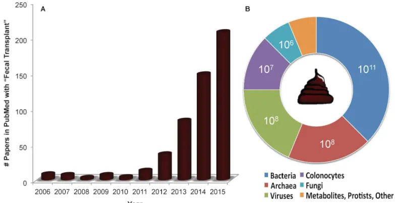
Fig 1
shows the
very recent growth of the term in PubMed references involving both human and model system
studies.
Several analyses report clinical resolution of
Clostridium difficile
infection (CDI) [
1
–
5
],
though the long-term effects of the transplants are unknown [
6
]. Preliminary results also
dem-onstrate positive outcomes for insulin sensitivity [
7
], multiple sclerosis [
8
], and Crohn
’
s disease
a11111
OPEN ACCESS
Citation:Bojanova DP, Bordenstein SR (2016) Fecal Transplants: What Is Being Transferred? PLoS Biol 14(7): e1002503. doi:10.1371/journal.pbio.1002503
Published:July 12, 2016
Copyright:© 2016 Bojanova, Bordenstein. This is an open access article distributed under the terms of the
Creative Commons Attribution License, which permits unrestricted use, distribution, and reproduction in any medium, provided the original author and source are credited.
Funding:This work was supported by National Science Foundation Awards DEB 1046149 and IOS 1456778. The funders had no role in study design, data collection and analysis, decision to publish, or preparation of the manuscript.
Competing Interests:The authors have declared that no competing interests exist.
[
9
]. The presumptive element connecting these conditions is the gut
’
s bacterial community,
and thus the treatment
’
s success enthusiastically revolves around intestinal bacteria that are
assumed mostly viable in feces. There are a few studies in mice and humans that validate the
positive effects of cultured bacteria on CDI [
10
,
11
] and mucosal barrier function [
12
].
Addi-tionally, the microbial portion of human stool can be highly enriched from other fecal material
through microfiltration [
13
,
14
], spore fractionation [
15
], and density gradients [
16
].
Here, we tentatively emphasize that viable bacteria may not be the only player in donor
feces that affect the recipient's biology, a fact that is well appreciated by experts. Viruses,
archaea, fungi, animal colonocytes, protists, and a number of metabolites that commensal
bac-teria make or are dependent upon can potentially occur in unprocessed feces. Here, we
accen-tuate the patterns seen in fecal composition analyses and various experiments that illuminate
functional effects of individual components of fecal matter. We also highlight important and
tractable questions for which further reductionism could help deconstruct the benefit of
con-stituent parts of fecal matter.
Fecal Composition
Human fecal composition has not been intensively studied. The studies that have examined
composition are mostly from the 1970s and 1980s and report varying results, perhaps because
of variation in diet and health. On average, adult fecal matter is estimated to be 75% water and
25% solid matter [
17
]. The vast majority of solid matter is organic material, whose makeup
consists of 25%
–
54% microbial cells (with a slight portion likely consisting of viruses) that may
Fig 1. The growth of fecal transplants as reflected in references in PubMed and the estimated composition of human feces.The charts show (A) the rapid rise in publications on fecal transplants in the National Library of Medicine's search service (PubMed), particularly between 2012 and 2015, and (B) the estimated upper concentration of the biological entity per gram of unprocessed human feces, as cited in the text. Estimates do not necessarily reflect the viable number of the biological entity, and the concentration of the archaea is estimated from a methanogen breath test that is not solely based on the presence of archaea. Concentrations of metabolites, protists, and other entities were not identified.be alive or dead [
18
]. As microbial counts were based on light microscopy and a modification
of the Gram stain, the microbial cells were presumed to be mostly bacteria [
18
], but quality
evi-dence is lacking. Several other components are found in significant concentration, including
archaea, fungi, and microbial eukaryotes. One particular methanoarchaeon species,
Methano-brevibacter smithii
, was detected in 95.7% of patients spanning infants, adults, and the elderly
[
19
], and it can comprise up to 10% of all fecal anaerobes [
20
]. Viable colonocytes are also
readily isolated from newborn and adult feces [
21
–
23
]. No analysis of their potential
contribu-tion to the success of fecal transplants has been reported. Independent validacontribu-tions of these
esti-mates are needed, particularly measurements that consider all of the entities at once.
While transplants can be highly effective treatment in certain cases, concerns remain about
the hypothetical co-transfer of pathogenic microbes [
24
]. Contamination by environmental
microbes is also a risk during the collection, storage, and handling of donor stool, as seen in the
early periods of blood storage for transfusions [
25
,
26
]. To standardize laboratory protocols and
enhance stability of fecal matter, one option is to use frozen donor material from rigorously
screened volunteers. Several studies compared the efficacy of frozen versus fresh stool on
recur-rent or refractory CDI and reported little to no difference [
14
,
27
,
28
]. Extensive longitudinal
screening of stool donors is essential to track the long-term success of treatment, and further
metagenomic studies of the transferred fecal material and transfer proficiency to recipients are
warranted.
Bacteria
It is well established that the gut contains the highest density of microbes in the human body,
with the bacteria-to-human cell ratio recently estimated to be 1.3:1 [
29
]. In feces, bacteria
consti-tute 25%
–
54% of solid matter [
18
] and thus between 6.3% and 13.5% of total fecal matter.
Aver-aged estimates from 14 studies yield a mean bacterial concentration of nearly 10
11bacteria per
gram of wet stool [
29
]. Yet, a clear distinction was shown in a study between the viable (49%),
injured (19%), and dead (32%) bacterial cells collected from fresh fecal samples under anaerobic
conditions [
30
]. These statistics indicate that only 3.0%
–
6.6% of total fecal matter may be
com-posed of viable bacteria. The percentage could conceivably be even lower if samples are handled
in aerobic conditions for lengthy amounts of time, although frequent aerobic preparation of
fecal material has resulted in high cure rates. Furthermore, and as previously noted, transplants
with frozen fecal samples that may have reduced viable bacteria can lead to an almost identical
resolution of CDI to transplants with fresh samples [
27
,
31
]. It should be noted, however, that
even bacterial DNA or dead cells might retain some immunostimulatory functions, as colitis
symptoms in a dextrose sodium sulfate
–
induced mouse model were strikingly alleviated by
introduction of probiotic DNA and unviable irradiated bacterial cells [
32
,
33
].
Other studies suggest that interactions between the host genotype and microbiota can
potentially affect transplant outcomes. Across a collection of studies, human fecal donations
from related donors showed slightly higher resolution in CDI cases (93%) compared to
unre-lated donors (84%) [
34
]. This observation is notable in light of the recent finding that human
genetic variation is significantly correlated with variation in bacterial community composition
[
35
,
36
]. However, a recent meta-analysis demonstrated no significant difference in efficacy
between related and unrelated donors [
37
]. Furthermore, a placebo-controlled trial resulted in
the successful treatment of seven of nine people who received a transplant from a single,
unre-lated, donor [
38
]. Thus, the evidence to date suggests that relatedness either has little or no
effect on treating CDI.
treating CDI in mice and humans. Use of a six-species cocktail therapy suppressed recurrent
CDI in 92% of mice [
10
] when approximately 10
10cells per bacterial species were gavaged into
recipients. In another mouse study, 10
8colony-forming units of a single bacterium isolate,
Lachnospiraceae
D4, caused over a 10-fold reduction in the number of
C
.
difficile
colony-form-ing units per gram of cecal contents [
39
]. A cocktail of nontoxigenic
C
.
difficile
spores was also
successfully used in suppressing CDI recurrence in a human trial [
40
]. At 26 weeks of
treat-ment, only 0%
–
5% of patients from various treatment groups had toxigenic
C
.
difficile
remain-ing in feces. These studies indicate that cultured bacteria can, in certain cases, be effective
contributors to CDI disease resolution.
Viruses
Viruses from eukaryotes, bacteria, and archaea are less studied components of the gut
micro-biota than bacteria. From five fecal samples, count estimates indicate that the viral abundance
ranges from 10
8to 10
9viruses per gram of feces (wet weight), and the average
virus-to-bacte-rium ratio is 0.13 [
41
]. These estimates are comparably low to those reported in other
environ-ments where the virus-to-microbial cell ratios range from 1.4 to 160 [
42
], which supports the
emerging view that viruses exhibit a more temperate lifestyle in the gut [
43
,
44
]. Additionally, a
recent metagenomic study demonstrated that numerous temperate phages are transferred
dur-ing fecal transplants [
24
]. Prophages often assist in controlling invading pathogens, modulating
community structure, and maintaining gut homeostasis [
44
]. The dominance of temperate
viruses is, however, typical of healthy control feces, as patients suffering from bowel diseases
can have increased amounts of virulent phages [
45
]. One of the most abundant, conserved, and
prevalent bacteriophages in the human gut is crAssphage [
46
], a finding that suggests some
phages may be highly conserved in the human population.
The impact of bacteriophages on human health is under active consideration. Phage therapy
entails the isolation and inoculation of phages (or their antibacterial enzymes) that target a
spe-cific bacterium. While not all phage treatments are effective [
47
], several in vitro and in vivo
experiments have been successful. As a treatment for CDI, 10
8plaque-forming units per mL of
a specific phage were introduced into a human colon model. Over a period of 35 days, the
treat-ment caused a significant decrease in vegetative
C
.
difficile
cells (albeit there was an increase in
C
.
difficile
spores) as well as toxin production to levels below the detection threshold of the
assay [
48
]. Control replicates contained high concentrations of both vegetative cells and toxin.
Phage therapy of CDI in a hamster model also significantly delayed bacterial colonization and
the onset of symptoms [
49
]. Specific phage cocktails could, in theory, allow commensal
bacte-ria that are in competition with
C
.
difficile
to reflourish in the gut [
50
]. While
C
.
difficile
phages
may eventually be developed into therapeutic agents, there is yet no evidence that phages
spe-cific to
C
.
difficile
are transferred in fecal transplants.
Archaea and Fungi
Archaea are well-recognized but relatively understudied members of the human gut microbiota
[
57
], with methanoarchaeon comprising up to 10% of fecal anaerobes [
20
]. Based on the
con-centration of methane in breath, estimates suggest a minimum presence of 10
7–
10
8methano-gens per gram of both dry and wet stool [
58
,
59
], though it is unresolved what percentage of
these methanogens are from archaea versus bacteria. Higher than normal concentrations of
intestinal archaea are associated with Crohn
’
s disease and multiple sclerosis [
60
]. Similarly,
fungi in the gut have been cultured in 70% of healthy adults [
61
]. They occur in estimated
con-centrations of up to 10
6microorganisms per gram of feces [
62
] and appear to comprise only
0.03% of all microbes in feces [
63
].
Candida albicans
is the most common and studied yeast,
but it is kept in check by competitive commensal bacteria in a healthy gut. When bacterial
homeostasis is disturbed, however,
C
.
albicans
increases its numbers drastically [
64
,
65
]. These
fungi may also help induce intestinal diseases by penetrating the intestinal colonocyte barrier
and driving inflammation [
66
]. Indeed, high concentrations of
C
.
albicans
occur in individuals
with inflammatory bowel diseases [
67
,
68
]. The contribution of archaea and fungi to changes in
function will be an important area of future research.
Human Colonocytes
Interestingly, viable epithelial cells of the large intestine, or colonocytes, can be isolated at a
concentration of up to 10
7per gram of wet fecal material [
23
]. Viable colonic cells have
effec-tively been isolated from newborn fecal samples (
>
80% viable) [
21
] and biopsy specimens
from colonic crypts (
>
98% viable) [
22
]. Isolation is possible due to the resilient ability of
colo-nocytes to take on a globular shape and survive once exfoliated into the fecal stream [
69
]. Thus,
their viability and partial functionality is likely retained in the course of some transplant
treat-ments, especially in animal models that utilize feeding or oral gavage of fecal material.
By acting as the physical barrier between bacteria and the host
’
s internal tissues and organs,
colonocytes allow host tolerance of the intestinal microbiota [
70
]. When high levels of
colono-cyte death occur, their mediating role disintegrates because of increased intestinal permeability
[
71
]. Indeed, major pathological conditions of the bowel are associated with changes in the
growth and functions of the colonic epithelium [
22
,
72
], similar to changes frequently observed
in microbiota studies. Their restoration is key in successful recovery from such conditions. A
recent study transplanted healthy viable colon stem cells into an immunodeficient mouse model
with superficial colon damage and found that cells readily integrated, and a single layer of
epi-thelium fully covered areas lacking colonocytes [
73
]. The presence of colonic stem cells in feces
has yet to be recorded, although one study recovered stem cells from the colonic epithelium that
often sheds into the fecal stream [
74
]. Should colonic stem cells be identified in feces in human
or animal models, they may affect the success of transplants if they can engraft in recipients.
In addition to colonocytes, molecules such as immunoglobulin A (IgA) can act as the first
line of defense for the intestinal epithelium [
75
]. IgA reinforces the intestinal barrier and
pro-tects host cells against pathogens and enteric toxins in the gut [
75
]. For instance, IgA
signifi-cantly inhibited
C
.
difficile
toxin binding to hamster intestinal brush border membranes
compared to the control [
76
]. Likewise, human epithelial cell lines with IgA added to their
sur-face showed a decrease in
C
.
difficile
–
associated pathology compared to cells lacking IgA [
77
].
It remains to be seen if introducing IgA directly into human subjects will be beneficial.
Metabolites
colon [
78
–
80
]. Fiber strongly contributes to fecal weight, and low fiber diets in mice can lead to
an irreversible loss in bacterial diversity [
81
]. While direct reintroduction of missing fiber in
this study did not restore the diversity, transplants from mice with a high fiber diet did.
Fur-thermore, low fiber diets lead to
“
microbial starving,
”
whereby once-commensal bacteria attack
the intestinal lining [
82
]. Fiber supplements used in a study with
C
.
difficile
–
infected hamsters,
however, managed to significantly modulate onset time of systemic symptoms [
83
]. Fiber
intake has also been linked to increased microbial diversity and reduced obesity in humans
[
84
,
85
].
Butyrate-producing bacteria or butyrate concentrations in feces can be lower in patients
with colorectal cancer and ulcerative colitis [
86
–
88
]. Preliminary studies of enemas with
buty-rate or SCFA cocktails (acetate, butybuty-rate, and propionate) show some resolution in patients
with distal ulcerative colitis [
89
–
93
]. Following these treatments, 35%
–
67% of patients
exhib-ited improvement. Furthermore, oral administration of sodium butyrate in a colitis mouse
model alleviated inflammation and mucosal damage [
94
], and propionate led to improvement
of symptoms in a multiple sclerosis mouse model by promoting regulatory T cell
differentia-tion [
95
]. No adverse side effects were noted in any of these studies, though some metabolite
enemas are malodorous. One review, however, cautions against the use of such metabolites
[
96
]. While butyrate acts as an energy source, increases colonocyte growth, and decreases
apo-ptosis of colonocytes under healthy conditions [
97
], excess butyrate accumulation around
human colonic carcinoma cells has been connected with increased apoptosis [
98
]. Finally,
esti-mates suggest there are nearly 900 gene clusters in human gut
–
associated bacteria that make
small molecules [
99
]. Determining functions may be important in understanding the
compos-ite nature of feces and its effects on fecal transplants in humans and/or animal models.
Summary
Here, we cautiously note that bacteria, either viable or unviable in transferred fecal material,
may not be the only player in donor feces that affects the recipient's biology. On the one hand,
the effects of bacteria on CDI or animal model traits such as obesity [
100
] and toxin tolerance
[
101
] appear well justified thus far. On the other hand, in a broader context where fecal
trans-plants are solely utilized in animal model studies and other human diseases, judicious
reduc-tionism seems warranted in light of a limited understanding of the complex nature of feces.
Deconstructing the benefit and interactions of constituent parts of fecal matter will clarify the
relative importance and causality of each of these components and the potential development
of specific therapies.
Key Points and Future Directions
•
A few studies using cocktails of bacteria in animal models and humans show
suppres-sion of CDI. However, these studies are preliminary and limited.
•
Through bacterial targeting, phage therapy can potentially eliminate virulent bacteria
in a diseased gut and allow commensal bacterial to reflourish.
Acknowledgments
We thank Sean Davies, Ken Lau, Vincent Young, Kevin Kohl, Mike Sadowski, and Joseph
Zackular for providing helpful feedback on the manuscript.
References
1. Kassam Z, Hundal R, Marshall JK, Lee CH (2012) Fecal transplant via retention enema for refractory or recurrent Clostridium difficile infection. Archives of Internal Medicine 172: 191–193. doi:10.1001/ archinte.172.2.191PMID:22271132
2. van Nood E, Vrieze A, Nieuwdorp M, Fuentes S, Zoetendal EG, et al. (2013) Duodenal infusion of donor feces for recurrent Clostridium difficile. N Engl J Med 368: 407–415. doi:10.1056/ NEJMoa1205037PMID:23323867
3. Yoon SS, Brandt LJ (2010) Treatment of refractory/recurrent C. difficile-associated disease by donated stool transplanted via colonoscopy: a case series of 12 patients. J Clin Gastroenterol 44: 562–566. doi:10.1097/MCG.0b013e3181dac035PMID:20463588
4. Aas J, Gessert CE, Bakken JS (2003) Recurrent Clostridium difficile colitis: case series involving 18 patients treated with donor stool administered via a nasogastric tube. Clin Infect Dis 36: 580–585. PMID:12594638
5. Rao K, Young VB (2015) Fecal microbiota transplantation for the management of Clostridium difficile infection. Infect Dis Clin North Am 29: 109–122. doi:10.1016/j.idc.2014.11.009PMID:25677705
6. Borody TJ, Khoruts A (2012) Fecal microbiota transplantation and emerging applications. Nat Rev Gastroenterol Hepatol 9: 88–96.
7. Vrieze A, Van Nood E, Holleman F, Salojarvi J, Kootte RS, et al. (2012) Transfer of intestinal micro-biota from lean donors increases insulin sensitivity in individuals with metabolic syndrome. Gastroen-terology 143: 913–916.e917. doi:10.1053/j.gastro.2012.06.031PMID:22728514
8. Borody TJ, Leis S, Campbell J, al. e (2011) Fecal microbiota transplantation (FMT) in multiple sclero-sis (MS) [abstract]. Am J Gastroenterol 106:S352.
9. Suskind DL, Brittnacher MJ, Wahbeh G, Shaffer ML, Hayden HS, et al. (2015) Fecal microbial trans-plant effect on clinical outcomes and fecal microbiome in active Crohn's disease. Inflamm Bowel Dis 21: 556–563. doi:10.1097/MIB.0000000000000307PMID:25647155
10. Lawley TD, Clare S, Walker AW, Stares MD, Connor TR, et al. (2012) Targeted restoration of the intestinal microbiota with a simple, defined bacteriotherapy resolves relapsing Clostridium difficile dis-ease in mice. PLoS Pathog 8: e1002995. doi:10.1371/journal.ppat.1002995PMID:23133377
11. Petrof EO, Gloor GB, Vanner SJ, Weese SJ, Carter D, et al. (2013) Stool substitute transplant therapy for the eradication of Clostridium difficile infection: 'RePOOPulating' the gut. Microbiome 1:3. doi:10. 1186/2049-2618-1-3PMID:24467987
•
Metabolites can nourish the colonocyte barrier and intestinal bacteria. Oral
adminis-tration of metabolites can alleviate inflammation, mucosal damage, and multiple
scle-rosis symptoms. However, only 5% of the SCFAs produced in the distal colon are
estimated to be excreted into feces [
102
]. Hence, metabolite concentrations are likely
to be much lower than concentrations used in oral administration studies.
•
Archaea and fungi are common in feces. Though high concentrations of intestinal
archaea and certain fungi have been correlated to both intestinal and autoimmune
dis-eases, their causative effects are unknown.
•
Human genetic relatedness has little to no influence on the effectiveness of human
fecal transplants, though genetic factors do shape bacterial community composition.
•
Individual components of fecal matter can yield health benefits and may work
12. Li M, Liang P, Li Z, Wang Y, Zhang G, et al. (2015) Fecal microbiota transplantation and bacterial con-sortium transplantation have comparable effects on the re-establishment of mucosal barrier function in mice with intestinal dysbiosis. Front Microbiol 6:692. doi:10.3389/fmicb.2015.00692PMID:
26217323
13. Hamilton MJ, Weingarden AR, Unno T, Khoruts A, Sadowsky MJ (2013) High-throughput DNA sequence analysis reveals stable engraftment of gut microbiota following transplantation of previously frozen fecal bacteria. Gut Microbes 4: 125–135. doi:10.4161/gmic.23571PMID:23333862
14. Hamilton MJ, Weingarden AR, Sadowsky MJ, Khoruts A (2012) Standardized frozen preparation for transplantation of fecal microbiota for recurrent Clostridium difficile infection. Am J Gastroenterol 107: 761–767. doi:10.1038/ajg.2011.482PMID:22290405
15. Khanna S, Pardi DS, Kelly CR, Kraft CS, Dhere T, et al. (2016) A Novel Microbiome Therapeutic Increases Gut Microbial Diversity and Prevents Recurrent Clostridium difficile Infection. The Journal of Infectious Diseases doi:10.1093/infdis/jiv766
16. Hevia A, Delgado S, Margolles A, Sánchez B (2015) Application of density gradient for the isolation of the fecal microbial stool component and the potential use thereof. Scientific Reports 5: 16807. doi:
10.1038/srep16807PMID:26581409
17. Rose C, Parker A, Jefferson B, Cartmell E (2015) The Characterization of Feces and Urine: A Review of the Literature to Inform Advanced Treatment Technology. Crit Rev Environ Sci Technol 45: 1827– 1879. PMID:26246784
18. Stephen AM, Cummings JH (1980) The microbial contribution to human faecal mass. J Med Microbiol 13: 45–56. PMID:7359576
19. Dridi B, Henry M, El Khéchine A, Raoult D, Drancourt M (2009) High Prevalence of Methanobrevibac-ter smithii and Methanosphaera stadtmanae Detected in the Human Gut Using an Improved DNA Detection Protocol. PLoS ONE 4: e7063. doi:10.1371/journal.pone.0007063PMID:19759898
20. Lurie-Weinberger MN, Gophna U (2015) Archaea in and on the Human Body: Health Implications and Future Directions. PLOS Pathog 11: e1004833. doi:10.1371/journal.ppat.1004833PMID:26066650
21. Chandel DS, Braileanu GT, Chen JH, Chen HH, Panigrahi P (2011) Live colonocytes in newborn stool: surrogates for evaluation of gut physiology and disease pathogenesis. Pediatr Res 70: 153– 158. doi:10.1038/pr.2011.378PMID:21544008
22. Fonti R, Latella G, Bises G, Magliocca F, Nobili F, et al. (1994) Human colonocytes in primary culture: a model to study epithelial growth, metabolism and differentiation. International Journal of Colorectal Disease 9: 13–22. PMID:8027618
23. Nair PP (2002) Isolated colonocytes. United States Patent US6335193.
24. Chehoud C, Dryga A, Hwang Y, Nagy-Szakal D, Hollister EB, et al. (2016) Transfer of Viral Communi-ties between Human Individuals during Fecal Microbiota Transplantation. mBio 7: e00322–00316. doi:10.1128/mBio.00322-16PMID:27025251
25. Bihl F, Castelli D, Marincola F, Dodd RY, Brander C (2007) Transfusion-transmitted infections. Jour-nal of TranslatioJour-nal Medicine 5: 25–25. PMID:17553144
26. Gabriel M, Silvio DP, David H, Michael A, Moshe D, et al. (1991) Transfusion Reactions Due to Bacte-rial Contamination of Blood and Blood Products. Reviews of Infectious Diseases 13: 307–314. PMID:
2041964
27. Lee CH, Steiner T, Petrof EO, et al. (2016) Frozen vs fresh fecal microbiota transplantation and clinical resolution of diarrhea in patients with recurrent Clostridium difficile infection: A randomized clinical trial. JAMA 315: 142–149. doi:10.1001/jama.2015.18098PMID:26757463
28. Youngster I, Sauk J, Pindar C, Wilson RG, Kaplan JL, et al. (2014) Fecal Microbiota Transplant for Relapsing Clostridium difficile Infection Using a Frozen Inoculum From Unrelated Donors: A Random-ized, Open-Label, Controlled Pilot Study. Clin Infect Dis 58: 1515–1522. doi:10.1093/cid/ciu135
PMID:24762631
29. Sender R, Fuchs S, Milo R (2016) Are We Really Vastly Outnumbered? Revisiting the Ratio of Bacte-rial to Host Cells in Humans. Cell 164: 337–340. doi:10.1016/j.cell.2016.01.013PMID:26824647
30. Ben-Amor K, Heilig H, Smidt H, Vaughan EE, Abee T, et al. (2005) Genetic diversity of viable, injured, and dead fecal bacteria assessed by fluorescence-activated cell sorting and 16S rRNA gene analysis. Appl Environ Microbiol 71: 4679–4689. PMID:16085863
31. Satokari R, Mattila E, Kainulainen V, Arkkila PE (2015) Simple faecal preparation and efficacy of fro-zen inoculum in faecal microbiota transplantation for recurrent Clostridium difficile infection—an observational cohort study. Aliment Pharmacol Ther 41: 46–53. doi:10.1111/apt.13009PMID:
32. Rachmilewitz D, Katakura K, Karmeli F, Hayashi T, Reinus C, et al. (2004) Toll-like receptor 9 signal-ing mediates the anti-inflammatory effects of probiotics in murine experimental colitis. Gastroenterol-ogy 126: 520–528. PMID:14762789
33. Rachmilewitz D, Karmeli F, Takabayashi K, Hayashi T, Leider-Trejo L, et al. (2002) Immunostimula-tory DNA ameliorates experimental and spontaneous murine colitis. Gastroenterology 122: 1428– 1441. PMID:11984528
34. Gough E, Shaikh H, Manges AR (2011) Systematic review of intestinal microbiota transplantation (fecal bacteriotherapy) for recurrent Clostridium difficile infection. Clin Infect Dis 53: 994–1002. doi:
10.1093/cid/cir632PMID:22002980
35. Blekhman R, Goodrich JK, Huang K, Sun Q, Bukowski R, et al. (2015) Host genetic variation impacts microbiome composition across human body sites. Genome Biology 16: 1–12.
36. Goodrich JK, Waters JL, Poole AC, Sutter JL, Koren O, et al. (2014) Human genetics shape the gut microbiome. Cell 159: 789–799. doi:10.1016/j.cell.2014.09.053PMID:25417156
37. Kassam Z, Lee CH, Yuan Y, Hunt RH (2013) Fecal Microbiota Transplantation for Clostridium difficile Infection: Systematic Review and Meta-Analysis. Am J Gastroenterol 108: 500–508. doi:10.1038/ ajg.2013.59PMID:23511459
38. Moayyedi P, Surette MG, Kim PT, Libertucci J, Wolfe M, et al. (2015) Fecal Microbiota Transplantation Induces Remission in Patients With Active Ulcerative Colitis in a Randomized Controlled Trial. Gastro-enterology 149: 102–109.e106. doi:10.1053/j.gastro.2015.04.001PMID:25857665
39. Reeves AE, Koenigsknecht MJ, Bergin IL, Young VB (2012) Suppression of Clostridium difficile in the gastrointestinal tracts of germfree mice inoculated with a murine isolate from the family Lachnospira-ceae. Infect Immun 80: 3786–3794. doi:10.1128/IAI.00647-12PMID:22890996
40. Gerding DN, Meyer T, Lee C, Cohen SH, Murthy UK, et al. (2015) Administration of spores of nontoxi-genic Clostridium difficile strain M3 for prevention of recurrent C. difficile infection: a randomized clini-cal trial. Jama 313: 1719–1727. doi:10.1001/jama.2015.3725PMID:25942722
41. Kim MS, Park EJ, Roh SW, Bae JW (2011) Diversity and abundance of single-stranded DNA viruses in human feces. Appl Environ Microbiol 77: 8062–8070. doi:10.1128/AEM.06331-11PMID:
21948823
42. Wigington CH, Sonderegger D, Brussaard CPD, Buchan A, Finke JF, et al. (2016) Re-examination of the relationship between marine virus and microbial cell abundances. Nature Microbiology 1: 15024. 43. Reyes A, Semenkovich NP, Whiteson K, Rohwer F, Gordon JI (2012) Going viral: next generation
sequencing applied to human gut phage populations. Nat Rev Microbiol 10: 607–617.
44. Reyes A, Haynes M, Hanson N, Angly FE, Heath AC, et al. (2010) Viruses in the faecal microbiota of monozygotic twins and their mothers. Nature 466: 334–338. doi:10.1038/nature09199PMID:
20631792
45. Norman JM, Handley SA, Baldridge MT, Droit L, Liu CY, et al. (2015) Disease-specific alterations in the enteric virome in inflammatory bowel disease. Cell 160: 447–460. doi:10.1016/j.cell.2015.01.002
PMID:25619688
46. Dutilh BE, Cassman N, McNair K, Sanchez SE, Silva GG, et al. (2014) A highly abundant bacterio-phage discovered in the unknown sequences of human faecal metagenomes. Nat Commun 5: 4498. doi:10.1038/ncomms5498PMID:25058116
47. Sarker SA, Sultana S, Reuteler G, Moine D, Descombes P, et al. (2016) Oral Phage Therapy of Acute Bacterial Diarrhea With Two Coliphage Preparations: A Randomized Trial in Children From Bangla-desh. EBioMedicine 4: 124–137. doi:10.1016/j.ebiom.2015.12.023PMID:26981577
48. Meader E, Mayer MJ, Steverding D, Carding SR, Narbad A (2013) Evaluation of bacteriophage ther-apy to control Clostridium difficile and toxin production in an in vitro human colon model system. Anaerobe 22: 25–30. doi:10.1016/j.anaerobe.2013.05.001PMID:23685029
49. Nale JY, Spencer J, Hargreaves KR, Buckley AM, Trzepinski P, et al. (2015) Bacteriophage Combina-tions Significantly Reduce Clostridium difficile Growth In Vitro and Proliferation In Vivo. Antimicrob Agents Chemother 60: 968–981. doi:10.1128/AAC.01774-15PMID:26643348
50. Hargreaves KR, Clokie MRJ (2014) Clostridium difficile phages: still difficult? Frontiers in Microbiology 5(184): doi:10.3389/fmicb.2014.00184
51. McCallin S, Alam Sarker S, Barretto C, Sultana S, Berger B, et al. (2013) Safety analysis of a Russian phage cocktail: from metagenomic analysis to oral application in healthy human subjects. Virology 443: 187–196. doi:10.1016/j.virol.2013.05.022PMID:23755967
53. Hill DA, Hoffmann C, Abt MC, Du Y, Kobuley D, et al. (2010) Metagenomic analyses reveal antibiotic-induced temporal and spatial changes in intestinal microbiota with associated alterations in immune cell homeostasis. Mucosal Immunol 3: 148–158. doi:10.1038/mi.2009.132PMID:19940845
54. Dethlefsen L, Huse S, Sogin ML, Relman DA (2008) The pervasive effects of an antibiotic on the human gut microbiota, as revealed by deep 16S rRNA sequencing. PLoS Biol 6: e280. doi:10.1371/ journal.pbio.0060280PMID:19018661
55. Labrie SJ, Samson JE, Moineau S (2010) Bacteriophage resistance mechanisms. Nat Rev Microbiol 8: 317–327. doi:10.1038/nrmicro2315PMID:20348932
56. Hyman P, Abedon ST (2010) Bacteriophage host range and bacterial resistance. Adv Appl Microbiol 70: 217–248. doi:10.1016/S0065-2164(10)70007-1PMID:20359459
57. Gaci N, Borrel G, Tottey W, O’Toole PW, Brugère J (2014) Archaea and the human gut: New begin-ning of an old story. World J Gastroenterol 20: 16062–16078. doi:10.3748/wjg.v20.i43.16062PMID:
25473158
58. Stewart JA, Chadwick VS, Murray A (2006) Carriage, quantification, and predominance of methano-gens and sulfate-reducing bacteria in faecal samples. Lett Appl Microbiol 43: 58–63. PMID:
16834722
59. Weaver GA, Krause JA, Miller TL, Wolin MJ (1986) Incidence of methanogenic bacteria in a sigmoid-oscopy population: an association of methanogenic bacteria and diverticulosis. Gut 27: 698–704. PMID:3721294
60. Jhangi Sushrut G R, Glanz Bonnie, Cook Sandra, Nejad Parham, Ward Doyle, Li Ning, Gerber Georg, Bry Lynn, Weiner Howard (2014) Increased Archaea Species and Changes with Therapy in Gut Microbiome of Multiple Sclerosis Subjects. Neurology 82(10): S24.001.
61. Schulze J, Sonnenborn U (2009) Yeasts in the gut: from commensals to infectious agents. Dtsch Arz-tebl Int 106: 837–842. doi:10.3238/arztebl.2009.0837PMID:20062581
62. Wang ZK, Yang YS, Stefka AT, Sun G, Peng LH (2014) Review article: fungal microbiota and diges-tive diseases. Aliment Pharmacol Ther 39: 751–766. doi:10.1111/apt.12665PMID:24612332
63. Ott SJ, Kuhbacher T, Musfeldt M, Rosenstiel P, Hellmig S, et al. (2008) Fungi and inflammatory bowel diseases: Alterations of composition and diversity. Scand J Gastroenterol 43: 831–841. doi:10.1080/ 00365520801935434PMID:18584522
64. Rosenbach A, Dignard D, Pierce JV, Whiteway M, Kumamoto CA (2010) Adaptations of Candida albi-cans for growth in the mammalian intestinal tract. Eukaryot Cell 9: 1075–1086. doi:10.1128/EC. 00034-10PMID:20435697
65. White SJ, Rosenbach A, Lephart P, Nguyen D, Benjamin A, et al. (2007) Self-Regulation of Candida albicans Population Size during GI Colonization. PLoS Pathog 3: e184. PMID:18069889
66. Albac S, Schmitz A, Lopez-Alayon C, d'Enfert C, Sautour M, et al. (2016) Candida albicans is able to use M cells as a portal of entry across the intestinal barrier in vitro. Cell Microbiol 18: 195–210. doi:
10.1111/cmi.12495PMID:26242223
67. Ksiadzyna D, Semianow-Wejchert J, Nawrot U, Wlodarczyk K, Paradowski L (2009) Serum concen-tration of interleukin 10, anti-mannan Candida antibodies and the fungal colonization of the gastroin-testinal tract in patients with ulcerative colitis. Adv Med Sci 54: 170–176. doi: 10.2478/v10039-009-0023-6PMID:19758974
68. Standaert-Vitse A, Sendid B, Joossens M, Francois N, Vandewalle-El Khoury P, et al. (2009) Candida albicans colonization and ASCA in familial Crohn's disease. Am J Gastroenterol 104: 1745–1753. doi:10.1038/ajg.2009.225PMID:19471251
69. Nair P, Lagerholm S, Dutta S, Shami S, Davis K, et al. (2003) Coprocytobiology: on the nature of cellu-lar elements from stools in the pathophysiology of colonic disease. J Clin Gastroenterol 36: S84–93; discussion S94-86. PMID:12702972
70. Maynard CL (2012) Reciprocal Interactions of the Intestinal Microbiota and Immune System. 489: 231–241. doi:10.1038/nature11551PMID:22972296
71. Magnusson M, Magnusson KE, Sundqvist T, Denneberg T (1991) Impaired intestinal barrier function measured by differently sized polyethylene glycols in patients with chronic renal failure. Gut 32: 754– 759. PMID:1855681
72. Vaziri ND, Zhao YY, Pahl MV (2015) Altered intestinal microbial flora and impaired epithelial barrier structure and function in CKD: the nature, mechanisms, consequences and potential treatment. Nephrol Dial Transplant 31: 737–746. doi:10.1093/ndt/gfv095PMID:25883197
74. Jung P, Sato T, Merlos-Suarez A, Barriga FM, Iglesias M, et al. (2011) Isolation and in vitro expansion of human colonic stem cells. Nat Med 17: 1225–1227. doi:10.1038/nm.2470PMID:21892181
75. Mantis NJ, Rol N, Corthésy B (2011) Secretory IgA's Complex Roles in Immunity and Mucosal Homeostasis in the Gut. Mucosal immunology 4: 603–611. doi:10.1038/mi.2011.41PMID:
21975936
76. Dallas SD, Rolfe RD (1998) Binding of Clostridium difficile toxin A to human milk secretory compo-nent. Journal of Medical Microbiology 47: 879–888. PMID:9788811
77. Olson A, Diebel LN, Liberati DM (2013) Effect of host defenses on Clostridium difficile toxin-induced intestinal barrier injury. J Trauma Acute Care Surg 74: 983–989; discussion 989–990. doi:10.1097/ TA.0b013e3182858477PMID:23511135
78. Maslowski KM, Vieira AT, Ng A, Kranich J, Sierro F, et al. (2009) Regulation of inflammatory responses by gut microbiota and chemoattractant receptor GPR43. Nature 461: 1282–1286. doi:10. 1038/nature08530PMID:19865172
79. Arpaia N, Campbell C, Fan X, Dikiy S, van der Veeken J, et al. (2013) Metabolites produced by com-mensal bacteria promote peripheral regulatory T-cell generation. Nature 504: 451–455. doi:10.1038/ nature12726PMID:24226773
80. Furusawa Y, Obata Y, Fukuda S, Endo TA, Nakato G, et al. (2013) Commensal microbe-derived buty-rate induces the differentiation of colonic regulatory T cells. Nature 504: 446–450. doi:10.1038/ nature12721PMID:24226770
81. Sonnenburg ED, Smits SA, Tikhonov M, Higginbottom SK, Wingreen NS, et al. (2016) Diet-induced extinctions in the gut microbiota compound over generations. Nature 529: 212–215. doi:10.1038/ nature16504PMID:26762459
82. Sonnenburg ED, Sonnenburg JL (2014) Starving our microbial self: the deleterious consequences of a diet deficient in microbiota-accessible carbohydrates. Cell Metab 20: 779–786. doi:10.1016/j.cmet. 2014.07.003PMID:25156449
83. Horner KL, LeBoeuf RC, McFarland LV, Elmer GW (2000) Dietary fiber affects the onset of Clostrid-ium difficile disease in hamsters. Nutrition Research 20: 1103–1112.
84. Nicolucci AC, Reimer RA (2016) Prebiotics as a modulator of gut microbiota in paediatric obesity. Pediatr Obes doi:10.1111/ijpo.12140
85. Nicolucci AC, Hume MP, Reimer RA (2015) Effect of Prebiotic Fiber Intake on Adiposity and Inflam-mation in Overweight and Obese Children: Assessing the Role of the Gut Microbiota. Canadian Jour-nal of Diabetes 39: S43.
86. Machiels K, Joossens M, Sabino J, De Preter V, Arijs I, et al. (2014) A decrease of the butyrate-pro-ducing species Roseburia hominis and Faecalibacterium prausnitzii defines dysbiosis in patients with ulcerative colitis. Gut 63: 1275–1283. doi:10.1136/gutjnl-2013-304833PMID:24021287
87. Sokol H, Seksik P, Furet JP, Firmesse O, Nion-Larmurier I, et al. (2009) Low counts of Faecalibacter-ium prausnitzii in colitis microbiota. Inflamm Bowel Dis 15: 1183–1189. doi:10.1002/ibd.20903PMID:
19235886
88. Weir TL, Manter DK, Sheflin AM, Barnett BA, Heuberger AL, et al. (2013) Stool microbiome and meta-bolome differences between colorectal cancer patients and healthy adults. PLoS ONE 8: e70803. doi:10.1371/journal.pone.0070803PMID:23940645
89. Vernia P, Marcheggiano A, Caprilli R, Frieri G, Corrao G, et al. (1995) Short-chain fatty acid topical treatment in distal ulcerative colitis. Aliment Pharmacol Ther 9: 309–313. PMID:7654893
90. Scheppach W, Sommer H, Kirchner T, Paganelli GM, Bartram P, et al. (1992) Effect of butyrate ene-mas on the colonic mucosa in distal ulcerative colitis. Gastroenterology 103: 51–56. PMID:1612357
91. Harig JM, Soergel KH, Komorowski RA, Wood CM (1989) Treatment of diversion colitis with short-chain-fatty acid irrigation. N Engl J Med 320: 23–28. PMID:2909876
92. Breuer RI, Soergel KH, Lashner BA, Christ ML, Hanauer SB, et al. (1997) Short chain fatty acid rectal irrigation for left-sided ulcerative colitis: a randomised, placebo controlled trial. Gut 40: 485–491. PMID:9176076
93. Patz J, Jacobsohn WZ, Gottschalk-Sabag S, Zeides S, Braverman DZ (1996) Treatment of refractory distal ulcerative colitis with short chain fatty acid enemas. Am J Gastroenterol 91: 731–734. PMID:
8677939
94. Vieira EL, Leonel AJ, Sad AP, Beltrao NR, Costa TF, et al. (2012) Oral administration of sodium buty-rate attenuates inflammation and mucosal lesion in experimental acute ulcerative colitis. J Nutr Bio-chem 23: 430–436. doi:10.1016/j.jnutbio.2011.01.007PMID:21658926
96. Fröhlich EE, Mayerhofer R, Holzer P (2015) Reevaluating the hype: four bacterial metabolites under scrutiny. European Journal of Microbiology & Immunology 5: 1–13.
97. Tremaroli V, Backhed F (2012) Functional interactions between the gut microbiota and host metabo-lism. Nature 489: 242–249. doi:10.1038/nature11552PMID:22972297
98. Lupton JR (2004) Microbial degradation products influence colon cancer risk: the butyrate contro-versy. J Nutr 134: 479–482. PMID:14747692
99. Donia MS, Fischbach MA (2015) Small Molecules from the Human Microbiota. Science 349(6246): 1254766(1–10). doi:10.1126/science.1254766PMID:26206939
100. Ridaura VK, Faith JJ, Rey FE, Cheng J, Duncan AE, et al. (2013) Gut microbiota from twins discordant for obesity modulate metabolism in mice. Science 341(6150): doi:10.1126/science.1241214
101. Kohl KD, Stengel A, Dearing MD (2015) Inoculation of tannin-degrading bacteria into novel hosts increases performance on tannin-rich diets. Environmental Microbiology doi:10.1111/1462-2920. 12841