What is in? Pneumoperitoneum after sexual
intercourse
Patricia Botelho,
1Ana Franky Carvalho,
1,2Helena Torrão,
3Pedro Leão
1,21Department of General Surgery, Hospital of Braga, Braga, Portugal
2School of Health Sciences, University of Minho, Braga, Portugal
3
Department of Radiology, Hospital of Braga, Braga, Portugal
Correspondence to
Dr Patricia Botelho, pscfb@yahoo.com
To cite:Botelho P, Carvalho AF, Torrão H,et al.
BMJ Case RepPublished online: [please includeDay Month Year] doi:10.1136/ bcr-2013-009667
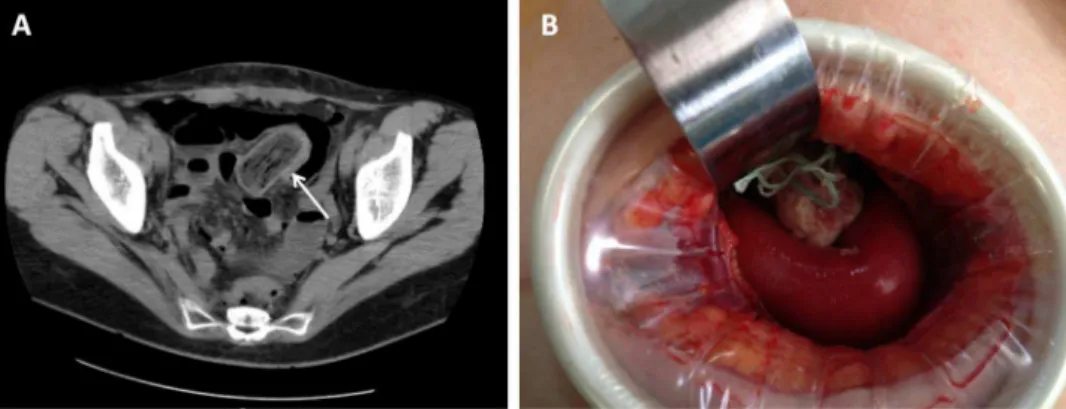
DESCRIPTION
A 49-year-old woman presented to the emergency
department with severe abdominal pain after
vigor-ous sexual intercourse. The patient had a history of
hysterectomy (due to cervix carcinoma) 3 months
before and due to postoperative haematic losses,
the patient frequently used tampons. The patient
claimed she had a tampon within the abdominal
cavity after sexual intercourse. An initial
gynaeco-logical examination showed no evidence of vagina
vault damage and foreign body. Since abdominal
pain persisted, the patient was submitted to an
abdominal
–
pelvic CT scan that revealed a
pneumo-peritoneum and the presence of a foreign body in
the abdominal cavity (arrow in
fi
gure 1A). The
patient underwent minilaparotomy, and a tampon
was found among intestine ansae (
fi
gure 1B); this
occurred due to the total rupture of the vaginal
vault, which was repaired through colporrhaphy.
There were no signi
fi
cant events in the
post-operative period. This case serves to highlight the
relevance of patient self-report, sometimes so
underestimated by clinicians and presents for the
fi
rst time in literature the CT imaging of a tampon
in the abdominal cavity.
Learning points
▸
Do not neglect patient self-report.
▸
Investigate patient's history.
▸
Always suspect a persistent abdominal pain.
Contributors PB evaluated the patient and wrote the manuscript. AFC and PL were involved in drafting the manuscript and revising it critically for important intellectual content. PB and PL performed the surgery. HT was involved in CT scanning and imaging evaluation.
Competing interests None.
Patient consent Obtained.
Provenance and peer reviewNot commissioned; externally peer reviewed.
Copyright 2013 BMJ Publishing Group. All rights reserved. For permission to reuse any of this content visit http://group.bmj.com/group/rights-licensing/permissions.
BMJ Case Report Fellows may re-use this article for personal use and teaching without any further permission.
Become a Fellow of BMJ Case Reports today and you can: ▸ Submit as many cases as you like
▸ Enjoy fast sympathetic peer review and rapid publication of accepted articles ▸ Access all the published articles
▸ Re-use any of the published material for personal use and teaching without further permission
For information on Institutional Fellowships contact consortiasales@bmjgroup.com
Visit casereports.bmj.com for more articles like this and to become a Fellow
Figure 1
(A) CT scan demonstrating the tampon body in the abdominal cavity. (B) In situ tampon during
minilaparotomy.
Botelho P,et al.BMJ Case Rep2013. doi:10.1136/bcr-2013-009667 1