in People with and without Diarrhea in Senegal
Bissoume Samb-Ba1,2, Catherine Mazenot1, Amy Gassama-Sow2, Gre´gory Dubourg1, Herve´ Richet1, Perrine Hugon1, Jean-Christophe Lagier1, Didier Raoult1, Florence Fenollar1*
1Unite´ de Recherche sur les Maladies Infectieuses et Tropicales Emergentes (URMITE) UM63, CNRS 7278, IRD 198, INSERM 1095, Aix-Marseille Universite´, Marseille, France and Dakar, Senegal,2Unite´ de Bacte´riologie Expe´rimentale, Institut Pasteur de Dakar, Dakar, Senegal
Abstract
Background:In Africa, there are several problems with the specific identification of bacteria. Recently, MALDI-TOF mass spectrometry has become a powerful tool for the routine microbial identification in many clinical laboratories.
Methodology/Principal Findings:This study was conducted using feces from 347 individuals (162 with diarrhea and 185 without diarrhea) sampled in health centers in Dakar, Senegal. Feces were transported from Dakar to Marseille, France, where they were cultured using different culture conditions. The isolated colonies were identified using MALDI-TOF. If a colony was unidentified, 16S rRNA sequencing was performed. Overall, 2,753 isolates were tested, allowing for the identification of 189 bacteria from 5 phyla, including 2 previously unknown species, 11 species not previously reported in the human gut, 10 species not previously reported in humans, and 3 fungi. 2,718 bacterial isolates (98.8%) out of 2,750 yielded an accurate identification using mass spectrometry, as did the 3 Candida albicans isolates. Thirty-two bacterial isolates not identified by MALDI-TOF (1.2%) were identified by sequencing, allowing for the identification of 2 new species. The number of bacterial species per fecal sample was significantly higher among patients without diarrhea (8.663) than in those with diarrhea (7.363.4;P= 0.0003). A modification of the gut microbiota was observed between the two groups. In individuals with diarrhea, major commensal bacterial species such asE. coliwere significantly decreased (85% versus 64%), as were severalEnterococcusspp. (E. faeciumandE. casseliflavus) and anaerobes, such asBacteroidesspp. (B. uniformisandB. vulgatus) andClostridiumspp. (C. bifermentans,C. orbiscindens,C. perfringens, andC. symbosium). Conversely, severalBacillus spp. (B. licheniformis,B. mojavensis, andB. pumilus) were significantly more frequent among patients with diarrhea.
Conclusions/Significance:MALDI-TOF is a potentially powerful tool for routine bacterial identification in Africa, allowing for a quick identification of bacterial species.
Citation:Samb-Ba B, Mazenot C, Gassama-Sow A, Dubourg G, Richet H, et al. (2014) MALDI-TOF Identification of the Human Gut Microbiome in People with and without Diarrhea in Senegal. PLoS ONE 9(5): e87419. doi:10.1371/journal.pone.0087419
Editor:Dipshikha Chakravortty, Indian Institute of Science, India
ReceivedAugust 15, 2013;AcceptedDecember 24, 2013;PublishedMay 1, 2014
Copyright:ß2014 Samb-Ba et al. This is an open-access article distributed under the terms of the Creative Commons Attribution License, which permits unrestricted use, distribution, and reproduction in any medium, provided the original author and source are credited.
Funding:This work was supported by the Institut Hospitalo-Universitaire Me´diterrane´e-Infection. The funders had no role in study design, data collection and analysis, decision to publish, or preparation of the manuscript.
Competing Interests:The authors have declared that no competing interests exist. * E-mail: [email protected]
Introduction
There are several problems in the specific identification of bacterial infections in Africa. Currently, bacterial identification is based on phenotypic tests, including Gram staining, bacterial culture, culture growth characteristics, and biochemical profiles. Even if culture processes are available in major hospitals in Africa, there are limitations to the performance of biochemical identifi-cation methods. Such traditional methods require the possession of many API strips including API-20E, API-20NE, API Staph kits, and API Anaerobe kits and many unique reagents that should be stocked under specific conditions and have expiration dates. Biochemical methods are time consuming. They often required knowledge about the type of microorganism being tested, and fail to accurately identify several bacteria species [1,2].
Five years ago, a revolution occurred in bacteriology with the advent of the routine identification of bacteria by matrix-assisted laser desorption ionization time-of-flight mass spectrometry (MALDI-TOF) [1,3–5]. Currently, this technique allows accurate
identification of bacteria without a priori knowledge of the type of microorganism. This technique is in widespread use in many clinical laboratories in Europe [1,3,4,6,7]. This method allows for the detection of bacteria in less than 1 hour and is cost effective. Thus, this technique has become a powerful tool for routine identification and could replace Gram staining and biochemical identification, but to this point, many studies using this technique have been mainly performed in Europe [2].
In this study, we evaluated the effectiveness of MALDI-TOF mass spectrometry on the identification of bacterial species isolated from feces from Senegalese patients with and without diarrhea by combining several culture conditions and rapid mass spectrometry identification.
Materials and Methods
Ethics Statement
All aspects of this study were approved by the National Ethical Committee (CNERS) of Senegal (SEN25/07). Written consent was obtained for all participants. For children, their parents or guardians provided also a written informed consent.
Patient Recruitment and Sample Management
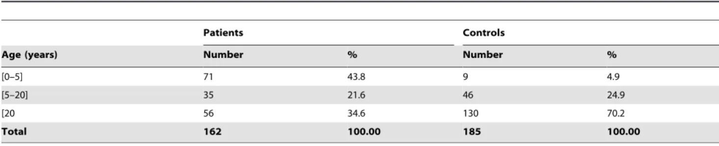
This study was based on 347 individuals, adults and children, sampled from March 2009 to January 2010:162 individuals with diarrhea and 185 without diarrhea (Table 1). Five health centers in Dakar, Senegal and its suburbs (Dominique-Pikine, Sicap Mbao, Roi Baudoin, Institut d’Hygie`ne Sociale, and Saint Martin) were included. Stool samples were collected from children and adults who attended these health centers. Control patients were hospitalized patients or outpatients without intestinal pathogens or recent treatment with antibiotics.
Stool specimens were collected in special sterile stool containers or with swabs for stool samples collected from infants. All stool samples were labeled and transported in cool boxes for examina-tion within 24 hours of collecexamina-tion to Institut Pasteur de Dakar (Senegal). At the laboratory, macroscopic and microscopic analyses were performed on fresh stool samples to look for enteric pathogens including eggs, cysts, and trophozoites of intestinal parasites as well as enteric viruses. Stool samples were preserved in two Nunc tubes (Fisher Thermo Scientific, Denmark) and stored at
220uC. They were transported from Dakar to Marseille, France in ice packs.
Culturomics Methods
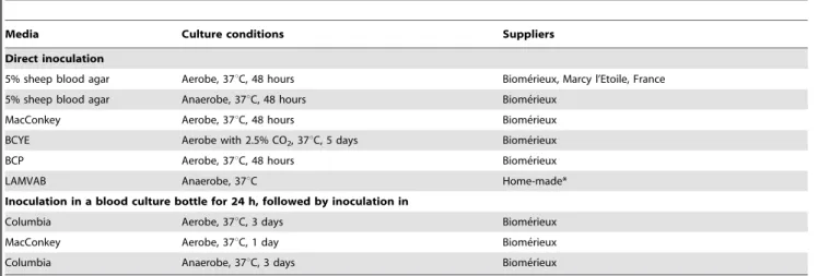
To enumerate the number of colony forming units (CFU) in the stool samples, 1 g of pasty stool was diluted in 9 ml of phosphate buffered saline (PBS), and 100ml of watery stool was diluted in 900ml of PBS. The diluted samples were introduced with a syringe for preincubation into aerobic and anaerobic blood culture bottles (BD Bactec Plus Lytic/10 Anaerobic, Aerobic, 39 Heidelberg, Germany) for 24 hours before being inoculated on agar plates as it has been previously reported that this strategy allowed the growth of bacterial species, mainly anaerobic, that were not detected by standard axenic culture, species [12,15]. Plates for anaerobic culture were pre-incubated for 24 h anaerobically. To identify the maximum number of bacterial species, stool samples were diluted from 1021 to 10210 and inoculated on agar plates using nine
different culture conditions that had been previously determined to be the most useful (Table 2) [12]. The microaerophilic and anaerobic incubations were carried out using microaerophilic bags (Oxoid, Basingstoke, England), anaerobic jars (Mitsubishi) and atmosphere generators (BD Diagnostics, Heidelberg, Germany). Each agar plate was carefully observed after 2 and 7 days of incubation. Any isolated colony was applied to mass spectrometry for identification.
Identification Using Mass Spectrometry
The isolated colonies were deposited on a MALDI-TOF target microflex (Bruker Daltonik, Wissembourg, France) and overlaid with matrix solution, a saturated solution of a- cyano-4-hydroxycinnamic acid in 50% acetonitrile and 2.5% trifluoroace-tic acid, after air-drying at room temperature for 5 minutes. Each colony was picked from an Eppendorf tube containing the Trypticase-Casein-Soy (AES) culture medium stored at 37uC. Broth culture-specific thioglycollate (BD Diagnostics) was used for anaerobes. Two spots were examined for each colony. Each deposit was covered with 2ml of the matrix solution. The Biotyper software was used to compare the protein profile of the bacteria obtained from a database (Bruker and the base of the Timone hospital) of protein profiles regularly updated based on the results of clinical diagnosis. This software takes into account a maximum of 100 mass peaks between 3,000 and 15,000 Da. A score.1.9 indicates a high-level identification of genus and species. A score.
1.7 indicates the identification of genus but not species, and a score lower than 1.7 indicates no identification of bacteria. If the species was still not accurately identified by MALDI-TOF after two attempts, the isolate was analyzed by 16S rRNA sequencing.
16S rRNA Amplification and Sequencing Identification
Bacterial DNA was extracted using the MagNA Pure LC kit DNA isolation kit III (Roche, France) with the MagNA Pure LC instrument, according to the manufacturer’s instructions. The 16S rRNA gene was amplified by PCR using the universal primer pair fd1and rp2and an annealing temperature of 52uC, as described elsewhere [16]. PCR products were purified using the PCR kit Nucleofast 96 (Macherey-Nagel, Hoerdt, France). Sequencing reactions were performed with the sequencing kit Big Dye Terminator version 1.1 (Perkin-Elmer, Coignieres, France) with primers 536F, 536R, 800F, 800R, 1050F, and 1050R(Table 3). Products of the sequencing reactions were purified and the sequences analyzed on an ABI PRISM 3130X Genetic Analyzer (Applied Biosystems, California, USA). The obtained sequences were compared with the GenBank database using BLAST software. A threshold value of similarity $98.7% was used for identification at the species level. Below this value, sequences were repeated to confirm the first obtained results. A new species was
Table 1.Population description.
Patients Controls
Age (years) Number % Number %
[0–5] 71 43.8 9 4.9
[5–20] 35 21.6 46 24.9
[20 56 34.6 130 70.2
Total 162 100.00 185 100.00
suspected when the similarity in the GenBank database with described bacteria was,98.7% [17,18].
Statistical Analyses
Statistical analyses were performed using EpiInfo6 software (http://www.cdc.gov/epiinfo/Epi6/EI6dnjp.htm). The results were concluded to be statistically significant whenP,0.05. The corrected chi-squared test or Fisher’s exact test was used where indicated.
Results
Culture
Overall, 2,753 isolates were tested, which allowed us to identify 189 bacterial species from 5 phyla, including an unknown species and 3 fungi (Table 4 and Figure 1) [19]. Two stool specimens from patients with diarrhea did not allow for the recovery of any bacteria. Candida albicans was detected from 3 patients with diarrhea (3/162 versus 0/185,P= 0.1). A total of 1,175 bacterial isolates were detected among patients with diarrhea and 1,575 were detected among patients without diarrhea. The number of different bacterial species per stool sample was significantly higher among patients without diarrhea (mean of 8.663, range 1 to 18) than among those with diarrhea (mean of 7.363.4, range 0 to 22; P= 0.0003). Finally, 59 out of the 153 bacterial species (38.6%)
identified among patients with diarrhea were specific for this group whereas 36 out of the 129 bacterial species (27.9%) identified among patients without diarrhea were specific for this group, although this difference is not significant (P= 0.059).
MALDI-TOF Mass Spectrometry Identification
Of the 2,750 bacterial isolates analyzed, 2,718 (98.8%) yielded an accurate identification using MALDI-TOF mass spectrometry (Table 4).
16S rRNA Amplification and Sequencing Identification
Thirty-two isolates out of the 2,750 (1.2%) were not identified by MALDI-TOF mass spectrometry. Among these isolates, 11 were identified using 16S rRNA sequencing: Bacteroides nordii, Bacillus clausii, Bacillus thuringiensis, Clostridium cadaveris, Clostridium neonatale, Paenibacillus polymyxa, Staphylococcus sciuri, Shigella boydii, Shigella sonnei, and two new species were identified: a new clostridial species that was called Clostridium dakarense sp. nov. (GenBank accession number KC517358) and a newBacillusspecies,Bacillus casamencensis sp. nov. (GenBank accession number AF519462.1). The 16S rRNA sequence of thisBacillusspecies has been already detected in rice soils in Senegal but no description of the bacterium has been yet reported. The full genome ofC. dakarensehas been recently sequenced and reported [20].
Table 2.Culture media and conditionings used in this study.
Media Culture conditions Suppliers
Direct inoculation
5% sheep blood agar Aerobe, 37uC, 48 hours Biome´rieux, Marcy l’Etoile, France
5% sheep blood agar Anaerobe, 37uC, 48 hours Biome´rieux
MacConkey Aerobe, 37uC, 48 hours Biome´rieux
BCYE Aerobe with 2.5% CO2, 37uC, 5 days Biome´rieux
BCP Aerobe, 37uC, 48 hours Biome´rieux
LAMVAB Anaerobe, 37uC Home-made*
Inoculation in a blood culture bottle for 24 h, followed by inoculation in
Columbia Aerobe, 37uC, 3 days Biome´rieux
MacConkey Aerobe, 37uC, 1 day Biome´rieux
Columbia Anaerobe, 37uC, 3 days Biome´rieux
BCYE: Buffered Charcoal Yeast Extract; BCP: Bromocresol Purple; LAMVAB: Lactobacillus Anaerobic MRS with Vancomycin and Bromocresol green. *from Harteminket al. [15].
doi:10.1371/journal.pone.0087419.t002
Table 3.Primers used for 16S rRNA PCR and sequencing.
Primers Sequences (59–39) Annealing temperature
FD1 AGAGTTTGATCCTGGCTCAG 52uC
RP2 ACGGCTACCTTGTTACGACTT 52uC
536F CAGCAGCCGCGGTAATAC 50uC
536R GTATTACCGCGGCTGCTG 50uC
800F ATTAGATACCCTGGTAG 50uC
800R CTACCAGGGTATCTAAT 50uC
1050F TGTCGTCAGCTCGTG 50uC
1050R CACGAGCTGACGACA 50uC
The other isolates identified by 16S rRNA sequence included 1 of 2 Parabacteroides goldsteiniiisolates detected in the study, 1 of 2 Aneurinibacillus migulanusisolates, 2 (11%) of 18Bacillus amylolique-faciensisolates, 1 of 2 Bacillus endophyticusisolates, 1 (7.7%) of 13 Bacillus licheniformisisolates, 2 (7.7%) of 26Bacillus pumilusisolates, 3 (8%) of 37Bacillus subtilis, 1 of 4Clostridium clostridioformeisolates, 1 of 13 (7.7%)Clostridium lituseburenseisolates, 1 of 13 (7.7%)Kurthia gibsonii isolates, 1 of 4 Lactococcus lactis isolates, 1 of 25 (4%) Lysinibacillus fusiformisisolates, 1 of 2Lysinibacillus sphaericusisolates, 1 of 2Ruminococcus gnavus isolates, 1 of 12 (8.3%)Weissella cibaria isolates, and 2 of 11 (18.2%)Acinetobacter baumanniiisolates. When the spectra of the aforementioned isolates were added to the Bruker database, further identifications of these organisms by MALDI-TOF were accurate.
Common bacteria. Seven bacterial species (3.7%) were identified more than 100 times in fecal samples (261 Escherichia coli isolates, 256 Enterococcus faecium isolates, 159 Clostridium bifermentansisolates, 153Enterococcus faecalisisolates, 152Clostridium perfringensisolates, 137 Bacillus cereus isolates, and 106 Enterococcus hiraeisolates). Surprisingly, several bacteria were more common in patients without diarrhea includingE. colithan those without (P#
1023), E. faecium (P#1023), C. bifermentans (P= 0.002), and C. perfringens(P#1023), see Table 4 and Figure 2.
Thirty-nine bacterial species (20.6%) from 18 different genera were identified from between 10 and 100 fecal samples (Table 4 and Figure 2). Several were more common in patients with diarrhea than those without, such asBacillus licheniformis(P= 0.02), Bacillus pumilus (P= 0.002), and Staphylococcus aureus (P= 0.01). In contrast, people without diarrhea had more commonly Lysiniba-cillus fusiformis(P =0.001),Clostridium orbiscindens(P= 0.01), Clostrid-ium symbiosum (P= 0.03), Enterococcus casseliflavus (P= 0.03), Kurthia gibsonii(P= 0.02), andCollinsella aerofaciens(P= 0.01),Eggerthella lenta (P= 0.004), Bacteroides uniformis(P =0.001), and Bacteroides vulgatus (P =0.03).
Rare bacterial species. Overall, 81 out of 189 bacterial species (43%) were identified from between 2 and 10 fecal samples
(Table 4). Among them,Bifidobacterium breve, Propionibacterium acnes, Bacillus mojavensis, Finegoldia magna, and Streptococcus anginosus were each detected in only 4 patients with diarrhea (P= 0.047). Staphylococcus haemolyticus was detected in only 5 patients with diarrhea (P =0.02).Staphylococcus epidermidiswas significantly more frequent among people with diarrhea (7/162) than among those without (1/185, P= 0.02). In contrast, Eubacterium limosum was identified only in 5 people without diarrhea (P= 0.04).
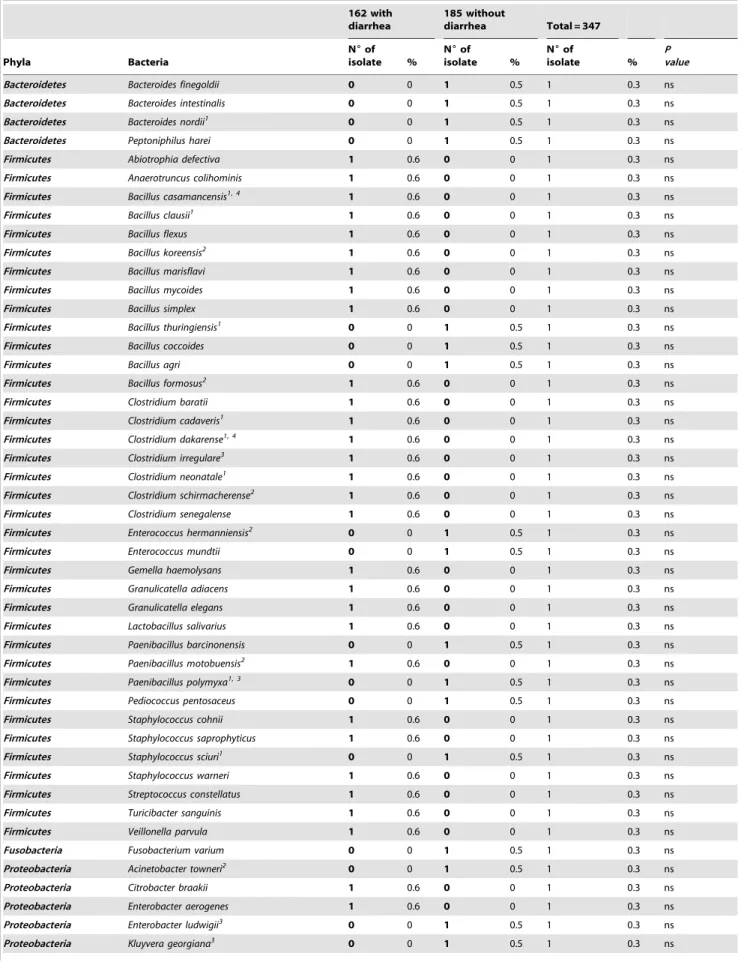
Bacterial species isolated only once. Overall, 51 bacterial species were identified only once (Table 4). Among them, 5 different bacterial species from the phylumActinobacteria, 5 from the generaBacillus,4 from the generaClostridium, and 2 from the genera Shigella were detected among patients with diarrhea. In contrast, several species of the generaBacteroides(4) andEnterococcus (2) were detected only among patients without diarrhea.
Bacterial identification depending of the age range. The isolates obtained from people with and without diarrhea depend-ing of the age range (less than 5 years, from 5 to 20 years, and more than 20 years) were compared. Only significant differences are presented (Table S1). For children from 0 to 5 year-old, 2 species of the generaClostridium were significantly more frequent among those without diarrhea, including 1 speciesC. glycolycum, for which the data were not significant when the entire population was analyzed. For adult of more than 20 year-old, 6 species (E. coli,E. faecium, B. uniformis, B. vulgatus, C. orbiscindens, and E. lenta), as previously observed in the entire population, were significantly more observed in people without diarrhea. In contrast, those with diarrhea had more commonlyS. aureus, F. magna, B. pumilus, as previously observed, as well as anotherBacillus species,B. subtilis. For people from 5 to 20 year-old,E. faecium,C. perfringens, andC. symbosium were significantly more detected in people without diarrhea, as observed in the entire population. Finally, the comparison of the isolates from people with diarrhea between them depending of the age range did not yield statistically significant results.
Figure 1. Isolates from individuals with diarrhea (D; top) and without diarrhea (ND; bottom).Each bacterial species corresponds to a node. The edge color represents the phylum (blue:Firmicutes; red:Proteobacteria; green:Bacteroidetes; yellow:Actinobacteria; pink:Fusobacteria). The common and specific bacteria detected from patients with diarrhea and those without are provided.
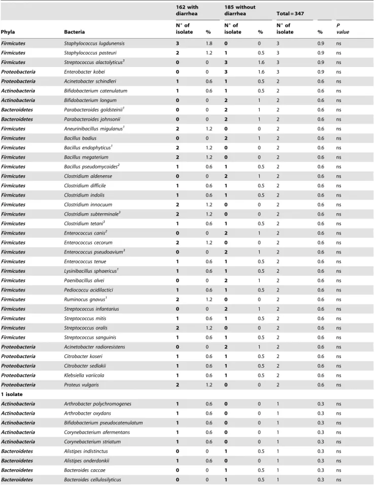
Table 4.Comparison between the prevalence of 189 bacterial species identified among 2,750 isolates from fecal samples of 162 individuals with diarrhea and 185 without diarrhea.
162 with diarrhea
185 without
diarrhea Total = 347
Phyla Bacteria
N6of
isolate %
N6of
isolate %
N6of
isolate %
P value
.100
Proteobacteria Escherichia coli 104 64.2 157 84.9 261 75.2 #1023 Firmicutes Enterococcus faecium 102 63 154 83.2 256 73.8 #1023 Firmicutes Clostridium bifermentans 60 37 99 53.5 159 45.8 0.002 Firmicutes Enterococcus faecalis 76 46.9 77 41.6 153 44 ns
Firmicutes Clostridium perfringens 53 32.7 99 53.5 152 43.8 #1023
Firmicutes Bacillus cereus 57 35.2 80 43.2 137 39.5 ns
Firmicutes Enterococcus hirae 48 29.7 58 31.3 106 30.5 ns
.10–100
Firmicutes Enterococcus gallinarum 34 21 52 28.1 86 24.8 ns
Proteobacteria Klebsiella pneumoniae 33 20.4 51 27.6 84 24.2 ns
Firmicutes Clostridium sordellii 29 17.9 48 25.9 77 22.2 ns
Firmicutes Lactococcus garvieae 23 14.2 34 18.4 57 16.4 ns
Bacteroidetes Bacteroides fragilis 20 12.5 35 18.9 55 15.8 ns
Firmicutes Enterococcus avium 20 12.3 32 17.3 52 14.5 ns
Firmicutes Clostridium orbiscindens 12 7.4 30 16.2 42 12.1 0.01 Proteobacteria Enterobacter cloacae 23 14.2 18 9.7 41 11.8 ns
Bacteroidetes Bacteroides uniformis 8 5 30 16.2 38 10.9 0.001 Firmicutes Bacillus subtilis1 22 13.6 15 8.1 37 10.7 ns Firmicutes Clostridium symbiosum 10 6.2 25 13.5 35 10 0.03 Firmicutes Enterococcus casseliflavus 10 6.2 25 13.5 35 10 0.03 Bacteroidetes Bacteroides thetaiotaomicron 9 5.5 21 11.3 30 8.6 ns
Firmicutes Streptococcus equinus 13 8 16 8.6 29 8.4 ns
Actinobacteria Collinsella aerofaciens 6 3.7 20 10.8 26 7.5 0.01 Firmicutes Bacillus pumilus1 19 11.7 7 3.8 26 7.5 0.002 Firmicutes Streptococcus lutetiensis 16 9.9 10 5.4 26 7.5 ns
Firmicutes Lysinibacillus fusiformis1 4 2.5 21 11.3 25 7.2 0.001
Bacteroidetes Bacteroides ovatus 10 6 14 7.6 24 6.9 ns
Firmicutes Streptococcus gallolyticus 16 9.9 8 4.3 24 6.9 ns
Proteobacteria Proteus mirabilis 9 5.6 15 8.1 24 6.9 ns
Actinobacteria Eggerthella lenta 4 2.5 19 10.3 23 6.6 0.004 Proteobacteria Comamonas kerstersii 8 4.9 12 6.5 20 5.8 ns
Firmicutes Clostridium butyricum 6 3.7 13 7 19 5.5 ns
Firmicutes Clostridium glycolycum 5 3 14 7.6 19 5.5 ns
Bacteroidetes Bacteroides vulgatus 2 1.2 16 8.7 18 5.2 #1023 Firmicutes Bacillus amyloliquefaciens1 10 6.2 8 4.3 18 5.2 ns
Firmicutes Clostridium tertium 11 6.8 7 3.8 18 5.2 ns
Firmicutes Clostridium cochlearium 4 2.5 12 6.5 16 4.6 ns
Bacteroidetes Parabacteroides distasonis 7 4.3 8 4.3 15 4.3 ns
Proteobacteria Morganella morganii 6 3.7 9 4.9 15 4.3 ns
Firmicutes Bacillus licheniformis1 10 6.2 3 1.6 13 3.7 0.02 Firmicutes Clostridium lituseburense1 4 2.5 9 4.9 13 3.7 ns
Firmicutes Kurthia gibsonii1 2 1.2 11 5.9 13 3.7 0.02
Firmicutes Clostridium ramosum 5 3 7 3.8 12 3.5 ns
Firmicutes Staphylococcus aureus 10 6.2 2 1 12 3.5 0.01
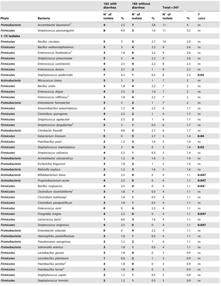
Table 4.Cont.
162 with diarrhea
185 without
diarrhea Total = 347
Phyla Bacteria
N6of
isolate %
N6of
isolate %
N6of
isolate %
P value
Proteobacteria Acinetobacter baumannii1 4 2.5 7 3.8 11 4 ns Firmicutes Streptococcus parasanguinis 8 4.9 3 1.6 11 3.2 ns
1–10 isolates
Firmicutes Bacillus circulans 5 3 5 2.7 10 2.9 ns
Firmicutes Bacillus weihenstephanensis 5 3 4 2.2 9 2.6 ns
Firmicutes Enterococcus thailandicus2 3 1.8 6 3.2 9 2.6 ns
Firmicutes Streptococcus pneumoniae 5 3 4 2.2 9 2.6 ns
Firmicutes Enterococcus canintestini 4 2.5 4 2.2 8 2.3 ns
Firmicutes Enterococcus durans 6 3.7 2 1 8 2.3 ns
Firmicutes Staphylococcus epidermidis 7 4.3 1 0.5 8 2.3 0.02
Actinobacteria Micrococcus luteus 5 3 2 1 7 2 ns
Firmicutes Bacillus siralis 3 1.8 4 2.2 7 2 ns
Firmicutes Enterococcus dispar 4 2.5 3 1.6 7 2 ns
Firmicutes Enterococcus raffinosus3 3 1.8 4 2.2 7 2 ns
Proteobacteria Enterobacter hormaechei 5 3 2 1 7 2 ns
Firmicutes Aneurinibacillus aneurinilyticus 2 1.2 4 2.2 6 1.7 ns
Firmicutes Clostridium sporogenes 4 2.5 2 1 6 1.7 ns
Firmicutes Streptococcus agalactiae 4 2.5 2 1 6 1.7 ns
Firmicutes Streptococcus dysgalactiae3 5 3 1 0.5 6 1.7 ns Proteobacteria Citrobacter freundii 1 0.6 5 2.7 6 1.7 ns
Firmicutes Eubacterium limosum 0 0 5 2.7 5 1.4 0.04
Firmicutes Paenibacillus pueri 2 1.2 3 1.6 5 1.4 ns
Firmicutes Staphylococcus haemolyticus 5 3 0 0 5 1.4 0.02 Firmicutes Streptococcus salivarius 4 2.5 1 0.5 5 1.4 ns
Proteobacteria Acinetobacter calcoaceticus 2 1.2 3 1.6 5 1.4 ns
Proteobacteria Escherichia fergusonii 3 1.8 2 1 5 1.4 ns
Proteobacteria Klebsiella oxytoca 2 1.2 3 1.6 5 1.4 ns
Actinobacteria Bifidobacterium breve 4 2.5 0 0 4 1.1 0.047 Actinobacteria Propionibacterium acnes 4 2.5 0 0 4 1.1 0.047
Firmicutes Bacillus mojavensis 4 2.5 0 0 4 1.1 0.047
Firmicutes Clostridium clostridioforme1 3 1.8 1 0.5 4 1.1 ns
Firmicutes Clostridium hathewayi 3 1.8 1 0.5 4 1.1 ns
Firmicutes Clostridium paraputrificum 3 1.8 1 0.5 4 1.1 ns
Firmicutes Enterococcus asini 0 0 4 2.2 4 1.1 ns
Firmicutes Finegoldia magna 4 2.5 0 0 4 1.1 0.047
Firmicutes Lactococcus lactis1 1 0.6 3 1.6 4 1.1 ns
Firmicutes Streptococcus anginosus 4 2.5 0 0 4 1.1 0.047 Proteobacteria Enterobacter asburiae 0 0 4 2.2 4 1.1 ns
Proteobacteria Haemophilus parainfluenzae 3 1.8 1 0.5 4 1.1 ns
Proteobacteria Pseudomonas aeruginosa 2 1.2 2 1 4 1.1 ns
Proteobacteria Salmonella enterica 3 1.8 1 0.5 4 1.1 ns
Firmicutes Lactobacillus gasseri 3 1.8 0 0 3 0.9 ns
Firmicutes Lactobacillus plantarum 1 0.6 2 1 3 0.9 ns
Firmicutes Paenibacillus jamilae2 3 1.8 0 0 3 0.9 ns
Firmicutes Paenibacillus larvae3 3 1.8 0 0 3 0.9 ns
Firmicutes Staphylococcus capitis 2 1.2 1 0.5 3 0.9 ns
Table 4.Cont.
162 with diarrhea
185 without
diarrhea Total = 347
Phyla Bacteria
N6of
isolate %
N6of
isolate %
N6of
isolate %
P value
Firmicutes Staphylococcus lugdunensis 3 1.8 0 0 3 0.9 ns
Firmicutes Staphylococcus pasteuri 2 1.2 1 0.5 3 0.9 ns
Firmicutes Streptococcus alactolyticus3 0 0 3 1.6 3 0.9 ns
Proteobacteria Enterobacter kobei 0 0 3 1.6 3 0.9 ns
Proteobacteria Acinetobacter schindleri 1 0.6 1 0.5 2 0.6 ns
Actinobacteria Bifidobacterium catenulatum 1 0.6 1 0.5 2 0.6 ns
Actinobacteria Bifidobacterium longum 0 0 2 1 2 0.6 ns
Bacteroidetes Parabacteroides goldsteinii1 0 0 2 1 2 0.6 ns Bacteroidetes Parabacteroides johnsonii 0 0 2 1 2 0.6 ns
Firmicutes Aneurinibacillus migulanus1 2 1.2 0 0 2 0.6 ns
Firmicutes Bacillus badius 0 0 2 1 2 0.6 ns
Firmicutes Bacillus endophyticus1 2 1.2 0 0 2 0.6 ns
Firmicutes Bacillus megaterium 2 1.2 0 0 2 0.6 ns
Firmicutes Bacillus pseudomycoides2 1 0.6 1 0.5 2 0.6 ns
Firmicutes Clostridium aldenense 0 0 2 1 2 0.6 ns
Firmicutes Clostridium difficile 1 0.6 1 0.5 2 0.6 ns
Firmicutes Clostridium indolis 1 0.6 1 0.5 2 0.6 ns
Firmicutes Clostridium innocuum 2 1.2 0 0 2 0.6 ns
Firmicutes Clostridium subterminale3 2 1.2 0 0 2 0.6 ns
Firmicutes Clostridium tetani3 1 0.6 1 0.5 2 0.6 ns
Firmicutes Enterococcus canis2 0 0 2 1 2 0.6 ns
Firmicutes Enterococcus cecorum 2 1.2 0 0 2 0.6 ns
Firmicutes Enterococcus pseudoavium3 0 0 2 1 2 0.6 ns
Firmicutes Enterococcus tenue 1 0.6 1 0.5 2 0.6 ns
Firmicutes Lysinibacillus sphaericus1 1 0.6 1 0.5 2 0.6 ns
Firmicutes Paenibacillus alvei 0 0 2 1 2 0.6 ns
Firmicutes Pediococcu acidilactici 1 0.6 1 0.5 2 0.6 ns
Firmicutes Ruminocus gnavus1 2 1.2 0 0 2 0.6 ns
Firmicutes Streptococcus infantarius 0 0 2 1 2 0.6 ns
Firmicutes Streptococcus mitis 1 0.6 1 0.5 2 0.6 ns
Firmicutes Streptococcus oralis 2 1.2 0 0 2 0.6 ns
Firmicutes Streptococcus sanguinis 1 0.6 1 0.5 2 0.6 ns
Proteobacteria Acinetobacter radioresistens 0 0 2 1 2 0.6 ns
Proteobacteria Citrobacter koseri 1 0.6 1 0.5 2 0.6 ns
Proteobacteria Citrobacter sedlakii 1 0.6 1 0.5 2 0.6 ns
Proteobacteria Klebsiella variicola 1 0.6 1 0.5 2 0.6 ns
Proteobacteria Proteus vulgaris 2 1.2 0 0 2 0.6 ns
1 isolate
Actinobacteria Arthrobacter polychromogenes 1 0.6 0 0 1 0.3 ns
Actinobacteria Arthrobacter oxydans 1 0.6 0 0 1 0.3 ns
Actinobacteria Bifidobacterium pseudocatenulatum 1 0.6 0 0 1 0.3 ns
Actinobacteria Corynebacterium afermentans 1 0.6 0 0 1 0.3 ns
Actinobacteria Corynebacterium striatum 1 0.6 0 0 1 0.3 ns
Bacteroidetes Alistipes indistinctus 0 0 1 0.5 1 0.3 ns
Bacteroidetes Alistipes onderdonkii 1 0.6 0 0 1 0.3 ns
Bacteroidetes Bacteroides caccae 0 0 1 0.5 1 0.3 ns
Table 4.Cont.
162 with diarrhea
185 without
diarrhea Total = 347
Phyla Bacteria
N6of
isolate %
N6of
isolate %
N6of
isolate %
P value
Bacteroidetes Bacteroides finegoldii 0 0 1 0.5 1 0.3 ns
Bacteroidetes Bacteroides intestinalis 0 0 1 0.5 1 0.3 ns
Bacteroidetes Bacteroides nordii1 0 0 1 0.5 1 0.3 ns
Bacteroidetes Peptoniphilus harei 0 0 1 0.5 1 0.3 ns
Firmicutes Abiotrophia defectiva 1 0.6 0 0 1 0.3 ns
Firmicutes Anaerotruncus colihominis 1 0.6 0 0 1 0.3 ns
Firmicutes Bacillus casamancensis1,4 1 0.6 0 0 1 0.3 ns
Firmicutes Bacillus clausii1 1 0.6 0 0 1 0.3 ns
Firmicutes Bacillus flexus 1 0.6 0 0 1 0.3 ns
Firmicutes Bacillus koreensis2 1 0.6 0 0 1 0.3 ns
Firmicutes Bacillus marisflavi 1 0.6 0 0 1 0.3 ns
Firmicutes Bacillus mycoides 1 0.6 0 0 1 0.3 ns
Firmicutes Bacillus simplex 1 0.6 0 0 1 0.3 ns
Firmicutes Bacillus thuringiensis1 0 0 1 0.5 1 0.3 ns
Firmicutes Bacillus coccoides 0 0 1 0.5 1 0.3 ns
Firmicutes Bacillus agri 0 0 1 0.5 1 0.3 ns
Firmicutes Bacillus formosus2 1 0.6 0 0 1 0.3 ns
Firmicutes Clostridium baratii 1 0.6 0 0 1 0.3 ns
Firmicutes Clostridium cadaveris1 1 0.6 0 0 1 0.3 ns
Firmicutes Clostridium dakarense1,4 1 0.6 0 0 1 0.3 ns
Firmicutes Clostridium irregulare3 1 0.6 0 0 1 0.3 ns
Firmicutes Clostridium neonatale1 1 0.6 0 0 1 0.3 ns
Firmicutes Clostridium schirmacherense2 1 0.6 0 0 1 0.3 ns
Firmicutes Clostridium senegalense 1 0.6 0 0 1 0.3 ns
Firmicutes Enterococcus hermanniensis2 0 0 1 0.5 1 0.3 ns
Firmicutes Enterococcus mundtii 0 0 1 0.5 1 0.3 ns
Firmicutes Gemella haemolysans 1 0.6 0 0 1 0.3 ns
Firmicutes Granulicatella adiacens 1 0.6 0 0 1 0.3 ns
Firmicutes Granulicatella elegans 1 0.6 0 0 1 0.3 ns
Firmicutes Lactobacillus salivarius 1 0.6 0 0 1 0.3 ns
Firmicutes Paenibacillus barcinonensis 0 0 1 0.5 1 0.3 ns
Firmicutes Paenibacillus motobuensis2 1 0.6 0 0 1 0.3 ns Firmicutes Paenibacillus polymyxa1,3 0 0 1 0.5 1 0.3 ns
Firmicutes Pediococcus pentosaceus 0 0 1 0.5 1 0.3 ns
Firmicutes Staphylococcus cohnii 1 0.6 0 0 1 0.3 ns
Firmicutes Staphylococcus saprophyticus 1 0.6 0 0 1 0.3 ns
Firmicutes Staphylococcus sciuri1 0 0 1 0.5 1 0.3 ns
Firmicutes Staphylococcus warneri 1 0.6 0 0 1 0.3 ns
Firmicutes Streptococcus constellatus 1 0.6 0 0 1 0.3 ns
Firmicutes Turicibacter sanguinis 1 0.6 0 0 1 0.3 ns
Firmicutes Veillonella parvula 1 0.6 0 0 1 0.3 ns
Fusobacteria Fusobacterium varium 0 0 1 0.5 1 0.3 ns
Proteobacteria Acinetobacter towneri2 0 0 1 0.5 1 0.3 ns
Proteobacteria Citrobacter braakii 1 0.6 0 0 1 0.3 ns
Proteobacteria Enterobacter aerogenes 1 0.6 0 0 1 0.3 ns
Proteobacteria Enterobacter ludwigii3 0 0 1 0.5 1 0.3 ns
Viral and Parasites Identification
Analyses in Dakar have allowed the detection of several viruses and parasites in feces. Ten rotaviruses (6.2%), 4 adenoviruses (2.7%), and 7 co-infections with both rotaviruses and adenoviruses (4.3%) were detected among diarrheic patients. SixteenEnterobius vermicularis(9.9%), 6Trichomonas intestinalis(3.7%), 5Cryptosporidium spp. (3%), 5 cysts of Entamoeba spp. (3%), 4 Schistosoma mansoni (2.7%), and 1Microsporidiumspp. (0.6%) were detected among 37 diarrheic people. Thirty-sixAscaris lumbricoides(among 24 diarrheic people and 12 without diarrhea), 8 Giardia duodenalis (among 6 diarrheic people and 2 without diarrhea), and 4Trichuris trichiura (among 1 diarrheic people and 3 without) were detected. Finally, 2 co-infections (Cryptosporidium spp. with Ascaris lumbricoides and Microsporidium spp. with Ascaris lumbricoides) were detected in patients with diarrhea and 1 co-infection (Trichuris trichiura with Ascaris lumbricoides) among a people without diarrhea.
Discussion
MALDI-TOF mass spectrometry coupled with culturomics has allowed for the identification of a large collection of bacterial species from specimens from Senegal and a preliminary compar-ison between the bacterial microbiota of people with and without diarrhea. This technique has allowed for the accurate identifica-tion of a large panel of anaerobes that are usually poorly identified by current phenotypic methods, which lack specificity and result in ambiguous or even erroneous identification [21,22]. For several bacterial species, their identification by MALDI-TOF failed because either the corresponding species missed in the database or either the number of spectra of the species was insufficient. Indeed, the continuous increases of the entries in database with the addition of our new spectra solved these problems and improved bacterial identification. In addition, the use of MALDI-TOF mass spectrometry detects the presence of previously rare bacteria that were difficult to identify using phenotypic methods [6,23–27].
Overall, the percentage of isolates from Senegal that were correctly identified at the genus and species level by mass spectrometry (98.2%) is nearly the same than the percentage (95.4%) observed in the first large scale experiment that used mass spectrometry in Marseille, France [1]. Both studies were performed using the same database. This study has allowed us
to test a large collection of isolated strains from Senegalese people. Only 3 bacterial species,Clostridium senegalense,Bacillus casamancensis, andClostridium dakarense, have been currently identified in Senegal. This confirms the high potential for culturomics approaches to result in the detection of new bacterial species associated with humans [28–39]. The increases in the database by the addition of more bacteria have allowed for improved bacterial identification by MALDI-TOF mass spectrometry. Thus, the current database seems accurate for the identification of bacteria in Senegal. This work allowed for the identification of 166 bacterial species already found in the human gut, 11 species previously detected in humans but not in the gut, 10 species detected in humans for the first time, and 2 unknown species.
The composition of the gut microbiota is complex [40]. A recent culturomics experiment using many culture conditions was performed on fecal samples from 2 healthy Senegalese individuals, 1 obese person, 1 person with resistant tuberculosis, and a patient with anorexia nervosa. This allowed the identification of 99, 219, 192, 39, and 133 different bacterial species per fecal sample, respectively [12–14]. Although the storage and transport condi-tions of the fecal samples were not optimal and many fewer culture conditions were used, this study demonstrates a modification of gut microbiota with several significant differences between the bacterial species identified among people with diarrhea and those without diarrhea. In people with diarrhea, major commensal bacterial species such as E. coli were significantly decreased, as were several Enterococcus spp. (E. faecium and E. casseliflavus); anaerobes, such as Bacteroides spp. (B. uniformis and B. vulgatus); andClostridiumspp. (C. bifermentans,C. orbiscindens,C. perfringens,C. symbosium,and C. glycolycum). Conversely, severalBacillus spp. (B. licheniformis, B. mojavensis, B. pumilus, and B. subtilis) were signifi-cantly more frequent among patients with diarrhea. In addition, the diversity ofBacillusspecies identified in patients with diarrhea is higher (19) than among those without diarrhea (11), but this difference was not significant (P= 0.055). Overall, a decrease of anaerobes in the gut flora, particularly Bacteroidetes, has already been reported during gastroenteritis using both culture and molecular methods [41,42]. Our data shows the occurrence of an imbalance of natural bacterial flora among patients with diarrhea.
Table 4.Cont.
162 with diarrhea
185 without
diarrhea Total = 347
Phyla Bacteria
N6of
isolate %
N6of
isolate %
N6of
isolate %
P value
Proteobacteria Neisseria flavescens 1 0.6 0 0 1 0.3 ns
Proteobacteria Proteus penneri 1 0.6 0 0 1 0.3 ns
Proteobacteria Pseudomonas luteola 1 0.6 0 0 1 0.3 ns
Proteobacteria Pseudomonas putida 0 0 1 0.5 1 0.3 ns
Proteobacteria Shigella boydii1 1 0.6 0 0 1 0.3 ns
Proteobacteria Shigella sonnei1 1 0.6 0 0 1 0.3 ns
P value is specified only when a significant difference was observed. Nuof isolate:Number of isolate; %: Percentage;ns: non significant value.
1Strains identified using a molecular analysis; 2Bacterial species that were never isolated in humans; 3Bacterial species isolated in humans but not in the human gut; 4New bacterial species.
For a long time, the high cost of a MALDI-TOF apparatus and the lack of specific reagent have limited the development of this technology. The expense of using MALDI-TOF mass spectrom-etry for identification now lies in the acquisition of a machine, which costs betweenJ100,000 andJ200,000 [21]. Recently, the cost per sample was calculated to be 1.35 euros for the Microflex system from Bruker [21]. The time required for bacterial identification has been improved to 1 minute 46 seconds using the Microflex system. In addition, MALDI-TOF mass spectrom-etry also has the potential for identification at the serotype level and antibiotic resistance profiling within minutes [43–51]. Thus, the rapid and accurate identification of routinely encountered bacterial species can be performed to improve the care of patients with infectious diseases. This technique will be a promising alternative for bacterial identification in Africa. Indeed, the main cost is based on the investment of purchasing the apparatus. The used reagents do not expire, do not require specific storage conditions, and are not expensive [1,6]. Finally, the protocol that involves directly deposited bacterial colonies onto the MALDI-TOF mass spectrometry plate regardless of the agar-based medium and without any subculture or colony preparation is very simple and can be widely used.
Overall, MALDI-TOF mass spectrometry is a potentially powerful tool for routine bacterial identification in Africa, as it
allows for the rapid identification of bacterial species, including those that are rare and difficult to identify using phenotypic methods. The next step will be to install MALDI-TOF mass spectrometers in African hospitals.
Supporting Information
Table S1 Summary of the significant differences ob-served between the prevalence of bacterial species from fecal samples of 347 individuals with and without diarrhea depending of the age range.
(DOCX)
Acknowledgments
We thank Denis Piak for his help.
Author Contributions
Conceived and designed the experiments: AGS FF DR. Performed the experiments: BSB JCL PH GD. Analyzed the data: CM HR FF DR. Contributed reagents/materials/analysis tools: BSB GD PH HR FF. Wrote the paper: FF JCL DR.
References
1. Seng P, Drancourt M, Gouriet F, La Scola B, Fournier PE, et al (2009) Ongoing revolution in bacteriology: routine identification of bacteria by matrix-assisted laser desorption ionization time-of-flight mass spectrometry. Clin Infect Dis 49: 543–551.
2. Patel R (2013) Matrix-Assisted Laser Desorption Ionization-Time of Flight Mass Spectrometry in Clinical Microbiology. Clin Infect Dis 57: 564–572. 3. van Veen SQ, Claas EC, Kuijper EJ (2010) High-throughput identification of
bacteria and yeast by matrix-assisted laser desorption ionization-time of flight mass spectrometry in conventional medical microbiology laboratories. J Clin Microbiol 48: 900–907.
4. Cherkaoui A, Hibbs J, Emonet S, Tangomo M, Girard M, et al (2010) Comparison of two matrix-assisted laser desorption ionization-time of flight mass spectrometry methods with conventional phenotypic identification for routine identification of bacteria to the species level. J Clin Microbiol 48: 1169–1175. 5. Stevenson LG, Drake SK, Shea YR, Zelazny AM, Murray PR (2010) Evaluation
of matrix-assisted laser desorption ionization-time of flight mass spectrometry for identification of clinically important yeast species. J Clin Microbiol 48: 3482– 3486.
6. Seng P, Abat C, Rolain JM, Colson P, Gouriet F, et al (2013) Emergence of rare pathogenic bacteria in a clinical microbiology laboratory: impact of MALDI-TOF mass spectrometry. J Clin Microbiol 51: 2182–2194.
7. Bizzini A, Durussel C, Bille J, Greub G, Prod’hom G (2010) Performance of matrix-assisted laser desorption ionization-time of flight mass spectrometry for identification of bacterial strains routinely isolated in a clinical microbiology laboratory. J Clin Microbiol 48: 1549–1554.
8. Lagier JC, Million M, Hugon P, Armougom F, Raoult D (2012) Human gut microbiota: repertoire and variations. Front Cell Infect Microbiol 2: 136. 9. Lozupone CA, Stombaugh JI, Gordon JI, Jansson JK, Knight R (2012)
Diversity, stability and resilience of the human gut microbiota. Nature 489: 220– 230.
10. De Filippo C, Cavalieri D, Di Paola M, Ramazzotti M, Poullet JB, et al (2010) Impact of diet in shaping gut microbiota revealed by a comparative study in children from Europe and rural Africa. Proc Natl Acad Sci U S A 107: 14691– 14696.
11. Lee S, Sung J, Lee J, Ko G (2011) Comparison of the gut microbiotas of healthy adult twins living in South Korea and the United States. Appl Environ Microbiol 77: 7433–7437.
12. Lagier JC, Armougom F, Million M, Hugon P, Pagnier I, et al (2012) Microbial culturomics: paradigm shift in the human gut microbiome study. Clin Microbiol Infect 18: 1185–1193.
13. Dubourg G, Lagier JC, Armougom F, Robert C, Hamad I, et al (2013) The gut microbiota of a patient with resistant tuberculosis is more comprehensively
studied by culturomics than by metagenomics. Eur J Clin Microbiol Infect Dis 32: 637–645.
14. Pfleiderer A, Lagier JC, Armougom F, Robert C, Vialettes B, et al (2013) Culturomics identified 11 new bacterial species from a single anorexia nervosa stool sample. Eur J Clin Microbiol Infect Dis 32: 1471–1481.
15. Hartemink R, Domenech VR, Rombouts FM (1997) LAMVAB–A new selective medium for the isolation of lactobacilli from faeces. J Microbiol Methods 29: 77– 84.
16. Drancourt M, Berger P, Raoult D (2004) Systematic 16S rRNA gene sequencing of atypical clinical isolates indentified 27 new bacterial species associated with humans. J Clin Microbiol 42: 2197–2202.
17. Stackebrandt E, Frederiksen W, Garrity GM, Grimont PA, Kampfer P, et al (2002) Report of the ad hoc committee for the re-evaluation of the species definition in bacteriology. Int J Syst Evol Microbiol 52: 1043–1047. 18. Weisburg WG, Barns SM, Pelletier DA, Lane DJ (1991) 16S ribosomal DNA
amplification for phylogenetic study. J Bacteriol 173: 697–703.
19. Shannon P, Markiel A, Ozier O, Baliga NS, Wang JT, et al (2003) Cytoscape: a software environment for integrated models of biomolecular interaction networks. Genome Res 13: 2498–2504. 1.
20. Lo CI, Mishra AK, Padhmanabhan R, Samb-Ba B, Gassama-Sow A, et al (2013) Non-contiguous finished genome sequence and description ofClostridium
dakarense sp. nov. Stand Genomic Sci 9: 14–27.
21. Seng P, Rolain JM, Fournier PE, La Scola B, Drancourt M, et al (2010) MALDI-TOF-mass spectrometry applications in clinical microbiology. Future Microbiol 5: 1733–1754.
22. La Scola B, Fournier PE, Raoult D (2011) Burden of emerging anaerobes in the MALDI-TOF and 16S rRNA gene sequencing era. Anaerobe 17: 106–112. 23. Tani A, Sahin N, Matsuyama Y, Enomoto T, Nishimura N, et al (2012)
High-throughput identification and screening of novelMethylobacteriumspecies using whole-cell MALDI-TOF/MS analysis. PLoS ONE 7: e40784.
24. Chan JF, Lau SK, Curreem SO, To KK, Leung SS, et al (2012) First report of spontaneous intrapartum Atopobium vaginae bacteremia. J Clin Microbiol 50: 2525–2528.
25. Gouriet F, Million M, Henri M, Fournier PE, Raoult D (2012)Lactobacillus rhamnosusbacteremia: an emerging clinical entity. Eur J Clin Microbiol Infect Dis 31: 2469–2480.
26. Dridi B, Raoult D, Drancourt M (2012) Matrix-assisted laser desorption/ ionization time-of-flight mass spectrometry identification of Archaea: towards the universal identification of living organisms. APMIS 120: 85–91. 27. Fernandez-Olmos A, Morosini MI, Lamas A, Garcia-Castillo M, Garcia-Garcia
L, et al (2012) Clinical and microbiological features of a cystic fibrosis patient chronically colonized withPandoraea sputorumidentified by combining 16S rRNA
Figure 2. Bacterial species for which significant differences have been observed between individuals with diarrhea and those without diarrhea.
sequencing and matrix-assisted laser desorption ionization-time of flight mass spectrometry. J Clin Microbiol 50: 1096–1098.
28. Lagier JC, El Karkouri K, Nguyen TT, Armougom F, Raoult D, et al (2012) Non-contiguous finished genome sequence and description of Anaerococcus senegalensissp. nov. Stand Genomic Sci 6: 116–125.
29. Kokcha S, Mishra AK, Lagier JC, Million M, Leroy Q, et al (2012) Non contiguous-finished genome sequence and description ofBacillus timonensis sp. nov. Stand Genomic Sci 6: 346–355.
30. Mishra AK, Lagier JC, Rivet R, Raoult D, Fournier PE (2012) Non-contiguous finished genome sequence and description ofPaenibacillus senegalensissp. nov. Stand Genomic Sci 7: 70–81.
31. Mishra AK, Lagier JC, Robert C, Raoult D, Fournier PE (2012) Non contiguous-finished genome sequence and description ofPeptoniphilus timonensis
sp. nov. Stand Genomic Sci 7: 1–11.
32. Kokcha S, Ramasamy D, Lagier JC, Robert C, Raoult D, et al (2012) Non-contiguous finished genome sequence and description ofBrevibacterium senegalense
sp. nov. Stand Genomic Sci 7: 233–245.
33. Lagier JC, Ramasamy D, Rivet R, Raoult D, Fournier PE (2012) Non contiguous-finished genome sequence and description ofCellulomonas massiliensis
sp. nov. Stand Genomic Sci 7: 258–270.
34. Mishra AK, Lagier JC, Robert C, Raoult D, Fournier PE (2012) Non-contiguous finished genome sequence and description ofClostridium senegalensesp. nov. Stand Genomic Sci 6: 386–395.
35. Ramasamy D, Kokcha S, Lagier JC, Nguyen TT, Raoult D, et al (2012) Genome sequence and description ofAeromicrobium massiliensesp. nov. Stand Genomic Sci 7: 246–257.
36. Lagier JC, Armougom F, Mishra AK, Nguyen TT, Raoult D, et al (2012) Non-contiguous finished genome sequence and description ofAlistipes timonensissp. nov. Stand Genomic Sci 6: 315–324.
37. Kokcha S, Mishra AK, Lagier JC, Million M, Leroy Q, et al (2012) Non contiguous-finished genome sequence and description ofBacillus timonensissp. nov. Stand Genomic Sci 6: 346–355.
38. Mishra AK, Hugon P, Robert C, Raoult D, Fournier PE (2012) Non contiguous-finished genome sequence and description ofPeptoniphilus grossensissp. nov. Stand Genomic Sci 7: 320–330.
39. Lagier JC, Gimenez G, Robert C, Raoult D, Fournier PE (2012) Non-contiguous finished genome sequence and description ofHerbaspirillum massiliense
sp. nov. Stand Genomic Sci 7: 200–209.
40. Gill SR, Pop M, Deboy RT, Eckburg PB, Turnbaugh PJ, et al (2006) Metagenomic analysis of the human distal gut microbiome. Science 312: 1355– 1359.
41. Albert MJ, Bhat P, Rajan D, Maiya PP, Pereira SM, et al (1978) Faecal flora of South Indian infants and young children in health and with acute gastroenteritis. J Med Microbiol 11: 137–143.
42. Balamurugan R, Janardhan HP, George S, Raghava MV, Muliyil J, et al (2008) Molecular studies of fecal anaerobic commensal bacteria in acute diarrhea in children. J Pediatr Gastroenterol Nutr 46: 514–519.
43. Hrabak J, Walkova R, Studentova V, Chudackova E, Bergerova T (2011) Carbapenemase activity detection by matrix-assisted laser desorption ionization-time of flight mass spectrometry. J Clin Microbiol 49: 3222–3227.
44. Kempf M, Bakour S, Flaudrops C, Berrazeg M, Brunel JM, et al (2012) Rapid detection of carbapenem resistance inAcinetobacter baumanniiusing matrix-assisted laser desorption ionization-time of flight mass spectrometry. PLoS ONE 7: e31676.
45. Edwards-Jones V, Claydon MA, Evason DJ, Walker J, Fox AJ, et al (2000) Rapid discrimination between methicillin-sensitive and methicillin-resistant
Staphylococcus aureus by intact cell mass spectrometry. J Med Microbiol 49: 295–300.
46. Walker J, Fox AJ, Edwards-Jones V, Gordon DB (2002) Intact cell mass spectrometry (ICMS) used to type methicillin-resistantStaphylococcus aureus: media effects and inter-laboratory reproducibility. J Microbiol Methods 48: 117–126. 47. Jackson KA, Edwards-Jones V, Sutton CW, Fox AJ (2005) Optimisation of
intact cell MALDI method for fingerprinting of methicillin-resistantStaphylococcus aureus. J Microbiol Methods 62: 273–284.
48. Du Z, Yang R, Guo Z, Song Y, Wang J (2002) Identification ofStaphylococcus aureusand determination of its methicillin resistance by matrix-assisted laser desorption/ionization time-of-flight mass spectrometry. Anal Chem 74: 5487– 5491.
49. Majcherczyk PA, McKenna T, Moreillon P, Vaudaux P (2006) The discriminatory power of MALDI-TOF mass spectrometry to differentiate between isogenic teicoplanin-susceptible and teicoplanin-resistant strains of methicillin-resistantStaphylococcus aureus. FEMS Microbiol Lett 255: 233–239. 50. Camara JE, Hays FA (2007) Discrimination between wild-type and
ampicillin-resistant Escherichia coliby matrix-assisted laser desorption/ionization time-of-flight mass spectrometry. Anal Bioanal Chem 389: 1633–1638.