Fibrosis Isolate CHA Shapes Its Virulence
Khady Mayebine Sall1,2,3,4, Maria Guillermina Casabona1,2,3,4, Christophe Bordi5, Philippe Huber1,2,3,4, Sophie de Bentzmann5, Ina Attre´e1,2,3,4, Sylvie Elsen1,2,3,4*
1INSERM, UMR-S 1036, Biology of Cancer and Infection, Grenoble, France,2CNRS, ERL 5261, Bacterial Pathogenesis and Cellular Responses, Grenoble, France,3 UJF-Grenoble 1, UJF-Grenoble, France,4CEA, DSV/iRTSV, Grenoble, France,5Laboratoire d’Inge´nierie des Syste`mes Macromole´culaires, UMR 7255 CNRS - Aix Marseille University, Marseille, France
Abstract
Pseudomonas aeruginosa, a human opportunistic pathogen, is capable of provoking acute and chronic infections that are associated with defined sets of virulence factors. During chronic infections, the bacterium accumulates mutations that silence some and activate other genes. Here we show that the cystic fibrosis isolate CHA exhibits a unique virulence phenotype featuring a mucoid morphology, an active Type III Secretion System (T3SS, hallmark of acute infections), and no Type VI Secretion System (H1-T6SS). This virulence profile is due to a 426 bp deletion in the 39 end of the gacSgene encoding an essential regulatory protein. The absence of GacS disturbs the Gac/Rsm pathway leading to depletion of the small regulatory RNAs RsmY/RsmZ and, in consequence, to expression of T3SS, while switching off the expression of H1-T6SS and Pel polysaccharides. The CHA isolate also exhibits full ability to swim and twitch, due to active flagellum and Type IVa pili. Thus, unlike the classical scheme of balance between virulence factors, clinical strains may adapt to a local niche by expressing both alginate exopolysaccharide, a hallmark of membrane stress that protects from antibiotic action, host defences and phagocytosis, and efficient T3S machinery that is considered as an aggressive virulence factor.
Citation:Sall KM, Casabona MG, Bordi C, Huber P, de Bentzmann S, et al. (2014) AgacSDeletion inPseudomonas aeruginosaCystic Fibrosis Isolate CHA Shapes Its Virulence. PLoS ONE 9(4): e95936. doi:10.1371/journal.pone.0095936
Editor:Min Wu, University of North Dakota, United States of America
ReceivedJanuary 9, 2014;AcceptedMarch 30, 2014;PublishedApril 29, 2014
Copyright:ß2014 Sall et al. This is an open-access article distributed under the terms of the Creative Commons Attribution License, which permits unrestricted use, distribution, and reproduction in any medium, provided the original author and source are credited.
Funding:KM Sall is a PhD student supported by the University of Grenoble. This work was in part supported by grants from Fondation innovations en infectiologie (FINOVI), Alliance nationale pour les sciences de la vie et de la sante´ (AVIESAN), Commissariat a` l’e´nergie atomique et aux e´nergies alternatives (CEA), Centre national de la recherche scientifique (CNRS) and Institut national de la sante´ et de la recherche me´dicale (INSERM). The funders had no role in study design, data collection and analysis, decision to publish, or preparation of the manuscript.
Competing Interests:The authors have declared that no competing interests exist.
* E-mail: sylvie.elsen@cea.fr
Introduction
Pseudomonas aeruginosa is an opportunistic Gram negative bacterium able to trigger either severe acute or chronic human infections, depending on the environmental signals it encounters. The persistence of the bacterium during decades in the lungs of individuals with cystic fibrosis (CF) is associated with massive and inefficient inflammation contributing to airway epithelium de-struction, decline of lung function and death due to respiratory failure [1]. During chronic infection, P. aeruginosahas to survive and adapt to the stressful environment encountered in the CF lungs where it is continuously exposed to antibiotics, oxidative and osmotic stresses as well as active host immune system. The CF respiratory mucus has been shown to directly impact bacterial gene transcription [2,3]. A CF mucoid strain was reported to trigger, in response to the CF niche, the synthesis of enzymes protecting the bacteria against oxidative stress and the activation of genes encoding the HSI-I Type VI Secretion System (H1-T6SS), known to play a role in bacterial competition [4,5,6]. In addition, the alginate production was unexpectedly repressed, and the expression of two small RNAs (PA2G_05393.1 and PA2G_03487.1) with putative regulatory roles was observed, pointing out the major effect of contact with CF mucus on bacterial physiology [3].
The CHA strain is a good example ofP. aeruginosamultifaceted adaptation in CF disease. It was originally isolated from a CF patient [14], 4 years after the first airways colonization by P. aeruginosa, and is thus not considered as an early infecting strain. This is an O6 serotype strain belonging to clone J, one common clonal group disseminated worldwide [15]. CHA is mucoid [16], a hallmark of chronicity, but at the same time, it is a highly efficient T3SS effector-producer fitting with acute infectious status [17,18,19,20]. Very recently, CHA has been included in the internationalP. aeruginosa reference panel, stressing the relevance to study its pathogenicity [21].
Using phenotypic and complementation experiments in parallel to genome analysis, we report in the present study that virulence properties of CHA result from an intrinsic genetic deletion leading to the absence of the histidine kinase (HK) GacS. GacS/GacA is a two-component regulatory system (TCS) that controls transcrip-tion of two small regulatory RNAs (sRNAs), RsmY and RsmZ. These two sRNAs prevent RsmA binding to its mRNA targets and consequently modulate, directly or indirectly, approximately 500 genes belonging to the RsmA regulon [22,23,24]. Activity of GacS is regulated in an opposite manner by two inner membrane sensors, RetS and LadS, in response to still unidentified stimuli [25,26]. The RetS/LadS/Gac/Rsm cascade is known to be a master regulator of the virulence factors ofP. aeruginosa, controlling the switch in their expression during the acute/chronic phase transition (reviewed in [27]). Hence, as illustrated here, mutations in this regulatory pathway profoundly impact the global virulence traits.
Materials and Methods
Bacterial strains
The P. aeruginosa and Escherichia coli strains, as well as the plasmids used in this study, are listed in Table 1. The genotype of the CHA strain was determined using the ArrayTube genotyping method [15]. Cells were grown aerobically in Luria Bertani (LB) medium at 37uC with agitation.P.aeruginosawas also cultured on Pseudomonas Isolation Agar plates (PIA; Difco). Antibiotics were added at the following concentrations (inmg/ml): 100 (ampicillin), 25 (gentamycin), 25 (kanamycin) and 10 (tetracyclin) for E. coli, 500 (CHA) or 200 (PAO1) (carbenicillin), 400 (CHA) and 200 (PAO1) (gentamycin) and 200 (tetracycline) forP. aeruginosa.
Animals
All protocols in this study were conducted in strict accordance with the French guidelines for the care and use of laboratory animals. The protocol for mouse infection was approved by the animal research committee of the institute (CETEA: Comite´ d’ E´ Thique en Expe´rimentation Animale). Pathogen-free BALB/c male mice (8-10 weeks) were obtained from Harlan Laboratories and housed in the CEA animal care facilities.
Genetic constructions
Deletion ofrsmAand retS– Fused uspstream and downstream flanking regions of rsmA and retS were amplified by Splicing by Overlap Extension-Polymerase Chain Reaction (SOE-PCR) pro-cedure using appropriate primer pairs (Table S1). The resulting fragments of 846 bp and 834 bp, respectively, were cloned into pCR-Blunt II-TOPO vector, sequenced and then subcloned into the SmaI site of the suicide plasmid pEX100-T. The resulting pEXDRsmA and pEXDRetS plasmids carry the counter-selectable sacB marker from Bacillus subtilis, which confers sensitivity to sucrose. Both plasmids were mobilized intoP. aeruginosastrain by triparental mating, using the conjugative properties of the helper
plasmid pRK2013. Co-integration events were selected on PIA plates containing carbenicillin. Single colonies were then plated on PIA medium containing 5% (w/v) sucrose to select for the loss of plasmid: the resulting sucrose-resistant strains were checked for carbenicillin sensitivity and forrsmA orretS(wild-type or deleted gene) genotype by PCR.
Fusion of VSV-G epitope to GacS - The VSV-G-Tag-coding sequence was fused to the 39end of chromosomal PAO1gacSor to that ofgacS*, the CHA naturally truncatedgacSgene, using SOE-PCR strategy. Upstream and downstream flanking regions ofgacS and gacS* were PCR amplified using PAO1 or CHA genomic DNA, respectively, and appropriate primer pairs (see Table S1). The resulting fragments of 813 bp and 752 bp, respectively, were cloned into pCR-Blunt II-TOPO vector, sequenced, then inserted into theSmaI site of pEX100-T, leading to pEX-GacS-VSV-G and pEX-GacS*-VSV-G plasmids. Allelic replacement was performed as described above. The presence of VSV-G-encoding sequences in the PAO1 and CHA genomes was assessed by PCR and confirmed by sequencing of PCR fragments.
Complementation - The rsmA and gacS genes were PCR amplified from PAO1 genomic DNA using appropriate primer pairs (Table S1). The PCR products were cloned into pCR-Blunt II-TOPO and sequenced. The 0.23-kb XbaI-HindIII restriction fragment containingrsmAwas cloned in pVLT31 plasmid under control of the IPTG-inducibleptacpromoter. The 2.85-kbSma I-XbaI restriction fragment containinggacSwas cloned into pJN105 plasmid under the arabinose-induciblepBADpromoter. The two expression vectors were introduced inP. aeruginosaby transforma-tion [39].
An integrative plasmid was also constructed to complementgacS mutation with one copy of the gene driven by its own promoter. A region comprising the 245 bp sequence upstream fromgacSand the entire gene was PCR amplified from PAO1 genomic DNA using appropriate primers (Table S1). After sequencing, theSma I-HindIII restriction fragment was cloned into pUC18-miniTn7T-Gm-LacZ20. The resulting plasmid was electroporated into the CHA strain along with the pTNS2, as described [34], leading to the CHA-GacS strain.
b-Galactosidase assays
b-Galactosidase activity was assayed as already described [40]. T3SS-dependent cytotoxicity assay
Bacteria were grown in LB to an A600 1.0 and added to the macrophage cell line J774 (J774A.1, catalog No. TIB-67, ATCC) at a multiplicity of infection (MOI) of 5. Cell death was assessed at 3 h post-infection by using a cytotoxicity detection kit (lactate dehydrogenase LDH; Roche) as described [17].
Sample preparation, antibodies and immunoblot analysis H1-T6SS Hcp1 (for haemolysin-coregulated protein 1) produc-tion and secreproduc-tion were assessed as described [41]. For GacS-VSV-G and GacS*-GacS-VSV-G analysis, 100ml of cultures atA600of 2.8-3.0 were harvested and analyzed as the total bacterial fraction. Membrane fractions were recovered after lysozyme treatment, sonication and ultracentrifugation. The samples were submitted to SDS-PAGE and immunoblotting analysis.
anti-guinea pig-HRP, anti-mouse-HRP) were used as recommended by the manufacturers.
Motility assays
Motilities were assayed on media as described [42]. All plates were inoculated with bacteria from overnight cultures on LB agar using sterile toothpicks.
Air-liquid biofilm
TheP. aeruginosaadherence assay was performed in individual glass tubes as previously described [43].
RT-PCR and RT-qPCR
The strains were grown at 37uC under agitation in LB, that was supplemented with 5 mM EGTA and 20 mM MgCl2(conditions ofin vitroT3SS induction) for Reverse Transcriptase (RT)-qPCR analysis. Total RNA was either extracted with the TRIzol Plus RNA Purification Kit (Invitrogen) then treated with DNase I (Amplification Grade, Invitrogen), or the PureYield RNA
Mid-iprep System (Promega), cleaned up and concentrated using the RNeasy kit (Qiagen). Yield, purity and integrity of RNA were further evaluated on Nanodrop and by agarose gel migration. Complementary DNA synthesis was carried out with SuperScript III First-Strand Synthesis System (Invitrogen) in presence or not of the SuperScript III RT enzyme to assess the absence of genomic DNA. For RT-PCR experiments, performed to assess the presence of GacS mRNA, PCR amplifications were performed using TaqPCRx DNA Polymerase (Invitrogen) and following the Basic PCR protocol described by the manufacturer. Calibration of the PCR amplification steps was done by varying number of cycles with primers targeting either gacS or 16S rRNA as a reference transcript. The RT-qPCR runs, used to quantify the effect of GacS on its target genes, were carried out on a CFX96 Real-Time System (Bio-Rad). Cycling parameters of the real time PCR were 98uC for 2 min, following by 45 cycles of 98uC for 5 s and 60uC for 10 s, ending with a melting curve from 65uC to 95uC to assess the specificity of the amplification. To determine the amplification kinetics of each product, the fluorescence derived from the incorporation of EvaGreen into the double-stranded PCR Table 1.Bacterial strains and plasmids used in this work.
Strain or plasmid Relevant characteristics Source/reference
P. aeruginosa
PAO1 Wound isolate, sequenced laboratory strain J. Mougous
TB (TBCF10839) Cystic fibrosis (CF) isolate [28]
LES400 CF epidemic strain [29]
KK1 CF isolate [30]
CF6 CF isolate [17]
PA7 Wound isolate [31]
PAK Clinical isolate D. Bradley
CHA Mucoid CF isolate [14]
CHA-GacS CHA with wild-type PAO1gacSin the chromosome This study
CHADrsmA CHA deleted of thersmAgene This study
CHADretS CHA deleted of theretSgene This study
CHADretS-GacS CHADretSwith PAO1gacSin the chromosome This study
Plasmids
pCR-Blunt II-TOPO Knr; commercial cloning vector Invitrogen
pEX100-T Apr; mobilisable vector, non-replicative in
P. aeruginosa [32]
pRK2013 Knr; helper plasmid with conjugative properties [33]
pUC18-mini-Tn7T- Gmr; translational fusion vector
lacZ20-Gm [34]
pUC18-mini-Tn7T- Promoter and entiregacSsequence of PAO1 cloned into
pGacS the integrative vector This study
pUC18-miniTn7T- pfha1-lacZtranslational fusion in
pUC18-mini-TN7T-PA0081-lacZ lacZ20-Gm [23]
pCTX-PA0081-lacZ Tcr;pfha1-lacZtranscriptional fusion in mini-CTX-lacZ [23]
pJN105 Gmr;
pBADtranscriptional fusion vector [35]
pJN-GacS pBAD-gacStranscriptional fusion in pJN105 This study
pVLT31 Tcr;plactranscriptional fusion vector [36]
pVLT-RsmA plac-rsmAtranscriptional fusion in pVLT31 This study
pMP220 Tcr;lacZtranscriptional fusion vector [37]
pMP220-rsmY-lacZ prsmY-lacZtranscriptional fusion in pMP220 [38]
pMP220-rsmZ-lacZ prsmY-lacZtranscriptional fusion in pMP220 [38]
products was measured at the end of each cycle using the SsoFast EvaGreen Supermix 2X Kit (Biorad). The results were analyzed using the Bio-Rad CFX Manager Software 3.0 (Bio-Rad). The relative mRNA quantity of each gene under GacS production as compared to absence of production was analyzed using the Relative Expression Software Tool REST2009 (Qiagen) with a pair wise fixed reallocation randomization test [44] coupled to a standard error (SE) calculated via a Taylor algorithm. The 16S rRNA was used as reference for normalization. The sequences of all primers are given in Table S1.
Results
H1-T6SS expression is off in CF strain CHA
The mucoid CF isolate CHA has been previously reported as harboring T3SS-dependent cytotoxicity in vitro [45,46]. As CF strains usually exhibit a decreased toxicity compared to strains triggering acute infection [9], we compared the virulence of CHA in mice to that of the PAO1 reference strain, which is a wound isolate. The CHA strain was clearly more virulent than the PAO1 strain in a murine acute model of lung infection (Figure S1A); this was associated with an increased dissemination of the bacteria both in blood and spleen compared to PAO1 (Figure S1B). The CHA T3SS-deficient strain exhibited a reduced toxicity in the model (data not shown) which is in agreement with a previous report pointing to T3SS being a major actor in CHA virulence [18].
We then examined the expression of H1-T6SS (HSI-I), a machinery expressed during chronic infection [4,5] and secreting bacterial toxins used for competing with other species in biofilm communities. Surprisingly, H1-T6SS production was undetectable in CHA as compared to other strains, notably CF strains TB, LES400, KK1 and CF6, or reference strains PAO1, PA7 and PAK, as assessed by the production of the Hcp1 component of the H1-T6SS apparatus (Figure 1A). The transcriptional fusion fha1-lacZ(PA0081) was expressed in CHA, albeit 2.4 fold less than in PAO1, while the corresponding translational fusion exhibited no reporter activity in CHA (Figure 1B). These results strongly suggested that CHA is incapable of H1-T6SS synthesis due to a default of mRNA stability and/or protein translation rather than an absence of HSI-I gene transcription.
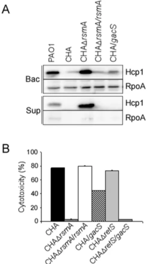
Levels of RsmA and RsmY/Z are perturbed in CHA T3SS and the H1-T6SS are known to be modulated in an opposite manner by the translational regulator RsmA, whose activity is antagonized by the two sRNAs, RsmY and RsmZ [23,25]. Thus, we examined the rsmY and rsmZ expression in CHA. ThersmYgene was not expressed in CHA, and only a slight expression of rsmZ was observed, mainly in overnight culture (Figure 2), while in PAO1, the two sRNAs were efficiently transcribed, with higher level ofrsmYexpression compared to that ofrsmZ(Figure 2), as already reported [3,24]. These data strongly suggested that, in CHA strain, amounts of RsmY and RsmZ sRNAs were not sufficient to efficiently titrate RsmA and to relieve its effect on its target mRNAs. Indeed, high amounts of Hcp1 were produced in the supernatant and whole cell extracts of the CHADrsmAmutant, a phenotype corrected by introduction of the rsmA wild-type gene in trans (Figure 3A). Additionally T3SS activity, assessed either by in vitro calcium-induced secretion of translocator proteins (data not shown) or T3SS-dependent cytotoxicity on macrophages (Figure 3B), was completely abol-ished by rsmA deletion and further re-induced by rsmA comple-mentation.
Taken together, the absence of H1-T6SS synthesis in the CHA strain, resulting from a constant translational inhibition exerted by RsmA on H1-T6SS mRNAs, and the highly active T3SS, due to RsmA-mediated positive post-transcriptional regulation of ExsA expression [47], are both related to a defective rsmY/Z gene expression.
A 426 bp chromosomal deletion led to a truncated and unstable GacS
We then investigated other regulatory players that could be involved in this deregulation by examining the recently sequenced genome of the CHA strain [48]. Indeed, a 426 bp deletion was found in the chromosome that affects the 39 end (last 146 nucleotides) ofgacSgene and the 59 end (first 278 nucleotides) of the downstream gene ldhA, encoding a lactate dehydrogenase (Figure 4A) [48]. ThisgacSdeletion generates a putative truncated protein that we named GacS*, possessing 19 unrelated residues in place of the 48 C-terminal residues of the PAO1 GacS protein (Figure 4B). The three phosphorylation sites (residues His-293, Asp-715 and His-859) are conserved (Figure 4C) but the fifth helix
Figure 1. H1-T6SS is not expressed in the CHA strain. (A)
Western blot analysis of Hcp1 from differentP. aeruginosaCF and non-CF strains. The cytoplasmic RpoA protein is used as a loading marker. (B)
b-galactosidase activities measured atA600of 1.5 from PAO1 and CHA
strains containing either a transcriptional or translationalpfha1-lacZ
of the alternative transmitter domain (H2), also called Histidine PhosphoTransfer (Hpt) domain, is missing (Figure 4D).
To test whether this deletion impacts the stability and/or functionality of GacS*, we first providedin transthe Hpt domain (H2 domain) of PAO1 GacS protein [49], which did not complement the GacS* function (not shown). Thus we assessed by immunoblot the presence of the GacS and GacS* proteins in PAO1 and CHA, respectively. To do so, we first added at the 39
end of the endogenous genes a sequence encoding the VSV-G Tag, generating GacS-VSV-G and GacS*-VSV-G proteins (see Materials and Methods). GacS*-VSV-G could not be detected in CHA, neither in the membranes nor in the whole cells, although GacS-VSV-G was clearly present in the membranes of the PAO1 strain (Figure 4E). We also ruled out the possibility that the absence of GacS* was a consequence of a transcription defect or an effect of the genomic deletion on mRNA stability by comparing the level ofgacStranscripts in both PAO1 and CHA strains using RT-PCR (Figure 4F). Altogether, these results strongly suggested that truncation of GacS C-terminus in CHA led to an unstable protein.
We next provided the PAO1gacSgene cloned under the control of the pBAD promoter to the CHA strain. Introduction of the functional gacS gene increased the amounts of rsmY and rsmZ transcripts in CHA as assessed by RT-qPCR (Figure 5), data corroborated by the restoration of rsmY/Z promoter activity (Figure 2). It also increased quantity ofPA0094transcripts,PA0094 being a part of the H1-T6SS-encoding cluster (Figure 5), as well as the synthesis of Hcp1 (Figure 3A). In addition, providinggacSgene to CHA strain significantly reduced the level ofexoStranscription, thus affecting the T3SS-dependent cytotoxicity on J774 macro-phages (Figure 3B). Furthermore, even though retS deletion in CHA did not affect T3SS activity (due to absence of a functional GacS protein), introducing gacS in the CHADretS background abolished it completely (Figure 3B). These features indicate that absence of a functional GacS may allow CHA to escape from the
negative control exerted by GacS/GacA TCS on T3SS during chronic infection.
Taken together, these results demonstrate that the truncation of GacS in CHA is responsible for defect inrsmY/Zgene expression as well as for alterations observed in T3SS and H1-T6SS synthesis.
The physiological absence of GacS shapes the phenotype of the clinical isolate
To get an overview of the impact of GacS absence in CHA, we investigated some of the major phenotypic traits of the bacterium. At first we studied its swimming and swarming motilities [42], as RsmA positively affects genes involved in formation of Type IVa pili and function of the flagellum [22]. These extracellular appendages are fundamental for motility, biofilm formation, and efficient injection of T3SS toxins into eukaryotic cells as this process requires cell adherence [50,51,52,53]. CHA clearly possesses functional flagellum and Type IVa pili as it is able to swim comparably to PAO1 and exhibits even better twitching motility (Figure 6A). As expected,rsmAmutation in CHA resulted in a decrease in the two motilities. Introduction of a functional copy of gacS in the chromosome of CHA did not substantially change its swimming motility, and only twitching was affected and down-regulated in our laboratory conditions (Figure 6A). Thus, unlike frequently reported for CF-adapted strains [7], CHA is a
Figure 2. Expression of the regulatory sRNAs is affected in the
CHA strains. The expression of prsmY::lacZ and prsmZ::lacZ was
measured in PAO1 and CHA at two differentA600. When indicated, the
pJN-GacS plasmid was introduced. The plasmid pMP220 is the promoter-freelacZplasmid used as a control. The reported values for enzyme activities are the average of at least two independent experiments performed in triplicate. The bars indicate the standard deviations. O/N: overnight culture.
doi:10.1371/journal.pone.0095936.g002
Figure 3. RsmA is responsible for absence of H1-T6SS and high
T3SS activity in CHA.(A) Hcp1 synthesis and secretion were analysed
by Western blot in the different strains as indicated. The cytoplasmic RpoA protein was used as a loading marker. Bac: bacteria, sup: supernatant. (B) T3SS-dependent cytotoxicity on J774 macrophages of wild-type, mutants and plasmid-complemented strains. Cytotoxicity was measured after 3 hours of infection and is expressed as a percentage of the total amount of LDH released from cells lysed with 1% Triton X-100. All tests were performed in triplicate.
motile CF isolate, with functional flagellum and Type IVa pili that may contribute to its cytotoxic phenotype.
Since synthesis of Pel and Psl exopolysaccharides is known to be under the control of the Gac/Rsm post-transcriptional cascade and to mediate biofilm formation [25,53], we examined the ability of CHA and different mutants to form biofilm and, besides
constitutive alginate production, synthesize Pel exopolysaccharide. CHA did not form biofilm rings in static growth conditions and inactivation ofrsmAgene in CHA led to a hyperbiofilm phenotype (Figure 6B). Introduction of a wild-typegacSgenein transrestored the ability of CHA to produce biofilm and triggered an 8.3
Figure 4. A genomic deletion in CHA affects the GacS regulator.(A) Genetic organization ofgacS(PA0928, 2778 bp) andldhA(PA0927,
990 bp) in PAO1. A 426 bp deletion in the CHA chromosome leads to 39truncatedgacS* and 59truncatedldhAgene. The two 11-tandem imperfect repeats located into the PAO1 genes are depicted as arrowheads. (B) Sequence alignment of the Hpt (Histidine phosphotransfer) and Hpt* (truncated domain) domains of PAO1 GacS and CHA GacS*, respectively. The 48 last amino acids of GacS Hpt are replaced by unrelated 19 amino acids in Hpt*, at the position indicated by brackets. The star points to the conserved phosphorylated Histidine 859 residue. (C) GacS* contains all the phosphorylation sites but lacks the C-terminal part of Hpt domain. The length in amino acids (aa) of the proteins is indicated in brackets. TM: transmembrane helix. (D) Modeling of Hpt and Hpt* domains of GacS and GacS*, respectively, using PyMol. One of the helices of the four-helix bundle motif featuring the Hpt domain, that is indicated by an arrow, is missing in the predicted Hpt* domain. (E) Western blot analysis of GacS-VSV-G and GacS-VSV-GacS*-VSV-GacS-VSV-G in whole bacteria (B) and in membrane fractions (Mb) of PAO1 and CHA. The blots were developed by antibodies specific to the VSV-G epitope and to the porin Opr86 (PA3648) (loading control) as indicated. Note the absence of GacS*-VSV-G. Localisation of a molecular weight marker is indicated at the left (in kDa). (F) RT-PCR analysis ofgacSgene transcript in PAO1 and CHA. The numbers of PCR cycles are indicated. 16S rRNA (16S) transcript levels indicate equal loading. Samples lacking SuperScript III RT enzyme during RT step (- RT) show that no DNA contamination was present.
increase in pelA transcript level (Figure 5), while it further enhanced biofilm production in a CHADretSstrain (Figure 6B).
Interestingly, different from the blue-green color of PAO1, the CHA strain exhibits a reddish color on PIA plate, likely resulting from a diffusible molecule (Figure 6C). This phenotype was not due to pyomelanin production, since inactivation of thehpdgene encoding the enzyme synthesizing HGA pyomelanin precursor [54] did not affect pigmentation (not shown). The production of this pigment is clearly under control of the Gac/Rsm pathway, since inactivation ofrsmAabolished the red pigmentation to a level
comparable to the one observed when wild-type gacS was introduced in a CHADretSbackground (Figure 6C). Complemen-tation of CHA with gacS gene affected the phenotype slightly. These data indicated that production of this red pigmentation, similar to T3SS, should escape from the control of Gac/RsmYZ during chronic infection. It is known that RsmA controls synthesis of pigments like pyocyanin [22,55], and P. aeruginosa is able to synthesize other phenazines, as two red pigments called aerugi-nosins A and B, whose biological relevance is still not known [56]. Therefore the red color may rely on specific combination and quantities of these pigments. It is important to point out that phenazines are pigments with redox properties that contribute to bacterial pathogenicity in both acute and chronic models of infection. As identity and amount of phenazines are crucial for toxicity as well as for biofilm formation ([57] and references herein), as illustrated for the precursor of pyocyanin called 5MPCA which is more efficient for yeast killing than pyocyanin itself [56], identifying the nature of the Gac/Rsm pathway-regulated pigment(s) produced by CHA could give new insights in the physiological impact of this pigmentation.
All these data indicated that a deletion in gacS gene shapes pathogenic ability of the CHA clinical isolate by blocking the Gac/ Rsm pathway. Indeed, CHA is a mucoid and motile strain expressing acute virulence factors that has lost the ability to switch from acute to chronic lifestyle.
Discussion
Particular environmental conditions found in chronic infection, such as in CF, forceP. aeruginosato adapt in order to survive. In this work, by combining genomic and extensive phenotypic analyses, we found that an intrinsic genomic deletion in the CF strain CHA equips bacteria with virulence factors that are considered to be more aggressive and are in general associated
Figure 5. Complementation of CHA withgacS impacts sRNAs
transcription as well as amount of major RsmA-target mRNAs,
as assessed by RT-qPCR analysis. Relative mRNA quantity
compares CHA/pJN-GacS to CHA/pJN105. Positive values indicate that mRNA levels are higher ingacS-complemented CHA. The p-values were calculated with REST2009 using Pair Wise Fixed Reallocation Randomi-sation Test. *: p,0.025,**: p,0.0015; ***: p,0.0002.
doi:10.1371/journal.pone.0095936.g005
Figure 6. Impact ofgacSinactivation in CHA on different virulence traits.(A) Swimming (left) and twitching (right) motilities of indicated
strains. CHA-GacS contains thegacSgene amplified from PAO1 integrated into the chromosome. The images of the plates with regard to both the extent of spreading and morphology of motile bacteria are representative. (B) Air-liquid biofilm assay of indicated strains, containing either the empty plasmid (control) or the plasmid expressing the protein of interest, as indicated. For the assays with GacS, 0.5% arabinose was added in the medium. Biofilm was stained with crystal violet after 24 h at 30uC and quantified. The means of each assay performed in quadruplicate and their standard deviation are indicated below the corresponding pictures. (C) Pigment production in CHA is strongly affected byrsmAmutation, andretSmutation whengacSis integrated into the chromosome, as observed on PIA plate.
with acute infections. Indeed, one of the key regulatory pathways, the RetS/LadS/Gac/Rsm pathway, proposed to control the transition between acute and chronic infection-associated pheno-types [25,26], is impaired by a genomic deletion that affects the gacSgene region leading to an unstable truncated form of GacS. The same signalling pathway was found affected by mutations either in ladS [58] or in retS [59] in two other clinical strains isolated from acute and chronic infection, respectively. However, inactivation of GacS, the master regulator of this pathway, impacts more strongly the read-outs than that of LadS and RetS, by blocking the switch orchestrated by the Gac/Rsm pathway.
Spontaneous mutations ingacSand/orgacArepresent the most important mechanism responsible for phenotypic variation of many pseudomonads, as observed in rhizosphere-associated Pseudomonas[60].Pseudomonassp. strain PCL1171 undergoes phase variation which is characterized by different production of secondary metabolites and exoenzymes. This phenotypic variation was reported to be caused by spontaneous and reversible mutations ingacS and gacA genes, probably randomly generated by an inefficient MutS-dependent repair of replication-related mismatches [60,61]. One of these gacS mutations, a 307 bp deletion event, implied beforehand a spontaneous mutation that created a perfect 10 bp-direct tandem repeat allowing the recombination rearrangement [61]. A similar mechanism might have occurred in CHA genome to generate the deletion between the two 11-tandem imperfect repeats (59-CGGCCTGCCA/GG) flanking the 39end of thegacSgene and the 59end of theldhAgene in PAO1 genome (Figure 5A).
NumerousP. aeruginosaphenotypic variants are retrieved from the CF lungs, as the genome constantly accumulates mutations [1,7] and even short-term growth in biofilm was shown to generate high genetic diversity inP. aeruginosa communities [62]. Interest-ingly,gacS mutants in PA14 are prone to generate stable SCVs when growing in biofilm or exposed to stresses [63] as well asin vivo[64]. When CHA was grown in static conditionsin vitro, we also observed the emergence of stable SCV-like colonies (not shown). These observations suggest that the absence of GacS might have conferred to the CHA bacterium further advantage for persistence in CF lung by providing it with the capacity to convert to stress-tolerant SCVs.
Clone CHA strains were isolated worldwide from rivers in Germany, soil in Japan, and from several CF patients in Central Europe [15]. In addition to the original strain used in this study, two other clone CHA strains, one isolated from the environment and another from the sputum of a CF patient with normal lung function, were sequenced [48]. Interestingly, only the here-characterized CHA strain harbors agacSdeletion that affects so strongly its virulence properties. Genome examination did not highlight any other genetic event that could explain such deregulation [48]. This indicates that strains of the same clonal group isolated from diverse environments do not necessarily share the same infective properties [48]. Analysis of numerous CF strains with the same T6SS (absence)/T3SS (presence) pattern of expression will help to determine whether this genetic mechanism of turning a bacterium to hypervirulence is an isolated phenom-enon or if it is a widespread alternative among CF lung isolates.
The CHA strain over-expresses alg genes and overproduces alginate [16]; this mucoid phenotype is due to one mutation in mucAleading to replacement of Ala-5 residue by Gly in the anti-sigma MucA protein [48] (Figure S2). Several reports indicated that mucA mutation negatively affects flagellum motility [11,65], leads to a reduced expression of T3SS [66] as well as of other traits of acute virulence like elastase production (T2SS) [67], suggesting that mucoidy correlates with reduced virulence [68,69]. However,
despite a mucoid phenotype, CHA is endowed with efficient T3SS and also motility appendages that may contribute to its aggressive phenotype. Two mechanisms couplingmucAmutation to reduced T3SS gene expression have been reported, one dependent and the other independent of the regulator Vfr [47,68]. Indeed, the cAMP/Vfr-dependent signalling (CVS) pathway, known to activate synthesis of the virulence factors associated with acute disease such as the T3SS, was shown to be turned-off inmucA -mutant strains by a mechanism involving AlgU and AlgR; this pathway thus activates alginate synthesis and reduces T3SS-dependent virulence [68]. However, for a still unknown reason, inactivation ofvfr does not affect T3SS in CHA (B. Toussaint, personal communication); this is surprising in regard to the high induction of CHA T3SS expression in response to calcium depletion, known to induce cAMP synthesis and, consequently, the CVS pathway [70]. Hence, neither the Gac/Rsm pathway nor the CVS pathway modulates the highly active T3SS in this isolate. The second mechanism linking mucoidy to T3SS expression implies a Vfr-independent pathway, in which mucA inactivation triggers RsmYZ transcription through activation of AlgZ/AlgR TCS, in a mechanism requiring GacS/GacA TCS [47]. In CHA strain, in which both mucA and gacS genes are inactive, this particular regulatory pathway can not control T3SS gene expression. Besides these pathways, other elements influence expression of virulence factors involved in acute and persistent infections, such as the second messenger c-di-GMP, which has been reported to inversely control T6SS and T3SS expression [71]. Interestingly, the c-di-GMP dependent-switch is linked, at an unknown level, to the RetS/Gac/Rsm pathway and requires the regulatory sRNAs. As no efficient amounts of sRNAs are synthesized in CHA, the role of c-di-GMP in the regulation of virulence factors of CHA requires additional analyses. Finally, a recent finding has added another level of complexity to the RetS/ Gac/Rsm pathway. Indeed, a new member of the RsmA/CsrA family, called RsmF/RsmN, has been identified in P. aeruginosa [72,73]. This new post-transcriptional regulator controls some of RsmA targets, notably T3SS and H1-T6SS mRNA, and its expression is negatively controlled by RsmA. Assessing its role in the virulence pattern of the CHA strain would be another challenge.
To conclude, these data clearly establish that adaptation in CF lungs generates clones with profound deregulation in the intertwined pathways controlling pathogenicity. Their study can allow identifying the different molecular links connecting the essential regulatory cascades and, thus, new therapeutic targets.
Supporting Information
Figure S1 Survival rates and bacterial dissemination in CHA-infected mice.Acute pneumonia was provoked in mice by nasal instillation of a bacterial suspension (56106CFU) of either CHA or PAO1. The reference PAO1 strain was provided by A. Rietsch as PAO1F (A) Kaplan-Meyer survival curves were established from 10 infected mice per strain. Statistical differences were calculated with LogRank test. (B) Mice were euthanized 15 hours post-infection; blood and spleen were withdrawn andP. aeruginosaCFU were determined in each tissue. Data represent the mean CFU + SEM calculated for total tissue (n = 5 mice per strain). Statistical differences between strain dissemination: p = 0.009 (*) in blood and spleen as established by Mann-Whitney test.
(TIF)
either pJN105 (empty vector) or pJN-MucA were plated on PIA plates containing gentamycin (400mg/ml) and 0.2% arabinose, as indicated, for 16 h at 37uC.
(TIF)
Materials and Methods S1 Used for generating Figures S1 and S2.
(DOC)
Table S1 Oligonucleotides used in this work. (DOC)
Acknowledgments
We are grateful to Stephen Lory for the two PA0081-lacZ plasmids, Ste´phanie Bouillot for technical assistance with mouse experiments and
Yann Denis (IMM transcriptome plateform, Marseille) for RT-qPCR technical skills. We also thank Sylvie Llopart and Elisabeth Blanc-Ferras, SCP-Biologics, and Guillaume Mondesert, TSU-Infectious Diseases (Sanofi-Aventis Research and Development, Toulouse, France) for providing the anti-Opr86 antibodies. Thanks also to Ce´line Miganeh from Genostar for the bioinformatic analysis on CHA genomes.
Author Contributions
Conceived and designed the experiments: CB PH SdB IA SE. Performed the experiments: KMS MGC CB PH SE. Analyzed the data: KMS MGC CB PH SdB IA SE. Wrote the paper: SdB IA SE.
References
1. Folkesson A, Jelsbak L, Yang L, Johansen HK, Ciofu O, et al. (2012) Adaptation of Pseudomonas aeruginosa to the cystic fibrosis airway: an evolutionary perspective. Nat Rev Microbiol 10: 841-851.
2. Wolfgang MC, Jyot J, Goodman AL, Ramphal R, Lory S (2004) Pseudomonas aeruginosa regulates flagellin expression as part of a global response to airway fluid from cystic fibrosis patients. Proc Natl Acad Sci U S A 101: 6664-6668. 3. Cattoir V, Narasimhan G, Skurnik D, Aschard H, Roux D, et al. (2013)
Transcriptional response of mucoid Pseudomonas aeruginosa to human respiratory mucus. MBio 3: e00410-00412.
4. Potvin E, Lehoux DE, Kukavica-Ibrulj I, Richard KL, Sanschagrin F, et al. (2003) In vivo functional genomics of Pseudomonas aeruginosa for high-throughput screening of new virulence factors and antibacterial targets. Environ Microbiol 5: 1294-1308.
5. Mougous JD, Cuff ME, Raunser S, Shen A, Zhou M, et al. (2006) A virulence locus of Pseudomonas aeruginosa encodes a protein secretion apparatus. Science 312: 1526-1530.
6. Hood RD, Singh P, Hsu F, Guvener T, Carl MA, et al. (2010) A type VI secretion system of Pseudomonas aeruginosa targets a toxin to bacteria. Cell Host Microbe 7: 25-37.
7. Rodriguez-Rojas A, Oliver A, Blazquez J (2012) Intrinsic and environmental mutagenesis drive diversification and persistence of Pseudomonas aeruginosa in chronic lung infections. J Infect Dis 205: 121-127.
8. Smith EE, Buckley DG, Wu Z, Saenphimmachak C, Hoffman LR, et al. (2006) Genetic adaptation by Pseudomonas aeruginosa to the airways of cystic fibrosis patients. Proc Natl Acad Sci U S A 103: 8487-8492.
9. Hogardt M, Heesemann J (2010) Adaptation of Pseudomonas aeruginosa during persistence in the cystic fibrosis lung. Int J Med Microbiol 300: 557-562. 10. Ramsey DM, Wozniak DJ (2005) Understanding the control of Pseudomonas
aeruginosa alginate synthesis and the prospects for management of chronic infections in cystic fibrosis. Mol Microbiol 56: 309-322.
11. Pulcrano G, Iula DV, Raia V, Rossano F, Catania MR (2012) Different mutations in mucA gene of Pseudomonas aeruginosa mucoid strains in cystic fibrosis patients and their effect on algU gene expression. New Microbiol 35: 295-305.
12. Mowat E, Paterson S, Fothergill JL, Wright EA, Ledson MJ, et al. (2011) Pseudomonas aeruginosa population diversity and turnover in cystic fibrosis chronic infections. Am J Respir Crit Care Med 183: 1674-1679.
13. Workentine ML, Sibley CD, Glezerson B, Purighalla S, Norgaard-Gron JC, et al. (2013) Phenotypic heterogeneity of Pseudomonas aeruginosa populations in a cystic fibrosis patient. PLoS One 8: e60225.
14. Toussaint B, Delic-Attree I, Vignais PM (1993) Pseudomonas aeruginosa contains an IHF-like protein that binds to the algD promoter. Biochem Biophys Res Commun 196: 416-421.
15. Wiehlmann L, Wagner G, Cramer N, Siebert B, Gudowius P, et al. (2007) Population structure of Pseudomonas aeruginosa. Proc Natl Acad Sci U S A 104: 8101-8106.
16. Delic-Attree I, Toussaint B, Froger A, Willison JC, Vignais PM (1996) Isolation of an IHF-deficient mutant of a Pseudomonas aeruginosa mucoid isolate and evaluation of the role of IHF in algD gene expression. Microbiology 142 (Pt 10): 2785-2793.
17. Dacheux D, Toussaint B, Richard M, Brochier G, Croize J, et al. (2000) Pseudomonas aeruginosa cystic fibrosis isolates induce rapid, type III secretion-dependent, but ExoU-insecretion-dependent, oncosis of macrophages and polymorpho-nuclear neutrophils. Infect Immun 68: 2916-2924.
18. Ader F, Le Berre R, Faure K, Gosset P, Epaulard O, et al. (2005) Alveolar response to Pseudomonas aeruginosa: role of the type III secretion system. Infect Immun 73: 4263-4271.
19. Fito-Boncompte L, Chapalain A, Bouffartigues E, Chaker H, Lesouhaitier O, et al. (2011) Full virulence of Pseudomonas aeruginosa requires OprF. Infect Immun 79: 1176-1186.
20. Shen DK, Filopon D, Kuhn L, Polack B, Toussaint B (2006) PsrA is a positive transcriptional regulator of the type III secretion system in Pseudomonas aeruginosa. Infect Immun 74: 1121-1129.
21. De Soyza A, Hall AJ, Mahenthiralingam E, Drevinek P, Kaca W, et al. (2013) Developing an international Pseudomonas aeruginosa reference panel. Micro-biologyopen 2: 1010-1023.
22. Burrowes E, Abbas A, O’Neill A, Adams C, O’Gara F (2005) Characterisation of the regulatory RNA RsmB from Pseudomonas aeruginosa PAO1. Res Microbiol 156: 7-16.
23. Brencic A, Lory S (2009) Determination of the regulon and identification of novel mRNA targets of Pseudomonas aeruginosa RsmA. Mol Microbiol 72: 612-632.
24. Brencic A, McFarland KA, McManus HR, Castang S, Mogno I, et al. (2009) The GacS/GacA signal transduction system of Pseudomonas aeruginosa acts exclusively through its control over the transcription of the RsmY and RsmZ regulatory small RNAs. Mol Microbiol 73: 434-445.
25. Goodman AL, Kulasekara B, Rietsch A, Boyd D, Smith RS, et al. (2004) A signaling network reciprocally regulates genes associated with acute infection and chronic persistence in Pseudomonas aeruginosa. Dev Cell 7: 745-754. 26. Ventre I, Goodman AL, Vallet-Gely I, Vasseur P, Soscia C, et al. (2006)
Multiple sensors control reciprocal expression of Pseudomonas aeruginosa regulatory RNA and virulence genes. Proc Natl Acad Sci U S A 103: 171-176. 27. Coggan KA, Wolfgang MC (2012) Global regulatory pathways and cross-talk control pseudomonas aeruginosa environmental lifestyle and virulence pheno-type. Curr Issues Mol Biol 14: 47-70.
28. Tummler B, Koopmann U, Grothues D, Weissbrodt H, Steinkamp G, et al. (1991) Nosocomial acquisition of Pseudomonas aeruginosa by cystic fibrosis patients. J Clin Microbiol 29: 1265-1267.
29. Salunkhe P, Smart CH, Morgan JA, Panagea S, Walshaw MJ, et al. (2005) A cystic fibrosis epidemic strain of Pseudomonas aeruginosa displays enhanced virulence and antimicrobial resistance. J Bacteriol 187: 4908-4920.
30. Bastonero S, Le Priol Y, Armand M, Bernard CS, Reynaud-Gaubert M, et al. (2009) New microbicidal functions of tracheal glands: defective anti-infectious response to Pseudomonas aeruginosa in cystic fibrosis. PLoS One 4: e5357. 31. Roy PH, Tetu SG, Larouche A, Elbourne L, Tremblay S, et al. (2010) Complete
genome sequence of the multiresistant taxonomic outlier Pseudomonas aeruginosa PA7. PLoS One 5: e8842.
32. Schweizer HP, Hoang TT (1995) An improved system for gene replacement and xylE fusion analysis in Pseudomonas aeruginosa. Gene 158: 15-22.
33. Figurski DH, Helinski DR (1979) Replication of an origin-containing derivative of plasmid RK2 dependent on a plasmid function provided in trans. Proc Natl Acad Sci U S A 76: 1648-1652.
34. Choi KH, Schweizer HP (2006) mini-Tn7 insertion in bacteria with single attTn7 sites: example Pseudomonas aeruginosa. Nat Protoc 1: 153-161. 35. Newman JR, Fuqua C (1999) Broad-host-range expression vectors that carry the
L-arabinose-inducible Escherichia coli araBAD promoter and the araC regulator. Gene 227: 197-203.
36. de Lorenzo V, Eltis L, Kessler B, Timmis KN (1993) Analysis of Pseudomonas gene products using lacIq/Ptrp-lac plasmids and transposons that confer conditional phenotypes. Gene 123: 17-24.
37. Spaink HP, Okker RJH, Wijffelman CA, Pees E, Lugtenberg BJJ (1987) Promoters in the Nodulation Region of the Rhizobium-Leguminosarum Sym Plasmid Prl1ji. Plant Molecular Biology 9: 27-39.
38. Bordi C, Lamy MC, Ventre I, Termine E, Hachani A, et al. (2010) Regulatory RNAs and the HptB/RetS signalling pathways fine-tune Pseudomonas aeruginosa pathogenesis. Mol Microbiol 76: 1427-1443.
39. Chuanchuen R, Narasaki CT, Schweizer HP (2002) Benchtop and microcen-trifuge preparation of Pseudomonas aeruginosa competent cells. Biotechniques 33: 760, 762-763.
41. Casabona MG, Silverman JM, Sall KM, Boyer F, Coute Y, et al. (2013) An ABC transporter and an outer membrane lipoprotein participate in posttranslational activation of type VI secretion in Pseudomonas aeruginosa. Environ Microbiol 15: 471-486.
42. Rashid MH, Kornberg A (2000) Inorganic polyphosphate is needed for swimming, swarming, and twitching motilities of Pseudomonas aeruginosa. Proc Natl Acad Sci U S A 97: 4885-4890.
43. Vallet I, Olson JW, Lory S, Lazdunski A, Filloux A (2001) The chaperone/usher pathways of Pseudomonas aeruginosa: identification of fimbrial gene clusters (cup) and their involvement in biofilm formation. Proc Natl Acad Sci U S A 98: 6911-6916.
44. Pfaffl MW, Horgan GW, Dempfle L (2002) Relative expression software tool (REST) for group-wise comparison and statistical analysis of relative expression results in real-time PCR. Nucleic Acids Res 30: e36.
45. Dacheux D, Attree I, Schneider C, Toussaint B (1999) Cell death of human polymorphonuclear neutrophils induced by a Pseudomonas aeruginosa cystic fibrosis isolate requires a functional type III secretion system. Infect Immun 67: 6164-6167.
46. Dacheux D, Goure J, Chabert J, Usson Y, Attree I (2001) Pore-forming activity of type III system-secreted proteins leads to oncosis of Pseudomonas aeruginosa-infected macrophages. Mol Microbiol 40: 76-85.
47. Intile PJ, Diaz MR, Urbanowski ML, Wolfgang MC, Yahr TL (2014) The AlgZR two-component system recalibrates the RsmAYZ posttranscriptional regulatory system to inhibit expression of the Pseudomonas aeruginosa type III secretion system. J Bacteriol 196: 357-366.
48. Bezuidt OK, Klockgether J, Elsen S, Attree I, Davenport CF, et al. (2013) Intraclonal genome diversity of Pseudomonas aeruginosa clones CHA and TB. BMC Genomics 14: 416.
49. Roux L, Filloux A, Sivaneson M, de Bentzmann S, Bordi C The LadS hybrid histidine kinase triggersPseudomonas aeruginosachronic infection by forming a multicomponent signal transduction system with the GacS/GacA two component system. Submitted.
50. Giraud C, Bernard C, Ruer S, De Bentzmann S (2010) Biological ‘glue’ and ‘Velcro’: molecular tools for adhesion and biofilm formation in the hairy and gluey bug Pseudomonas aeruginosa. Environ Microbiol Rep 2: 343-358. 51. O’Toole GA, Kolter R (1998) Flagellar and twitching motility are necessary for
Pseudomonas aeruginosa biofilm development. Mol Microbiol 30: 295-304. 52. Klausen M, Heydorn A, Ragas P, Lambertsen L, Aaes-Jorgensen A, et al. (2003)
Biofilm formation by Pseudomonas aeruginosa wild type, flagella and type IV pili mutants. Mol Microbiol 48: 1511-1524.
53. Mikkelsen H, Sivaneson M, Filloux A (2011) Key two-component regulatory systems that control biofilm formation in Pseudomonas aeruginosa. Environ Microbiol 13: 1666-1681.
54. Hunter RC, Newman DK (2010) A putative ABC transporter, hatABCDE, is among molecular determinants of pyomelanin production in Pseudomonas aeruginosa. J Bacteriol 192: 5962-5971.
55. Pessi G, Williams F, Hindle Z, Heurlier K, Holden MT, et al. (2001) The global posttranscriptional regulator RsmA modulates production of virulence determi-nants and N-acylhomoserine lactones in Pseudomonas aeruginosa. J Bacteriol 183: 6676-6683.
56. Gibson J, Sood A, Hogan DA (2009) Pseudomonas aeruginosa-Candida albicans interactions: localization and fungal toxicity of a phenazine derivative. Appl Environ Microbiol 75: 504-513.
57. Mavrodi DV, Bonsall RF, Delaney SM, Soule MJ, Phillips G, et al. (2001) Functional analysis of genes for biosynthesis of pyocyanin and phenazine-1-carboxamide from Pseudomonas aeruginosa PAO1. J Bacteriol 183: 6454-6465. 58. Mikkelsen H, McMullan R, Filloux A (2011) The Pseudomonas aeruginosa reference strain PA14 displays increased virulence due to a mutation in ladS. PLoS One 6: e29113.
59. Cramer N, Klockgether J, Wrasman K, Schmidt M, Davenport CF, et al. (2011) Microevolution of the major common Pseudomonas aeruginosa clones C and PA14 in cystic fibrosis lungs. Environ Microbiol 13: 1690-1704.
60. van den Broek D, Bloemberg GV, Lugtenberg B (2005) The role of phenotypic variation in rhizosphere Pseudomonas bacteria. Environ Microbiol 7: 1686-1697.
61. van den Broek D, Chin AWTF, Bloemberg GV, Lugtenberg BJ (2005) Molecular nature of spontaneous modifications in gacS which cause colony phase variation in Pseudomonas sp. strain PCL1171. J Bacteriol 187: 593-600. 62. Boles BR, Thoendel M, Singh PK (2004) Self-generated diversity produces ‘‘insurance effects’’ in biofilm communities. Proc Natl Acad Sci U S A 101: 16630-16635.
63. Davies JA, Harrison JJ, Marques LL, Foglia GR, Stremick CA, et al. (2007) The GacS sensor kinase controls phenotypic reversion of small colony variants isolated from biofilms of Pseudomonas aeruginosa PA14. FEMS Microbiol Ecol 59: 32-46.
64. Nelson LK, Stanton MM, Elphinstone RE, Helwerda J, Turner RJ, et al. (2010) Phenotypic diversification in vivo: Pseudomonas aeruginosa gacS- strains generate small colony variants in vivo that are distinct from in vitro variants. Microbiology 156: 3699-3709.
65. Tart AH, Wolfgang MC, Wozniak DJ (2005) The alternative sigma factor AlgT represses Pseudomonas aeruginosa flagellum biosynthesis by inhibiting expres-sion of fleQ. J Bacteriol 187: 7955-7962.
66. Wu W, Badrane H, Arora S, Baker HV, Jin S (2004) MucA-mediated coordination of type III secretion and alginate synthesis in Pseudomonas aeruginosa. J Bacteriol 186: 7575-7585.
67. Mohr CD, Rust L, Albus AM, Iglewski BH, Deretic V (1990) Expression patterns of genes encoding elastase and controlling mucoidy: co-ordinate regulation of two virulence factors in Pseudomonas aeruginosa isolates from cystic fibrosis. Mol Microbiol 4: 2103-2110.
68. Jones AK, Fulcher NB, Balzer GJ, Urbanowski ML, Pritchett CL, et al. (2010) Activation of the Pseudomonas aeruginosa AlgU regulon through mucA mutation inhibits cyclic AMP/Vfr signaling. J Bacteriol 192: 5709-5717. 69. Rau MH, Hansen SK, Johansen HK, Thomsen LE, Workman CT, et al. (2010)
Early adaptive developments of Pseudomonas aeruginosa after the transition from life in the environment to persistent colonization in the airways of human cystic fibrosis hosts. Environ Microbiol 12: 1643-1658.
70. Yahr TL, Wolfgang MC (2006) Transcriptional regulation of the Pseudomonas aeruginosa type III secretion system. Mol Microbiol 62: 631-640.
71. Moscoso JA, Mikkelsen H, Heeb S, Williams P, Filloux A (2011) The Pseudomonas aeruginosa sensor RetS switches type III and type VI secretion via c-di-GMP signalling. Environ Microbiol 13: 3128-3138.
72. Marden JN, Diaz MR, Walton WG, Gode CJ, Betts L, et al. (2013) An unusual CsrA family member operates in series with RsmA to amplify posttranscriptional responses in Pseudomonas aeruginosa. Proc Natl Acad Sci U S A 110: 15055-15060.