___________________________
Corresponding author: Imen Rekik, Faculty of sciences of Sfax, Sfax, Tunisia. Tel: 0021674676616, Fax: 0021674274437, email: [email protected]
UDC 575 DOI: 10.2298/GENSR1303837R
Original scientific paper
MOLECULAR CLONING AND IN SILICO ANALYSIS OF THREE SOMATIC EMBRYOGENESIS RECEPTOR KINASE mRNA FROM DATE PALM
Imen REKIK, Amine ELLEUCH, Walid KRIAA, Noureddine DRIRA
Laboratory of plant biotechnology, Faculty of sciences of Sfax, University of Sfax. Tunisia
Rekik I., A.. Elleuch, W. Kriaa and N. Drira (2013): Molecular cloning and in silico analysis of three somatic embryogenesis receptor kinase mRNA from date palm. Genetika, Vol 45, No. 3, 837-853.
We report here the isolation and characterizations of three somatic embryogenesis receptor kinase (PhSERK) genes from palm date by a rapid amplification of cDNA ends (RACE) approach. PhSERKs belong to a small family of receptor kinase genes, share a conserved structure and extensive sequence homology with previously reported plant SERK genes. Sequence analysis of these genes revealed the sequence size of 11051 pb (PhSERK1), 7981 pb (PhSERK2) and 10510 pb (PhSERK3). The open reading frames of PhSERK1, PhSERK2 and PhSERK3 are 1914 pb, 1797 pb and 1719 pb respectively. PhSERKs belongs to the LRR-type cell surface RLKs, which possess a number of characteristic domains. These include an extracellular domain (EX) containing a variable number of LRR units, signal pepetide (SP) immediately followed by a single transmembrane domain (TM) and an intracellular kinase domain. The phylogenetic tree shows that the protein PhSERK1, PhSERK2 and PhSERK3 clustered within monocots SERKs proteins groups. We also predicted the secondary and tertiary with ligand binding sites structure of the protein PhSERKs.
Key words: Phoenix dactylifera; Phylogeny; Protein; Somatic embryogenesis receptor kinase; Structure
INTRODUCTION
domain (TOR et al., 2009). Based on the criteria of extracellular domain structure and kinase domain phylogeny, RLKs are divided into subfamilies (SHIU et al., 2003). The Somatic Embryogenesis Receptor Kinase (SERK) gene family belongs to the leucine rich repeat (LRR) subfamily of RLKs. These RLKs contain varying numbers of LRRs in their extracellular receptor domain. SERK genes belong to subgroup II (LRRII) and contain five LRR domains (SHIUet al., 2003). The family has been defined according to several factors. The first is the presence of 11 exons with conserved splicing boundaries and the tendency for each exon to encode a specific protein domain. Secondly the SERK amino acid sequence contains a particular order of domains from N to C-terminal: Signal peptide (SP), leucine zipper (ZIP), 5 LRRs, a proline-rich domain (SPP), transmembrane, kinase and C-terminal domains. The SPP domain, containing the SPP motif and the C terminal domain are considered to be the characteristic domains of SERK proteins (BAUDINO et al., 2001; HECHTet al., 2001). Although this is largely correct for annotated SERK genes there is some divergence from the set criteria. The first SERK genes identified were linked to competence of cultured cells to form somatic embryos in carrot (Daucus carota), orchard grass (Dactylis glomerata) and Arabidopsis thaliana species (SCHMIDT et al., 1997; Somleva et al., 2000; Hecht et al., 2001). Since that time SERK gene expression has been associated with somatic embryogenesis (SE) and organogenesis in numerous species (NOLAN et al., 2003; ZAKIZADEH et al., 2010). In Arabidopsis five SERK genes have been identified (HECHT et al., 2001) (AtSERKs 1-5) and the gene functioning in SE is AtSERK1 (locus At1g71830). As understanding of the roles of the different members of the SERK gene family has increased, it has become apparent that these genes function in diverse signaling pathways with roles from development to defense. The Arabidopsis SERK gene family is subdivided into two subfamilies, generated from an ancestral gene duplication event. The first subfamily consists of AtSERKs 1 and 2 (SERK1/2) and the second subfamily, AtSERKs 3, 4 and 5 (SERK3/ 4/5) (HECHTet al., 2001; HEet al., 2007; ALBRECHTet al., 2008).
2007). Rice SERK, OsSERK1, shows activity in both somatic embryogenesis and fungal defence (HUet al., 2005).
In this study, we isolated and characterized three new SERKs mRNA from date palm and we performed protein structure prediction of date palm. Our analyses suggested that these SERKs was orthologous to other plant SERKs, and the possible role of PhSERKs would be crucial for the explanation fungal defense of Phoenix dactylifera.
MATERIALS AND METHODS Fungal material and elicitor preparation
F. oxysporum Alternaria isolate used in this study was isolated from healthy date palm tissues. Fungal cultures were routinely grown in the dark on malt extract agar (Sigma product) medium at 26±/2 ˚C. For elicitor preparation, 100 ml of sterile malt extract liquid medium in an Erlenmeyer flask was inoculated with agar plugs (5 mm) from 8 days old fungal cultures grown on malt extract agar medium. Cultures in liquid medium were incubated for 15 days at 26±/2 ˚C on a gyratory shaker at 125 rpm and then centrifuged for 15 min 4000 rpm to remove spores and mycelium. The supernatant was aseptically collected and represented the fungal elicitor preparation that was directly used for callus elicitation.
In vitro culture conditions
The immature inflorescences of Phoenix dactylifera cv deglet Nour were sterilized with 0.01% (w/v) mercuric chloride (HgCl2) for 1 h. The explants were cultured on the callus induction media. The latter consisted of basal Murashige and Skoog (MS) salts supplemented with KH2PO4 (120 mg/l), myo-inositol (100 mg/l), glycine (2 mg/ l), L-glutamine (100 mg/l), adenine (25 mg/l), nicotinic acid (0.5 mg/l), HCL-pyrodoxin (0.5 mg/l), sucrose (50 g/l), and agar (8 g/l). All the cultures were maintained in darkness at 28 ˚C to obtain embryogenic or organogenic calli for 6 to 12 months. The latter were transferred to the culture room at 26-28˚C and 16 hour day-light condition. The embryogenic calli obtained were incubated in a half strength MS liquid test medium supplemented with activated charcoal (0.3 g/l) and 2, 4-D (1 mg/l) (Fki et al., 2003; Kriaa et al., 2012). Test medium representing the elicitation treatment consisted of MS medium where water was replaced by the fungal culture medium extract. They were then kept on a rotary shaker set at 120 rpm. The callus of date palm were collected separately and frozen immediately in liquid nitrogen and stored at -80°C for RNA extraction.
cDNA library construction (mRNA isolation, cDNA synthesis and cloning)
Isolation of PhSERKs cDNA
Sequencing and BlastX (ALTSCHULet al., 1997) of some clone reveled partial homology with a region of SERKs genes. Completion of the others domains and 5’untranslated region (UTR) of putative PhSERKs was done using the rapid amplification of 5’cDNA ends (5’ RACE) kit (Invitrogen) according to the manufacturer’s instructions. The first strand cDNAs was synthesized from mRNAs using gene-specific primers and gene specific nested primers (Tab. 1). In brief, the first strand cDNA synthesis was done with SuperscriptTM II RT, RNA template removed by RNase mix, purified in the SNAP columns, followed by homopolymeric tailing of cDNA and then amplification of the target cDNA. The PCR product was cloned in pGEMTeasy vector and sequenced using M13 modified primers (Tab. 1). All parts of PhSERKs cDNA are combined to obtain a full length cDNA.
Table 1. Sequence of primers used in 5’ RACE PCR
Prediction of Putative Signal Peptide and Protein and Phylogenetic Analysis
The signal peptide was predicted by using the “SignalP 4.0 Server” tool (http://www.cbs.dtu.dk/servicesSignalP/). The transmembrane region (TM) was predicted by RYHTHM (http://proteinformatics.charite.de/rhythm/). We predicted the secondary and tertiary structure with ligand binding sites of the proteins PhSERKs using the programs Phyre2 (http://www.sbg.bio.ic.ac.uk/phyre2).
We have used interPro (http://www.ebi.ac.uk/interpro/) and ScanProsite (http://us.expasy.org/prosite/) to analyze the function structure of PhSERKs proteins. Detection of potential recombinant between PhSERK mRNA sequences was carried out using RDP (Recombination Detection Program) version 2.0. Sequences were aligned using CLUSTALW and the resulting alignments were visually inspected and manually edited. Thirty nine SERK protein sequences were retrieved from GenBank using BLASTP and TBLASTN with PhSERK1, PhSERK2 and PhSERK3 as query. The tree of SERK proteins was constructed with MEGA 5.1 (TAMURA et al., 2011) using the Maximum Likelihood methodbased on the Poisson correction model. All positions containing gaps and missing data were eliminated. The bootstrap consensus tree inferred from 500 replicates is taken to represent the evolutionary history of the taxa analyzed. Branches corresponding to partitions reproduced in less than 50% bootstrap replicates are collapsed.
PhSERK1_R 5’TCATCTCAACTTCCGTCTGAAA 3’
PhSERK1_NR 5’TGGTGTGCGCTCTTCTTTTA 3’
PhSERK2_R 5’-TCATCTTGGGCCAGACAGTTCCACGGC-3’
PhSERK2_NR 5’-CTCCTGAGAGGTTGTTGTTTGAGAGATC-3’
PhSERK3_R 5’-CAGATGGCACTAAGATAGCCGTAAAGCGGTTAAC-3’
RESULTS AND DISCUSSION
Molecular Cloning of PhSERKs cDNA and genes prediction
In monocots, presence of the basal meristem in the leaf base makes it an ideal material for studies on somatic embryogenesis. We sequenced the fifty above-mentioned cDNA clone and nucleotide sequences were searched against GenBank databases using the BLASTX algorithm. We found that a 1kb, 1.2Kb and 1.1 pb fragments have a strong homology with protein kinase SERK1, SERK2 and SERK3 respectively. These parts corresponds to kinase domain, C terminal domain and 3 ' untranslated region of SERKs proteins. Completion of the others domains and 5’untranslated region (UTR) of putative PhSERKs was done using the rapid amplification of 5’cDNA ends using RT PCR (5’ RACE) to obtain complete PhSERKs cDNAs. A full-length SERKs cDNA was isolated from a Phoenix dactylifera F. oxysporum induced cDNA library. The sequences were then verified by obtaining full-length cDNAs in a single RT- PCR reaction, independently, and resequencing.
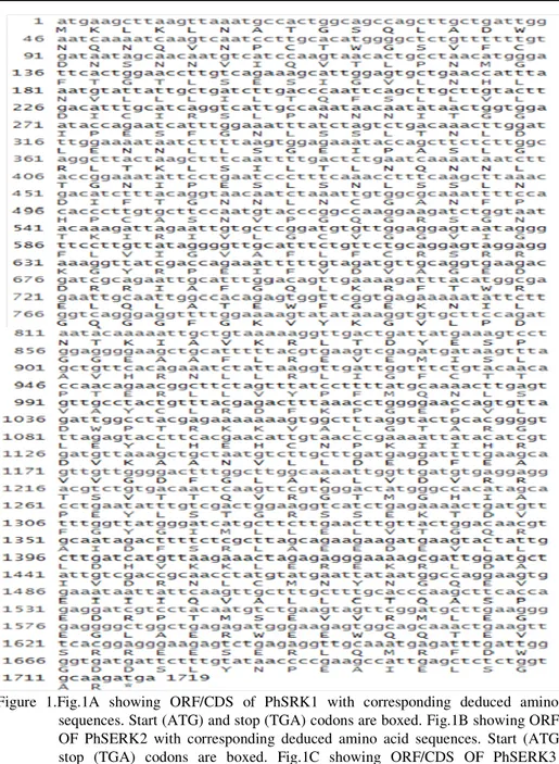
The mRNA or ORF sequence lengths of PhSERK1 and PhSERK2 are 1914pb and 1797pb respectively. The 3’UTR are 384 and 376 bp and 5’UTR are 315 bp and 390 bp, with a predicted amino acid length of 637 and 598 for PhSERK1 and PhSERK2, respectively. PhSERK3 mRNA is 1734 bp in length, with 185 and 165 bp long 5’ and 3’UTR, respectively, and a predicted amino acid length of 577aa (Fig. 1A; 1B; 1C). The predicted molecular mass of PhSERK1, PhSERK2 and PhSERK3 is 73, 67 and 64 kDa, respectively. Using the program RDP V 2, the recombination hypothesis was tested and the highest probabilities were found between the three PhSERKs. These probabilities were well supported by all six methods tested in RDP2. Results with GENECONV program revealed that PhSERK1 may result from a hypothetic recombination between PhSERK2 and PhSERK3 in position (135 pb- 308 pb) and (391 pb- 431 pb). Since there is not enough genomic sequences of Phoenix dactylifera cv deglet Nour in the NCBI website, we sought the homologue PhSERKs gene family in the Khalas variety. Furthermore, the exon–intron structure of PhtSERKs gene was deduced. We compared the PhSERK1, PhSERK2 and PhSERK3 cDNAs and whole genome shotgun sequence of Khalas variety witch reference in GenBank are ACYX02000007.1, ACYX02043306.1 and ACYX02050452.1 respectively using the BLASTN program (Al-Dous et al., 2011). This comparison and gene prediction using FGENESH web site (http://linux1.softberry.com/) showed that the PhSERK1 gene (11051 pb) (Fig.1A) and PhSERK2 (7981 pb) (Fig.1B) organized into 10 introns and 11 exons. All SERK genes of monocots studied so far have conserved structures (11 exons and 10 introns) (Hu et al., 2005; Jun Ma et al., 2012). However the PhSERK3 (10510 pb) (FIG. 1C) gene has 10 exons and 9 introns unlike the other genes SERKS.
PhSERKs proteins prediction
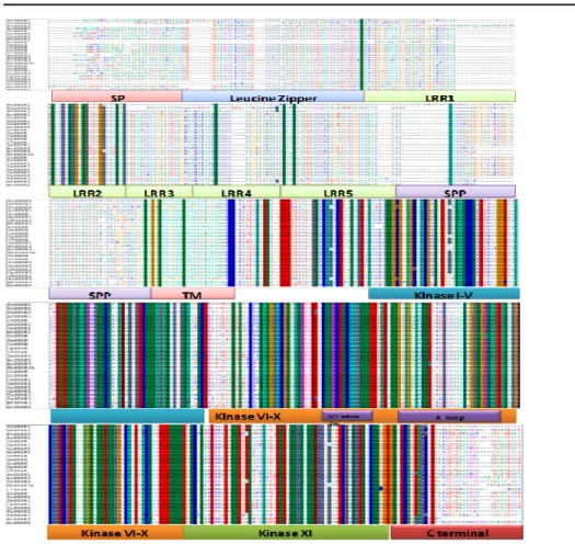
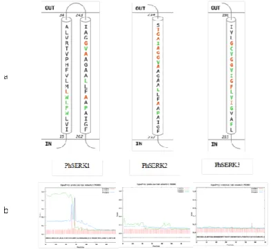
Surprisingly, PhSERK2 and PhSERK3 lack the signal peptide domain, which are also absent in the DcSERK and TaSERK2 and TaSERK3 (Triticum aestivum) proteins (Fig. 2, Fig. 3b) (SCHMIDT et al., 1997) and the similarity between both proteins is 70%. At the protein level, PhSERK1, PhSERK2 and PhSERK3 showed highest identity with AcSERK1, CnSERK and TaSERK3 respectively (Baudino et al., 2001; Ito et al., 2005). It is clear from the amino acid sequence alignment that the PhSERKs align closely with SERK genes from other species (Fig. 2). More detailed analysis was done using Prosite for the identification of regions characteristic of SERK proteins (HOFMANN et al., 1999). A hydrophobic amino acid signal peptide sequence with a possible cleavage site between positions 29 and 30 is present in PhSERK1, which is conserved in rice, maize, Arabidopsis and Medicago (Fig.3). This is followed by a leucine-zipper (LZ) sequence in PhSERK1, PhSERK2 and PhSERK3. The LZ domain are present in the AtSERK1 (Hecht et al., 2001), ZmSERK1 (Zea mays) and OsSERK1 (Oryza sativa) (Baudinos et al., 2001) and MtSERK (Medicago truncatula ) (NOLAN et al., 2003), but not in DcSERK (Daucus carota ) (SCHMIDTet al., 1997). The leucine-zipper sequence is followed by five LRRs in PhSERK2, as defined earlier by HECHTet al., (2001) for AtSERK in Arabidopsis thaliana. PhSERK1 protein contains four LRR domains with glycosylation sites with delated LRR3 (Fig.2) (HECHT et al., 2001). These repeats are thought to from a horseshoe-shaped cavity that promotes protein-protein interactions with small globular domains during rather diverse molecular recognition processes in animals and plants (KOBE and DEISENHOFER, 1995). The Pro-rich domain with SPP motif, located between the LRRs and transmembrane region, has been suggested to act like a hinge providing flexibility to the extracellular part of the receptor or as a region for interaction with the cell wall (HECHTet al., 2001). The PhSERK3 protein shows the presence of characteristic four LRRs with delated LRR5, followed by the transmembrane domain and the C-terminal region but lacks the SPP motif. For PhSERK1 there was also a strongly preferred models of two TM helices from position 15 to 34 and 243 to 262 (Fig.3a) predicted by RHYTHM server. PhSERK2 and PhSERK3 contain one TM helices at positions 217 to 237 and 196 to 215 respectively. These results are confirmed by secondary structure prediction using the program Phyre2. The intracellular kinase domain contains 11 sub-domains of conserved amino acid sequences in all the three PhSERK proteins, as described by HANKS Et al (1988).
LRR4, one in LRR5, two in SPP and one in domain V of the kinase region in almost PhSERK1 and PhSERK3, but in PhSERK2 there are only five N-glycosylation sites. For TaSERK1 and TaSERK2 (Triticum aestivum), there are N-glycosylation sites, two in LRR2, two in LRR4, one in LRR5 and one in domain V of the kinase region (BHUMICA et al., 2008). In LRR5, the glycosylation is O-linked instead of N-linked in PhSERKs as shown in Arabidopsis and Medicago (SHAH et al., 2001; NOLANet al., 2003). In extensions also, usually all prolines in the SPP repeat are hydroxylated and are considered to be targets for O-linked glycosylation (SCHMIDTet al., 1997). PhSERK3 does not share one of the characteristic features of the SERK proteins, i.e. it lacks SPP motif but contains the C-terminal region which has also been suggested to be a SERK-specific feature (SCHMIDT et al., 1997; BAUDINOet al., 2001; ALBERTINI et al., 2005) (Fig.3). Further works on the expression level of this gene should be done.
Figure 3. PhSERKs sequences analysis. a: The transmembrane region predicted by RHYTHM tool. b: Prediction of signal peptide sequence for PhSERK1, PhSERK2 and PhSERK3.
Phylogenetic relationships of the SERK proteins
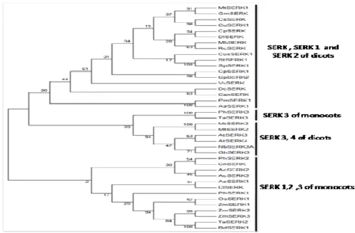
organization (CRONQUIST 1988). The phylogenetic tree of SERK protein (Fig. 4) reveals that organized into four major monophyletic groups. The SERK, SERK1 and SERK2 of the dicots are monophyletic with 30 % bootstrap support. The PhSERK3 is more closely related to TaSERK3 than the other species tested with 100% bootstrap support. The SERK 3 is joined to SERK4 of dicots except MtSERK2 at a bootstrap value of 94 %. The SERK1, SERK2 and SERK3 of the monocots are also monophyletic with 2 % bootstrap support. PhSERK2 was clustered on monophyletic group with CnSERK, AcSERK2 and AcSERK3 with 30% bootstrap. It appears that PhSERK1 is weakly united with ZmSERK1, ZmSERK2, ZmSERK3, OsSERK1, TaSERK2 and BdSERK1 with weak bootstrap support (25%).
The PhSERK3 is closest to TaSERK3 as is evident from the phylogenetic tree while TaSERK3 was grouped with the SERK proteins which lack the SPP motif (Fig.4).
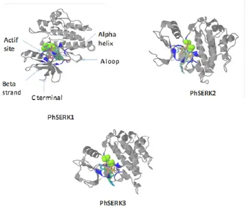
Three-dimensional structure of PhSERKs proteins
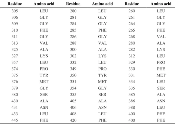
For three-dimensional structure with ligand binding sites structure of PhSERK1, 2 and 3, we used the web site that also provides a display that shows the division of family proteins into domains via 3D LigandSite program (Fig. 5), which uses an effective consensus strategy both in template selection, which combines PSI-BLAST and IMPALA and T-Coffee. The model based on template for the three SERK is proto-oncogene tyrosine-protein kinase abl1 (Nagar et al., 2006).The PhSERK1 (297- 575 pb) sequence the PhSERK2 (272- 550 pb) and PhSERK3 (235- 531pb) have been modeled with 100.0% confidence by the single highest scoring template. The others residues of PhSERK sequence could be modeled at >90% confidence using multiple-templates. This region corresponds to the ATP-binding region signature, activation loop domain, and kinase domain (serine/threonine protein kinases active-site). All PhSERKs amino acids, which are involved in fixing the ligand, are shown in table 2. The results found by 3DSiteLigand program are proven using the site Prosite. The proteins PhSERK1 and PhSERK2 have the same amino acid ligand binding since both proteins have the same function in somatic embryogenesis, anther development and male gametophyte maturation (ALBRECHT et al., 2005; COLCOMBET et al., 2005). For protein PhSERK3, we noticed that there are differences in the amino acid ligand binding compared to PhSERK1 and PhSERK2 (Tab.2) this is normal since the function of SERK1 and SERK2 protein is the programmed cell death pathway and defense responses (CHINCHILLA et al., 2007; HEESE et al., 2007). The OsSERK3 and OsSERK1, showed an activity in both somatic embryogenesis and fungal defense (HUet al., 2005).
In this study, we isolated PhSERK1, PhSERK2, PhSERK3 genes from an F. oxysporum induced cDNA library of palm date. Our phylogenetic and genetic evolution analysis confirmed that PhSERKs belongs to a gene family in Phoenix dactylifera , which leads to the conclusion that more studies must be carried out.
Tabe 2. PhSERKs amino acids which are involved in fixing of ligand
ACKNOWLEDGEMENT
This work was supported by grants from the Tunisian minister of Higher Education and Scientific Research
Received August 26th, 2013
Accepted October 05th, 2013
REFERENCES
ALBRECHT, C., E. RUSSINOVA, V.HECHT, E. BAAIJENS, and S. DE VRIES (2005): The Arabidopsis thaliana somatic embryogenesis receptor-like kinases1 and 2 control male sporogenesis. Plant Cell, 17: 3337-3349.
ALBRECHT, C., E. RUSSINOVA, B. KEMMERLING, M. KWAAITAAL, and S.C DE VRIES (2008): Arabidopsis somatic embryogenesis receptor kinase proteins serve brassinosteroid-dependent and independent signaling pathways. Plant Physiol, 148: 611–619.
Residue Amino acid Residue Amino acid Residue Amino acid
305 LEU 280 LEU 260 LEU
306 GLY 281 GLY 261 GLY
309 GLY 284 GLY 264 GLY
310 PHE 285 PHE 265 PHE
311 GLY 286 GLY 268 VAL
313 VAL 288 VAL 280 ALA
325 ALA 300 ALA 282 LYS
327 LYS 302 LYS 312 LEU
357 LEU 332 LEU 329 PRO
374 PRO 349 PRO 330 PHE
375 TYR 350 TYR 331 MET
376 MET 351 MET 334 LEU
379 GLY 354 GLY 335 SER
380 SER 355 SER 385 ALA
430 ALA 405 ALA 386 ASN
431 ASN 406 ASN 388 LEU
433 LEU 408 LEU 400 PHE
AL-DOUS, E. K., GEORGE BINU, E. AL-MAHMOUD. MARYAM, Y. AL-JABER. MONEERA, A.JOEL MALEK (2011): De novo genome sequencing and comparative genomics of date palm (Phoenix dactylifera). Nat Biotechnol, 29: 521– 527.
ALTSCHUL, S. W.GISH, W. MILLER, E. MYERSAND, and D. LIPMAN (1997): Basic logical alignement search tool. Journal of Molecular Biology, 215: 403-410.
BAUDINO, S., S. HANSEN, R.BRETTSCHNEIDER, V.HECHT, T.DRESSELHAUS, H. LORZ, C. DUMAS, and P. ROGOWSKY (2001): Molecular characterization of two novel maize LRR receptor-like kinases, which belong to the SERK gene family. Planta, 213: 1–10.
BHUMICA, S., P. JITENDRA, and K. PARAMJIT (2008): Characterization of three somatic embryogenesis receptor kinase genes from wheat, Triticum aestivum. Plant Cell Rep, 27: 833–843.
CATHALA, J. F., B. SAVOURET, B. MENDEZ, M. KARIN, J. A. MARTIAL, and J. D. BAXTER (1983): A method for isolation of intact translationally active ribonucleic acid. DNA, 2:329-335.
CHINCHILLA, D., C. ZIPFEL, S. ROBATZEK, B. KEMMERLING, T. NURNBERGER, J. D. G. JONES, G. FELIX, and T. BOLLER (2007): A flagellin-induced complex of the receptor FLS2 and BAK1 initiates plant defence. Nature, 448: 497-412.
COLCOMBET, J., D.A. BOISSON, R. ROS-PALAU, C.E VERA, and J.I SCHROEDER (2005): Arabidopsis somatic embryogenesis receptor kinases1 and 2 are essential for tapetum development and microspore maturation. Plant Cell, 17 :3350-3361.
FKI, L., MASMOUDI, R., N. DRIRA,and A. RIVAL (2003): An optimised protocol for plant regeneration from embryogenic suspension cultures of date palm, Phoenix dactylifera L. cv. Deglet Nour . Plant Cell Reports, 21: 517–524. HANKS, S. K., A.M. QUIN, and T. HIMTER (1988): The protein kinase family: Conserved features and deduced phylogeny of
the catalytic domains. Science, 241: 42-52.
HE, K., X.GOU, T.YUAN, H. LIN, T.ASAMI, S. YOSHIDA, S.D. RUSSELL, and J. LI (2007): BAK1, BKK1 regulate brassinosteroid-dependent growth and brassinosteroid independent cell-death pathways. Curr Biol, 17 :1109-1115.
HECHT, J.P., C.VIELLE, M.V. HARTOG, D.L. SCHMIDT, K.BOUTILIER, U. GROSSNICKLAUS, and S.C DE VRIES (2001): The Arabdopsis somatic embryogenesis receptor kinase 1 gene is expressed in developing ovules and embryos and enhances embryogenic competence in culture.Plant Physiol, 127:803–816.
HEESE, A., D.R. HANN, S.GIMENEZ-IBANEZ, A.M.E. JONES, K.HE, J. LI, J.I. SCHROEDER, S.C PECK, and J.P RATHJEN (2007): The receptor-like kinase SERK3/BAK1 is a central regulator of innate immunity in plants. Proc Natl Acad Sci USA, 104:12217-12222.
HOFMANN, K., P. BUCHER, L. FALQUET, and A. BAIROCH (1999): The PROSITE database: its status in 1999. Nucl Acids Res, 27: 215–219.
HU, H., L. XIONG, and Y. YANG (2005): Rice SERK1 gene positively regulates somatic embryoenesis of cultured cell and host defense response against fungal infection. Planta, 222: 107–117.
ITO, Y., K. TAKAYA, and N. KURATA (2005): Expression of SERK family receptor-like protein kinase genes in rice. Biochim Biophys Acta, 1730: 253–258.
JUN, M., H.YEHUA, W.CHENGHOU, L. HEPING, H. ZHONGYI, and S. GUANGMING (2012): Cloning and Molecular Characterization of a SERK Gene Transcriptionally Induced during Somatic Embryogenesis in Ananas comosus cv. Shenwan. Plant Mol Biol Rep, 30:195–203.
KEMMERLING, B. A.SCHWEDT, P.RODRIGUEZ, S.MAZZOTTA, M.FRANK, S.A. QAMAR, T. MENGISTE, S.BETSUYAKU, J.E. PARKER, C. MUSSIG (2007): The BRI1-associated kinase 1, BAK1, has a brassinolide-independent role in plant cell-death control. Curr Biol 17: 1116-1122.
KOBE, B and J. DEISENHOFER (1995): A structural basis of the interactions between leucine-rich repeats and protein ligands. Nature, 374:183–186.
KRIAA, W., B.SGHAIER-HAMMAMI, F. MASMOUDI-ALLOUCHE, R. BENJEMAA-MASMOUDI,and N. DRIRA (2012):The date palm (Phoenix dactylifera L.) micropropagation using completely mature female flowers. CR Biol, 335: 194-2044.
LEBRUN, F. RENTIER-DELRUE, L. MERCIER (1988): A simple method for the preparation of the covalently closed circular form of plasmid DNA. Biotechniques, 6: 3-5.
LI, J., J.WEN, K.A. LEASE, J.T. DOKE, F.E. TAX, J.C WALKER (2002): BAK1, an Arabidopsis LRR receptor-like protein kinase, interacts with BRI1 and modulates brassinosteroid signaling. Cell, 110: 213-222.
NAGAR, B., O.HANTSCHEL, M.SEELIGER, J.M. DAVIES, W.I. WEIS, G. SUPERTI-FURGA, J. KURIYAN (2006): Organization of the SH3-SH2 unit in active and inactive forms of the c-Abl tyrosine kinase. Mol Cell, 21: 787–798.
NOLAN, K.E., R.R IRWANTO, and R.J ROSE (2003): Auxin up-regulates MtSERK1 expression in both Medicago truncatula root-forming and embryogenic cultures. Plant Physiol, 133: 218-230.
SCHMIDT, F., M.A.J. GUZZO, S.C TOONEN, and S.DE VRIES (1997): A leucine rich repeat containing receptor-like kinase marks somatic plant cells competent to form embryos. Development, 124: 2049-2062.
SHAH, K., J. VERVOORT, and S.C DE VRIES (2001): Role of threonines in the AtSERK1 activation loop in phosphorylation. J Biol Chem, 276: 41263-41269.
SHIU, S.H and A.B BLEECKER (2003): Expansion of the Receptor-Like Kinase/Pelle Gene Family and Receptor-Like Proteins in Arabidopsis. Plant Physiol, 132: 530-543.
SOMLEVA, M.N. E.D.L SCHMIDT, and S.C DE VRIES (2000): Embryogenic cells in Dactylis glomerata L. (Poaceae) explants identified by cell tracking and by SERK expression. Plant Cell Rep, 19:718-726.
TOR, M. M.T LOTZE, and N. HOLTON (2009): Receptor-mediated signalling in plants: molecular patterns and programmes. J Exp Bot, 60: 3645-3654.
MOLEKULARNO KLONIRANJE I in silico ANALIZA TRI mRNK RECEPTORA KINAZE U TOKU SOMATSKE EMBRIOGENEZE KOD PALME
Imen REKIK, Amine ELLEUCH, Walid KRIAA, Noureddine DRIRA
Laboratorija za biljnu biotehnologiju, Fakultet za naukuLaboratory of plant biotechnology, Univerzitet u Sfaksu, Tunis
Izvod
Izvršeno je izolovanje i karakterizacija tri gena receptor kinaze (PhSERK) u somatskoj embriogenezi palme brzom amplifikacijom cDNK krajeva (RACE). PhSERKs pripadaju maloj familiji receptor kinaze gena koji imaju konzerviranu strukturu i ekstenzivnu homologiju sekvenci sa ranije izolovanim SERK genima. Analizom je utvr ena veli ina sekvenci i to: 11051 pb (PhSERK1), 7981 pb (PhSERK2) i 10510 pb (PhSERK3). ORF kod PhSERK1, PhSERK2 i PhSERK3 su 1914 pb, 1797 pb I 1719 pb.. PhSERKs pripada LRR-tipa površine elije RLKs, koja ima brojne karakteristike domena. Filogenetsko stablo pokazuje da se proteini PhSERK1, PhSERK2 i PhSERK3 grupišu u klaster unutar SERKs grupe proteina.
Primljeno 26. VIII 2013.