Morpho-anatomical characterization of embryogenic calluses from
immature zygotic embryo of peach palm during somatic
embryogenesis
Simone de Alencar Maciel1, Paulo Cesar Poeta Fermino Junior2, Ricardo Alexandre da Silva3 and Jonny Everson Scherwinski-Pereira4*
1
Programa de Pós-graduação em Agronomia, Universidade Federal do Acre, Rio Branco, Acre, Brazil. 2Universidade Federal do Acre, Centro de Ciências Biológicas e da Natureza, Rio Branco, Acre, Brazil.3Programa de Pós-graduação em Biotecnologia Vegetal, Departamento de Biotecnologia Vegetal, Universidade Federal do Rio de Janeiro, Rio de Janeiro, Rio de Janeiro, Brazil. 4Embrapa Recursos Genéticos e Biotecnologia, Empresa Brasileira de Pesquisa Agropecuária, Av. W5 Norte (final), s/n, Cx. Postal 2372, 70770-917, Brasília, Distrito Federal, Brazil. *Author for correspondence. E-mail: [email protected]
ABSTRACT. The objective of this study was to morpho-anatomically characterize nodular embryogenic calluses from zygotic embryos of peach palm during the induction of somatic embryogenesis. Immature zygotic embryos were pre-treated in MS medium added to Picloram and 2,4-D (25 μM) and BAP (0, 5, 10 μM). After three months, primary calluses were transferred to MS induction medium added to Picloram and 2,4-D (450 μM). After six months, the embryogenic calluses were then histologically analyzed and cultivated in the maturation medium. The competent tissues of the zygotic embryos differentiated embryogenic calluses under action of both Picloram and 2,4-D auxins (450 μM), where the presence of multi-granular structures were observed. Histological observations showed that in the nodular embryogenic calluses, the outlying parenchymal cells exhibit cellular characteristics of high mitotic activity. Differentiation of tracheal elements exists in embryogenic calluses connecting the callus to the explant. The evaluated cytokinin/auxin interaction influences the development of embryogenic calluses and globular structures.
Key words: Bactris gasipaes, micropropagation, histology, morphology, callus anatomy, somatic embryos.
RESUMO. Caracterização morfoanatômica de calos embriogênicos originados de embriões zigóticos imaturos de pupunheira durante a embriogênese somática. O objetivo deste trabalho foi caracterizar morfoanatomicamente calos nodulares embriogênicos originados de embriões zigóticos de pupunheira durante a indução da embriogênese somática. Embriões zigóticos imaturos de pupunha foram inicialmente pré-tratados em meio de cultura MS, solidificado com 2,5 g L-1 de phytagel® e suplementado com Picloram e 2,4-D na concentração de 25 μM e BAP (0, 5, 10 μM). Após três meses, os calos primários foram transferidos para meio de indução, com Picloram e 2,4-D (450 μM). Após seis meses, os calos nodulares embriogênicos formados foram então analisados histologicamente e repicados para o meio de maturação para a progressão das estruturas multigranulares embriogênicas. Verificou-se que os tecidos competentes dos embriões zigóticos imaturos diferenciaram nódulos embriogênicos pela ação de ambas as auxinas (Pi e 2,4-D) em 450 μM. Observações histológicas mostraram que, nos nódulos embriogênicos, as células parenquimáticas mais periféricas exibem características celulares de alta atividade mitótica. Existe diferenciação de elementos traqueais nos calos embriogênicos conectando o calo ao explante. A interação citocinina/auxina influencia o desenvolvimento dos calos embriogênicos e das estruturas globulares.
Palavras-chave: Bactris gasipaes, micropropagação, histologia, morfologia, anatomia de calos, embriões somáticos.
Introduction
The Peach palm is an Arecaceae that is suitable for the production of fruits and heart of palm. According to Clement and Santos (2002), the peach palm was domesticated so that its fruit could be used in the indigenous economy, in the forms of flour, a fermented drink and boiled fruit. In today’s economy,
both urban and rural, it is used as boiled fruit.
seeds can lead to high genetic variability in these crops. Propagation in vitro of the plants can therefore become an important tool for overcoming these problems inherent to the species, as it enables large-scale clonal and mass multiplication of individuals, provided that efficient protocols are developed (SCHERWINSKI-PEREIRA; FORTES, 2003; STEINMACHER et al., 2007; NOMURA et al., 2008).
Propagation in vitro, via somatic embryogenesis, offers good potential for clonal multiplication, where an isolated cell can be induced to produce first an embryo, and then a complete plant (TAHIR; STASOLLA, 2006). Success in initiation and establishment of embryogenic cultures basically depends on the type and physiological stage of the explants (NAMASIVAYAM, 2007). This is the first important step in the transition of the somatic cells into embryonic cells (DE JONG et al., 1993). Therefore, it is essential to recognize the explants that have morphogenic competence (GELDNER et al., 2000; CARAMORI et al., 2001; PHILLIPS, 2004; TELLES; BIASI, 2005).
In peach palm, attempts have been made at clonal multiplication of the species using different types of explants, particularly using protocols of somatic embryogenesis (ALMEIDA; ALMEIDA, 2006; STEINMACHER et al., 2007). In general, although the results are consistent, there have still been relatively few studies on the structural aspects linked to the morphology and histology of the embryogenic callus induced during somatic embryogenesis, given the preference of authors for quantitative data in their attempts to establish commercial protocols.
The histological alterations associated with the position and activity of the competent cells are normally basic requirements when studying somatic embryogenesis. Maheswaran and Williams (1985) observed that somatic embryogenesis of Trifolium
repens, obtained from immature zygotic embryos,
originated cells of the epidermis of the hypocotyl, which proliferated to produce somatic embryos in one phase of callus. The initiation of development of somatic embryogenesis of Carya illinoinensis, induced by different auxins revealed, through the morphological and anatomic analysis, that the auxins ANA and 2,4-D induced accentuated cell division in the subepidermal layer of the cotyledons of immature embryos (RODRIGUEZ; WETZSTEIN, 1998). Steinmacher et al. (2007) observed that during somatic embryogenesis in peach palm, the first events of cell division of the mesocotyl of the zygotic embryo occur from the subepidermal cells, particularly in the cells adjacent to the vascular tissues.
The objective of this work was to evaluate, morpho-anatomically, nodular embryogenic calluses originating from zygotic embryos of peach palm during the induction of somatic embryogenesis.
Material and methods
Immature zygotic peach palm embryos were used as the source of explant. These were obtained from peach palm plants of the Germplasm Bank of Embrapa Acre, Rio Branco, in the state of Acre (9°58'22" S e 67°48'40" W), while they were still dark green, at approximately eight weeks, after anthesis. First, the seeds were extracted from the depulped fruits then, using a manual press, the zygotic embryos were removed and disinfected in a laminar flow chamber, by immersion in alcohol (70%) for 15 seconds, and then in sodium hypochlorite solution (1.25%) for 10 min. Next, the embryos were washed three consecutive times, with distilled water, autoclaved and placed in culture medium for pre-treatment.
The explants were pre-treated in MS culture medium (MURASHIGE; SKOOG, 1962), to which was added 30 g L-1 sucrose, 2.5 g L-1 Phytagel®, Morel
and Wetmore (1951) vitamins, and supplemented with Picloram and 2,4-D at a concentration of 25 μM and BAP at concentrations of 0, 5 and 10 μM. For the inoculation, 110 mL glass bottles were used, containing approximately 20 mL of culture medium. After 104 days of pre-treatment, the primary calluses formed were transferred to the MS induction medium, supplemented with Picloram and 2,4-D, both at a concentration of 450 μM, according to the results obtained for peach palm production by Steinmacher et al. (2007). The embryonic formations obtained in this treatment were transferred, after 179 days of cultivation, to the maturation medium comprised of MS salts supplemented with 45 μM Picloram and 2,4-D with 11.25 μM of 2-iP. The composition of the culture media in the different phases of somatic embryogenesis induction in peach palm can be seen in Table 1.
The pH of the culture media was adjusted to 5.8±0.1 before the addition of Phytagel®, and
subsequently autoclaved at 121°C, for 15 min. in 1.3 atm. The experiments were maintained in a growth room at a temperature of 25±2°C, in the absence of light. The experiment was installed in a completely randomized design, with eight repetitions and five embryos per replicate.
James et al. (1994), with modifications. These were fixed in glutaraldehyde solution 2.5%, in phosphate buffer 0.1 M (pH 7.2), for 24h, at room temperature. After fixing, the samples were washed three times in phosphate buffer and dehydrated in increasing ethanol series (20 to 100%). After dehydration, the samples were imbedded in acrylic resin, LR White medium grade (London, UK) for a period of seven days, during which time they were kept in a refrigerator for resin infiltration. The samples were then embedded in transparent gelatine capsules containing resin, and placed in 55oC to
polymerize, for 18h. From this material, sections of 1 μm were obtained, using an ultramicrotome (Leica), stained with blue toluidine solution 1%, and examined under an optical microscope (Zeiss).
Table 1. Components of the culture media used in the pre-treatment, induction and maturation of somatic embryos induced in immature peach palm embryos.
Components Pre-treatment Induction Maturation
Culture media MS MS MS
Myo-inositol (mg L-1) 100 100 100
Picloram (μM) 25 450 45
2,4-D (μM) 25 450 45
BAP (μM) 0, 5, 10 - -
2iP (μM) - - 11.25
Phytagel® (g L-1) 2.5 2.5 2.5
Results and discussion
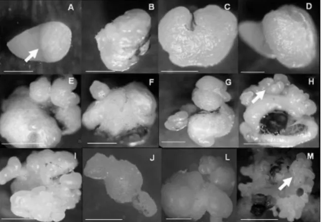
After 41 days of cultivation, the immature zygotic peach palm embryos began to present intumescence in the mesocotyl region (Figure 1A). Callogenesis was observed in all the treatments evaluated, at a rate of between 20 and 34% of the explants cultivated with 2,4-D and Picloram, respectively, which varied in size and embryonic potential, depending on the auxin used (Figure 1A-M). The addition of BAP to the pre-induction culture medium did not cause any difference in the formation of primary calluses. Similar results for the addition of BAP to the culture medium used for the induction of primary calluses in peach palm were also observed by Steinmacher et al. (2007), who cite that it is not necessary to add this cytokinine in the initial stages of callogenesis in peach palm. According to the observations of George and Sherrington (1984), the calluses present distinct morphogenic expressions, depending to the explant and nutritive medium used. The same authors report that in general, the type of callus formed in a determined genotype, its degree of cell differentiation and its morphogenic potential depend on the species, the type of explant and the constituents of the culture medium.
It is observed that from the region of the mesocotyl (Figure 1A), the auxin Picloram was the one that provided the bests responses in terms of development primary and embryogenic calluses. However, the growth of primary calluses was slow and gradual, and after 164 days in cultivation, presented significant increases in cell masses due the action of Picloram (Figure 1B-G), when in the somatic embryo induction medium. According to Karp (1995), the differences observed in callus proliferation occur because the explants can vary in their sensitivity to growth regulators and/or due to the differences in endogenous content of the hormones.
In general, the competent tissues in zygotic embryos differentiate embryogenic calluses under the action of the auxins Picloram and 2,4-D in 450 μM, and the presence of granular structures was observed (Figure 1H and I), especially in the medium with Picloram. Similar results were obtained in studies with the induction of somatic embryos in oil palm, in which the authors observed that the presence of 2,4-D stimulated the morphogenic responses of flower buds (TEIXEIRA et al., 1994). The induction of the embryogenic or organogenic route is influenced and determined by the type of explant, genotype, growth regulators, culture medium, and cultivation conditions (HOU; JIA, 2004; PARAMAGEETHAM et al., 2004). According to Namasivayam (2007), the success of the initiation and establishment of embryogenic cultures basically depends on the type of physiological stage of the explants. Guerra and Handro (1998) observed that the route of the somatic embryogenesis in Euterpe edulis occurs as a response to the interaction between the physiological stage of the explant, and the type and concentration of growth regulators present in the culture medium.
Morphogenesis in vitro results in the interaction between the processes of induction, cell competence, determination and cell differentiation (CHRISTIANSON; WARNICK, 1983), which culminate in the obtaining of somatic organs or embryos.
Figure 1. Morphological phases of somatic embryogenesis in peach palm: (A) Intumescence of the mesocotyl region of the zygotic embryo (arrow). Bar: 2 mm; (B and C) Start of development of the primary callus induced by the auxins Picloram and 2,4-D with 25 μM. Bars: 2 and 3 mm; (D) Growth of the primary callus in the zygotic embryo after 41 days of cultivation. Bar: 2 mm; (E and F) Granular structures on the surface of the primary callus obtained with 450 μM of 2,4-D. Bars: 4 and 3 mm; (G) Development of globular structures on the surface of the primary callus. Bar: 2 mm; (H and I) Embryogenic callus obtained after 164 days (arrow). Bar: 1 mm; (J) Detail of the isolated somatic embryo. Bar: 1 mm; (L) Differentiated structures at the start of maturation of the somatic embryos. Bar: 2 mm; (M) Maturation and multiplication of the somatic embryos (arrow). Bar: 3 mm.
The addition of the auxins 2,4-D and Picloram, in the induction media of somatic embryogenesis favored the development of nodular embryogenic callus with differentiated cell characteristics. The callus present regions with external granular morphology of heterogeneous cellular composition, with coating cells, similar to the epidermis, parenchyma cells, similar to the fundamental parenchyma, and tracheal elements (Figures 2.1 and 2.2).
The coating cells and the cell layer underlying the granular structures in the nodular embryogenic callus present less dense cytoplasm and a non-evident nuclei. The internal cell layers (centripetally) show a dense cytoplasm and prominent nuclei, alternating with more internal cells without these characteristics. The embryogenic nodules are therefore arranged in the form of concentric rings with cells with meristematic characteristics (Figure 2.1). In the base of the granular structures of the callus, in contact with the explant, there are differentiated tracheal elements of the xylem (Figures 2.3 and 2.4), with ring thickenings of the secondary walls (Figure 2.4).
Therefore, in peach palm, the initiation of the somatic embryogenesis occurs by the multiplication of internal cells in the embryogenic nodules. Similar results were observed by Rodriguez and Wetzstein
(1998), in which the start of development of somatic embryogenesis of Carya illinoinensis (Wagenh) C. Koch,induced by different auxins revealed, through morphological and anatomic analysis, that the auxins NAA and 2,4-D induce accentuated cell division in the subepidermal layer of the cotyledons of immature embryos.
Figure 2. Nodular embryogenic callus of Bactris gasipaes Kunth
.
(peach palm): 1. Longitudinal section of the embryogenic nodule from the zygotic embryo (star), with concentric rings with meristematic characteristics (circles). Bar = 50 μm. 2. Detail of the peripheral region of the embryogenic nodule in transversal section. Bar = 10 μm. 3. Formation of xylem at the base of the callus. Bar = 10 μm. 4. Detail of the tracheal elements of the xylem at the base of the callus. Bar = 10 μm. PC = parenchymal cells; CC = coating cells; PC = parenchymal cells with dense cytoplasm; TE = tracheal elements.Conclusion
Combination of BAP with the auxins Picloram and 2,4-D is not necessary in the pre-treatment of the explants for the formation of primary calluses during the somatic embryogenesis in peach palm.
High concentrations of the auxins 2,4-D and Picloram in the culture media are necessary for the induction of nodular embryonic structures in pech palm during the induction of somatic embryogenesis.
The use of Picloram provides better formation of embryogenic structures than 2,4-D, for the induction of somatic embryogenesis in peach palm.
The initiation of somatic embryogenesis in peach palm from zygotic embryo occurs by the multiplication of internal cells in the embryogenic nodules.
Acknowledgements
References
ALMEIDA, M.; ALMEIDA, C. V. Somatic embryogenesis and in vitro plant regeneration from pejibaye adult plant leaf primordia. Pesquisa Agropecuária Brasileira, v. 41, n. 9, p. 1449-1452, 2006.
CARAMORI, L.; FÁVARO, S.; VIEIRA, L. Thidiazuron as a promoter of multiple shoots in cotton explants (Gossypium hirsutum L.). Acta Scientiarum. Agronomy, v. 23, n. 5, p. 1195-1197, 2001.
CHRISTIANSON, M. L.; WARNICK, D. A. Competence and determination in the process of in vitro shoot organogenesis. Developmental Biology, v. 95, n. 2, p. 288-293, 1983.
CLEMENT, C. R.; SANTOS, L. Pupunha no mercado de Manaus: preferências de consumidores e suas implicações. Revista Brasileira de Fruticultura, v. 24, n. 3, p. 778-779, 2002.
CLEMENT, C. R.;URPÍ, J. E. M. Pejibaye palm (Bactris gasipaes, Arecaceae): Multi-use potential for the lowland humid tropics. Economic Botany, v. 42, n. 1, p. 302-311, 1987.
DE JONG, A. J.; SCHMIDT, E. D. L.; VRIES, S. C. Early events in higher-plant embryogenesis. Plant Molecular Biology, v. 22, n. 2, p. 367-377, 1993. GELDNER, N.; HAMANN, T.; JÜRGENS, G. Is there a role for auxin in early embryogenesis? Journal of Plant Growth Regulation, v. 32, n. 2-3, p. 187-191, 2000. GEORGE, E. F.; SHERRINGTON, P. D. Plant Propagation by tissue culture. Eversley: Exegetics, 1984.
GUERRA, M. P.; HANDRO, W. Somatic embryogenesis and plant regeneration in different organs of Euterpe edulis Mart. (Palmae): Control and structural features. Journal of Plant Research, v. 111, n. 1, p. 65-71, 1998.
HOU, S. W.; JIA J. F. High frequency plant regeneration from Astragalus melilotoides hypocotul and stem explants via somatic embryogenesis and organogenesis. Plant Cell, Tissue and Organ Culture, v. 79, n. 1, p. 95-100, 2004. JAMES, E. K.; REIS, V. M.; OLIVARES, F. L.; BALDANI, J. I.; DÖBEREINER, J. Infection of sugar cane by the nitrogen-fixing bacterium Acetobacter diazotrophicus. Journal of Experimental Botany, v. 45, n. 6, p. 757–766, 1994.
KARP, A. Somaclonal variation as a tool for crop improvement. Euphytica, v. 85, n. 3, p. 295-302, 1995. MAHESWARAN, G.; WILLIAMS, E. G. Origin and development of the embryoids formed directly on immature embryos of Trifolium repensin vitro. Annals of Botany, v. 56, n. 5, p. 619-630, 1985.
MOREL, G.; WETMORE, R. H. Tissue culture of monocotyledons. American Journal of Botany, v. 38, n. 2, p. 138-140, 1951.
MURASHIGE, T.; SKOOG, F. A revised medium for rapid growth and bio assays with tabacco tissue culture. Physiologia Plantarum, v.15, n. 3, p. 473-497, 1962. NAMASIVAYAM, P. Acquisition of embryogenic competence during somatic embryogenesis. Plant Cell, Tissue and Organ Culture, v. 90, n. 1, p. 1-8, 2007. NOMURA, E. S.; LIMA, J. D.; GARCIA, V. A.; RODRIGUES, D. S. Crescimento de mudas micropropagadas da bananeira cv. Nanicão, em diferentes substratos e fontes de fertilizante. Acta Scientiarum. Agronomy, v. 30, n. 3, p. 359-363, 2008.
PARAMAGEETHAM, C.; BABU, G. P.; RAO, J. V. S. Somatic embryogenesis in Centella asiatica L. an important medicinal and neutraceutical plant of India. Plant Cell, Tissue and Organ Culture, v. 79, n. 1, p. 19-24, 2004. PHILLIPS, G. C. In vitro morphogenesis in plants: recent advances. In vitro cellular and Developmental Biology Plant, v. 40, n. 4, p. 342-345, 2004.
RODRIGUEZ, A. P. M.; WETZSTEIN, H. Y. A morphological and histological comparison of the initiation and development of pecan (Carya illinoinensis) somatic embryogenic cultures induced with naphthaleneacetic acid or 2,4-dichlorophenoxyacetic acid. Protoplasma, v. 204, n. 1-2, p. 71-83, 1998.
SCHERWINSKI-PEREIRA, J. E.; FORTES, G. R. L. Protocolo para produção de material propagativo de batata em meio líquido. Pesquisa Agropecuária Brasileira, v. 38, n. 9, p. 1035-1043, 2003.
STEINMACHER, D. A.; CANGAHUALA-INOCENTE, G. C.; CLEMENT, C. R.; GUERRA, M. P. Somatic embryogenesis from peach palm zygotic embryos. In Vitro cellular and Developmental Biology Plant, v. 43, n. 2, p. 124-132, 2007.
TAHIR, M.; STASOLLA, C. Shoot apical development during in vitro embryogenesis. Canadian Journal of Botany, v. 84, n. 11, p. 1650-1659, 2006.
TEIXEIRA, J. B.; SÖNDAHL, M. R.; KIRBY, E. G. Somatic embryogenesis from immature inflorescence of oil palm. Plant Cell Reports, v. 13, n. 5, p. 247-250, 1994.
TELLES, C.; BIASI, L. Organogênese do caquizeiro a partir de ápices meristemáticos, segmentos radiculares e foliares. Acta Scientiarum. Agronomy, v. 27, n. 4, p. 581-586, 2005.
Received on May 5, 2008. Accepted on October 3, 2008.