Recebido em 14.04.2000. / Received in April, 14thof 2000.
Aprovado pelo Conselho Consultivo e aceito para publicação em 02.04.2002. / Approved by the Consultive Council and accepted for publication in April, 2ndof 2002. * Trabalho realizado no IPAC - Instituto de Patologia Geral e Cutânea. / Work done at “IPAC - Instituto de Patologia Geral e Cutânea”.
1Patologista; Mestre; Doutor em Medicina; Pesquisador Titular da Fundação Oswaldo Cruz. / Pathologist; Masters and Ph.D. in Medicine; Titular Researcher at the Fundação Oswaldo Cruz.
2Professor Adjunto de Dermatologia da Universidade Federal da Bahia; Dermatopatologista. / Adjunct Professor of Dermatology at the Federal University of Bahia; Dermatopathologist.
3Especialista em Dermatologia pela SBD. Mestre em Medicina. Médica do Serviço de Dermatologia do HUPES-UFBa. / Specialist in Dermatology by the Brazilian Dermatology Society (SBD). Doctor at the Service of Dermatology at HUPES-UFBa (Professor Edgar Santos University Hospital – Federal University of Bahia). 4Especialista em Dermatologia pela SBD / Specialist in Dermatology by the Brazilian Dermatology Society (SBD)
©2002by Anais Brasileiros de Dermatologia
Hanseníase associada a granuloma elastolítico
*Leprosy combined with elastolytic granuloma
*Aryon de Almeida Barbosa Jr
1Newton Sales Guimarães
2Ivonise Follador
3Leila Santos Sarno
4Constança Pithon Pereira
4Resumo: São descritos dois casos de Hanseníase combinados com granuloma elastolítico de células gigantes. Embora uma ocorrência concomitante não possa ser excluída, uma possível relação pato-genética entre as duas condições é postulada. É possível que um mecanismo imunológico desempenhe um papel no processo elastolítico, que poderia também ser causado por dano actínico na pele alterada pela Hanseníase.
Palavras-chave: Granuloma; hanseníase
Summary:Two cases of leprosy combined with elastolytic giant cell granuloma are reported. Though a coincidental occurrence cannot be excluded, a possible pathogenetic relationship between the two conditions is postulated. It is possible that an immunological mechanism plays a role in the elastolytic process, which could also be caused by actinic damage in the skin altered by leprosy.
Key Words: Granuloma; leprosy
Caso Clínico /
Case Report
INTRODUÇÃO
Nos últimos anos, houve aumento de interesse nas doenças que apresentam degeneração do tecido elástico da derme1, apesar do sistema elástico da pele não ser bem
entendido. Algumas condições inflamatórias e não infla-matórias associadas à elastólise da derme foram descritas.
A coexistência de hanseníase e granuloma elastolí-tico de células gigantes, para o qual usaremos o termo “LEGG” (Leprosy and Elastolytic Giant Cell Granuloma), até onde chega o nosso conhecimento, não foi até hoje reconhecido nem descrito.
O presente estudo documenta dois casos raros de hanseníase associada ao granuloma elastolítico de células gigantes em áreas expostas da pele e discute sua possível relação.
INTRODUCTION
In recent years there has been increased interest in diseases showing degeneration of dermal elastic tissue1, in spite of the fact that the skin elastic system is not well understood. Some inflammatory and non-inflammatory conditions associated with dermal elastolysis have been described.
The coexistence of leprosy and elastolytic giant cell granuloma, for which we use the term “LEGG”, so far as we are aware, has not hitherto been recognized nor described.
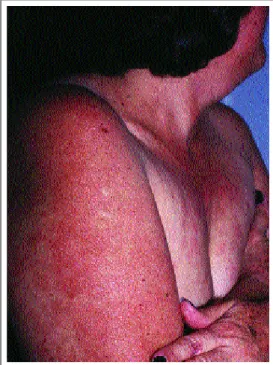
Figura 1: Lesões avermelhadas com hipopigmentação e pequena atrofia no centro. A pele circundante mostra afinamento e envelhecimento sugestivos de
elastose solar. Paciente 1.
Figure 1: Reddish lesions with hypopigmentation and slight atrophy of the centers. The surrounding skin shows thinning and wrinkling sugges-tive of solar elastosis. Patient 1.
alteração de sensibilidade. Não tomava nenhuma medica-ção. Os exames laboratoriais de rotina, incluindo exames para HTLVI e II, foram normais. Contudo, o índice bacterio-lógico dos esfregaços foi de 3,5. A suspeita clínica, antes dos exames laboratoriais, incluía hanseníase borderline, micose fungóide e lupus eritematoso.
Caso 2: mulher branca de 63 anos, de Salvador,
Bahia. Desenvolveu uma lesão cutânea na extremidade superior esquerda, que havia começado há 2 meses. A his-tória clínica não era relevante. Aparentava estar clinica-mente bem, exceto pela presença de pequenas pápulas mal definidas, discretamente brilhantes, com bordas irregula-res e aspecto anular na superfície flexora do antebraço esquerdo (Figura 2). A lesão media aproximadamente 7x5 cm de diâmetro e era da mesma cor da pele, mas mostran-do pequenas áreas de hipopigmentação. Apresentava diminuição da sensibilidade térmica. Os achados e resulta-dos resulta-dos exames de laboratório foram negativos, ou dentro dos limites normais, inclusive o índice bacteriológico. A suspeita clínica era hanseníase tuberculóide.
Achados Patológicos
Somente uma biópsia - biópsia incisional em fuso - foi feita de cada paciente. As biópsias foram fixadas em formalina a 10% por um dia. Preparados histológicos corados com H&E e Fite-Faraco, seccionados a 5
µm, foram usados para classificação e demonstração de M.Leprae. Ademais, foram realizados exames com orceína ácida e alcian blue (pH 2,5).
Caso 1:a biópsia cutânea de
patient had no subjective symptoms. The cutaneous lesions showed no sensibility alterations. She took no medication. Routine laboratory investigation, including tests for HTLVI and II, was normal. However, the average Bacterial Index (BI) of skin smears was 3.5. The clinical suspicion, before the laboratory examinations, included Borderline Leprosy, Mycosis Fungoides and Erythematous Lupus.
Case 2:a 63-year-old white woman from Salvador,
Bahia, developed a cutaneous lesion on her left upper extre-mity that had begun to develop for two months. Her medi-cal background was unremarkable. Clinimedi-cally she appeared to be quite well except for the presence of ill defined slightly shinny area with small papules and irregular edges of annular aspect on the flexor surface of her left forearm (Figure 2). The lesion measured about 7x5 Cm in diameter and was the same color as the skin, but showing small areas of hypo pigmentation. There was local thermal sensibility decrease. Findings and results of laboratory examinations were negative or within normal limits, including the BI, which was negative. The clinical suspicion was Tuberculoid Leprosy.
Pathological Findings
Only one biopsy of each patient was taken in the form of an elliptical biopsy. The biopsies were fixed in 10% formalin for one day.
H&Eand Fite-Faraco stained histo-logical preparations, sectioned at 5
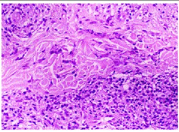
Figura 4: Biópsia cutânea da lesão do antebraço (lesão anu-lar). Fragmentos engolfados de fibras elásticas são vistas nas células gigantes multinucleadas entre as fibras de colágeno. H&E, 250X. Paciente 1.
Figure 4: Skin biopsy from the lesion of forearm (annular lesion). Engulfed fragments of elastic fibers are seen within multinuclear giant cells among collagen fibers. H&E, 250X. Patient 1.
Case 1:skin biopsy from one of the lesions revealed
inflammation of neurovascular bundles and skin appenda-ges. Macrophages and lymphocytes were the predominant cell-types. Large number of acid-fast bacilli (AFB) could be seen in the cytoplasm of macrophages, often with foamy aspect (Figure 3). In focal areas of the superficial and mid dermis, sometimes in close proximity to the marked leprosy tissue reaction there was a patchy lymphohistiocytic infil-trate, with many giant cells without vacuoles nor AFB
(Figure 4). Elastic fibers were less frequently found in these infiltrates. The few isolated bundles of fibers observed in this area were short and thin. Fragments of elastic fibers were demonstrated within the cytoplasm of a few of the giant cells and macrophages. In the deep reticular dermis, the elastic fibers appeared to be normal. The diagnosis of subpolar lepromatous leprosy combined with elastolytic giant cell granuloma was made.
Case 2: Histological examination of the lesion
revealed coexistence of macrophages and multinucleated giant cells that phagocytosed elastic fibers in the upper der-mis, causing them to disappear (Figure 5). Besides this lesion, there was perineural inflammation composed mainly of lymphocytes and histiocytes (Figure 6) present either in the deep dermis or in the vicinity of sweat
uma das lesões revelou inflamação dos feixes neurovascu-lares e anexos cutâneos. Macrófagos e linfócitos eram os tipos de células predominantes. Podia-se ver grande núme-ro de bacilos ácido-resistentes (BAAR), no citoplasma dos macrófagos, freqüentemente de aspecto espumoso (Figura 3) Em áreas focais da derme superficial e média, por vezes próximo da área do tecido hanseníaco reacional, havia um infiltrado linfohistiocitário com várias células gigantes sem vacúolo nem bacilos ácido-resistentes (figura 4) Estes infil-trados apresentavam menos fibras elásticas do que encon-tradas normalmente. Os poucos feixes de fibras observados nesta área eram curtos e finos. Foram demonstradas fibras elásticas no citoplasma de algumas células gigantes e macrófagos. Na derme reticular profunda, as fibras elásticas pareciam normais. Foi feito o diagnóstico de hanseníase virchowiana subpolar associada ao granuloma elastolítico de células gigantes.
Caso 2:exame histológico da lesão revelou
coexis-tência de macrófagos e células gigantes multinucleadas que fagocitaram as fibras elásticas da derme superior, cau-sando seu desaparecimento (Figura 5). Além desta lesão, havia inflamação perineural,
composta basicamente de linfócitos e histiócitos (Figura 6), presentes na derme profunda ou próximo às glândulas sudoríparas.
Figura 2: Lesões eritematosas, levemente infiltradas no antebraço. Paciente 2. / Figure 2: Erythematous, slightly infiltrated annular
lesions on the forearm. Patient 2.
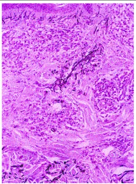
Figure 6: Perineural inflammatory infiltrate, composed mainly of lymphocytes and histiocytes is present in the mid-dermis. H&E 120X. Patient 2.
observadas alterações significativas na epiderme.
Tratamento e Curso Clínico
Uma vez feito o diagnóstico, foi iniciado tratamento para hanse-níase em ambos os casos. Terapia multidrogas, como as usadas em
pacientes com hanseníase multibacilar, foi recomendada para a paciente do caso 1. A paciente do caso 2 recebeu dose única de quimioterapia de curta duração. Nos 12 meses subseqüentes, não apareceram novas lesões nem foi observada recorrência, e ambas as pacientes mostravam melhora significativa.
DISCUSSÃO
A degeneração das fibras elásticas ou elastólise, aspec-to de algumas doenças cutâneas, constitui um grupo de doenças caracterizadas por diminuição ou desaparecimento do tecido elástico dérmico. A elastólise foi classificada como localizada ou generalizada e pode ser congênita ou adquirida, com ou sem manifestações sistêmicas.2Apesar da elastina – principal
proteí-na constitutiva das fibras elásticas – abranger 2% do total de proteínas da derme3, ela é importante fisiologicamente,
propor-cionando elasticidade à pele. Existem evidências bioquímicas de que a elastina é produzida pelos fibroblastos da pele.4
Alterações na estrutura ou metabolismo da elastina foram implicadas em diversas
doen-ças cutâneas adquiridas ou hereditárias. Embora a base bioquímica para as mudanças observadas na estrutura da elastina não seja conhecida, pensa-se que a elastase – uma enzima proteolítica – esteja
ved in the epidermis. The diagnosis of indeterminate leprosy combined with elastolytic giant cell granulo-ma was granulo-made.
Treatment and Clinical Course
Once the diagnosis was made, the treatment for leprosy was initiated in both cases. Multidrug Therapy (MDT), as used for multibacillary patients was recommended for patient 1. Patient 2 received a single dose of short-term chemotherapy (ROM). No new lesions appeared in the sub-sequent twelve months, nor was recurrence observed, during this time the lesions of both patients showed marked improvement.
DISCUSSION
Elastic fiber degradation or elastolysis, a feature of some cutaneous diseases, constitutes a group of disorders characterized by a decrease or disappearance of dermal elastic tissue. It has been classified as either localized or generalized and may be congenital or acquired with or without systemic manifestations.2
Although elastin, the prin-cipal protein constituent of the elastic fibers, comprises only about 2% of the total protein in dermis,3
it is physiolo-gically important, providing the resiliency of skin. There are biochemical evidences that elastin is produced by skin
fibroblasts.4
Alterations in the elastin structure or meta-bolism have been implicated in a number of heritable and acquired cutaneous diseases. Although the biochemical basis for the observed chan-ges in the elastin structure is
not known, elastase–a proteolytic enzyme– has been thought to be involved in the process, sometimes stimulated by cathepsin G.5
Moreover, the interaction of elastase with elastin depends on electrostatic forces.6
The pathogenic mechanisms of elastolysis are poorly understood. Defects in synthesis of elastic tissue, release of elastase by inflamma-tory tissue, decrease in serum copper, and immune mecha-nisms have been postulated as possible mechamecha-nisms. Although it is not clear whether inflammation is a primary event or if it occurs as a phenomenon secondary to the elas-tolytic process, there is some evidence that inflammation is important in various elastolysis.2
Dermal histiocytic and giant cell phagocytosis of elastic tissue (elastoclasis) is seldom found in several inflammatory dermatoses that may be considered to belong to a clinical spectrum of diseases characterized by a granu-lomatous infiltrate with elastolysis. Multinucleated giant cells containing elastic fibers are also found in annular granuloma (then known as annular elastolytic giant cell granuloma), actinic keratoses, persistent insect-bite reac-tions, elastosis perforans serpiginosa, granulomatous syphilis, foreign body granuloma, keratoacanthoma, basal cell carcinoma and certain variants of cutaneous T-cell dyscrasia i.e. granulomatous slack skin and mycosis fun-goides;7,8
as well in Adult T cell leukemia,9,10
necrobiosis lipoidica and senil purpura.11
Recently we have seen elasto-clasis in a cutaneous lesion from a patient with tegumentar leishmaniasis (unpublished data). Other conditions that need to be considered in the histopathologic differential diagnosis include cutaneous sarcoidosis and deep fungal infections. All these diagnoses may be excluded in our patients. Our specimens showed neither epithelioid tuber-cles nor numerous lymphocytes and the pathological pro-cess also spared the subcutis.
Elastoclasis may occur non-specifically, at least in some cases, in sun-protected areas, as well as in sun-expo-sed skin.12
Elastic tissue phagocytosis may be, however, a secondary event in several inflammatory dermatoses, with degradation of the elastic fibers that are present within the infiltrate.7,13
Alternatively, the primary event might be the granulomatous inflammatory reaction directed against elastic fibers, with actinic damage14
or not.15
However, the process of elastolysis by multinucleated giant cells has not yet been elucidated and is still uncertain. In elastolytic giant cell granuloma, collagen fibers are not affected.16
Elastic fibers are, however, digested by histiocytes and mul-tinucleated giant cells, therefore, in the post reactive central zone, the collagen fibers are intact and the elastic fibers are absent.14,17,18,19
Yanagihara et al, 1987,16
suggested that the elastolytic process in this disease proceeds in two steps: an extracellular digestion and an intracellular digestion of the elastic fibers.
The purely descriptive term elastolytic giant cell granuloma (EGCG), was introduced to overcome inade-quacies in the previous terminology.15
The lack of
unifor-envolvida no processo, às vezes, estimulada por catepsina G.5
Além do mais, a interação de elastase com elastina depende de forças eletrostáticas.6Os mecanismos patogênicos da elastólise
são pouco conhecidos. Defeitos na síntese do tecido elástico, liberação de elastase por tecido inflamatório, diminuição dos níveis séricos de cobre e mecanismos imunológicos são postu-lados como possíveis mecanismos. Apesar de não estar claro se a inflamação é um evento primário ou se ocorre como fenôme-no secundário ao processo elastolítico, existem algumas evidên-cias de que a inflamação é importante em várias elastólises.2
Histiocite dérmica e fagocitose das células gigantes do tecido elástico (elastoclasia) são eventualmente achados em várias dermatoses inflamatórias, que se pode considerar que pertençam a um espectro clínico de doenças caracterizadas por infiltrados granulomatosos com elastólise. Células gigan-tes multinucleadas contendo fibras elásticas também são encontradas no granuloma anular (então conhecido como gra-nuloma anular de células gigantes elastolíticas), ceratose actí-nica, reação persistente à picada de insetos, elastose perfuran-te serpiginosa, sífilis granulomatosa, granuloma de corpo estranho, queratoacantoma, carcinoma basocelular e certas variantes de discrasia de células T, i.e.pele laxa granulomato-sa e micose fungóide;7,8como também, leucemia de células T
do adulto, 9,10 necrobiose lipoídica e púrpura senil.11
Recentemente vimos elastoclasia em uma lesão cutânea de um paciente com leishmaniose tegumentar (dados não publi-cados). Outras condições que precisam ser consideradas no diagnóstico diferencial histopatológico incluem sarcoídose cutânea e infecções profundas por fungos. Todos estes diag-nósticos podem ser excluídos nas nossas pacientes. Nossos espécimes não mostraram tubérculos epitelióides nem linfóci-tos numerosos e o processo patológico poupou o subcutâneo. Elastoclasia pode ocorrer de forma não especifica, ao menos em alguns casos, em áreas protegidas do sol, assim como, em áreas da pele expostas ao sol.12A fagocitose do
teci-do elástico pode ser, no entanto, evento secundário em várias dermatoses inflamatórias, com degeneração das fibras elásticas que se encontram presentes no infiltrado.7,13Alternativamente,
é possível que o evento primário seja a reação inflamatória gra-nulomatosa dirigida contra as fibras elásticas, com dano actíni-co14ou não.15No entanto, o processo de elastose por células
gigantes multinucleadas ainda não foi elucidado e permanece incerto. No granuloma elastolítico de células gigantes, as fibras de colágeno não são afetadas.16Todavia, as fibras elásticas são
digeridas por histiócitos e células gigantes multinucleadas, e por essa razão, as fibras de colágeno na zona central pós-reati-va estão intactas e as fibras elásticas encontram-se ausen-tes.14,17,18,19Yanaguihara e colegas, em 1987,16 sugeriram que,
nessa doença, o processo elastolítico age em duas etapas: uma digestão extracelular e uma intracelular das fibras elásticas.
O termo descritivo granuloma elastolítico de células gigantes (“EGCG”) foi introduzido para suprir inadequações na terminologia anterior.15A falta de uniformidade na
tosas. Conseqüentemente, um componente adicional do algoritmo de diagnóstico, na abordagem da dermatite granu-lomatosa, é determinar se a fagocitose das fibras elásticas é uma característica proeminente da resposta histiocitária.8
O interesse especial nos casos apresentados é que os achados clínicos, incluindo a localização e os achados patológicos, são raros. No caso da paciente 2, o aspecto clí-nico da lesão, correspondente ao granuloma elastolítico superficial, induziu o médico a suspeitar de hanseníase tuberculóide. É amplamente reconhecido que o diagnóstico de hanseníase é muitas vezes difícil. Porém, a coexistência de hanseníase e granuloma elastolítico de células gigantes pode ser fonte de preocupação e pode representar um desa-fio maior para os patologistas. Apesar de diagnosticada a hanseníase, ainda assim, poderia ser erroneamente classifi-cada. A aparência granulomatosa da lesão, com células gigantes elasto-fagocitárias, pode sugerir o diagnóstico de hanseníase paucibacilar (tuberculóide ou dimorfa tuber-culóide) e, com isso, induzir a tratamento não apropriado.
Até onde sabemos, não há descrição clinico-patológi-ca de clinico-patológi-casos similares com o diagnóstico de elastoclasia. Rueda & Rodriguez relataram, em 1979,21um grupo de 16 pacientes,
de ambos os sexos, com hanseníase virchowiana com células gigantes, alguns deles com corpos asteróides. Apesar de terem se referido a estes casos como “hanseníase virchowiana de células gigantes”, e de não terem demonstrado, através de coloração para fibras elásticas, a incorporação de fibras elás-ticas dérmicas no citoplasma dos histiócitos e das células gigantes, é provável, de acordo com um membro da nossa equipe - AABJr-, que pelo menos alguns destes pacientes se encaixem no mesmo padrão clinico-patológico do “LEGG”.
As fibras elásticas da derme de pacientes com hanse-níase apresentam traços característicos, dependendo do tipo de hanseníase, período de duração da condição ativa e estru-turas dérmicas destruídas.22A questão que surge é saber se a
associação de hanseníase com granuloma elastolítico de células gigantes é mais do que simples coincidência. Embora a possibilidade de que as duas lesões tenham ocor-rido por acaso não possa ser totalmente excluída, o granulo-ma elastolítico de células gigantes é raro, e a ocorrência simultânea com hanseníase na mesma lesão cutânea sugere uma relação mais íntima. As duas pacientes apresentavam
it be confused with other forms of granulomatous dermati-tis. Accordingly, an additional component of the diagnostic algorithm in approaching granulomatous dermatitis is to determine whether elastic fiber phagocytosis is a promi-nent feature of the histiocytic response.8
The special interest of the presented cases is that the clinical findings, including the localization and the patho-logical findings, were uncommon. In patient 2, the clinical aspect of the lesion, corresponding to the superficial elas-tolytic granuloma, induced the clinician to suspect of Tuberculoid Leprosy. It is widely recognized that the diag-nosis of leprosy is often difficult. But the coexistence of leprosy and elastolytic giant cell granuloma (LEGG), could be a source of worry and may pose further a challenge for pathologists. In spite of the recognition of leprosy, one could well misinterpretate the leprosy form. The granulo-matous appearance of the lesion, with elastophagocytosing giant cells, might suggest a diagnosis of paucibacillary leprosy (tuberculoid or borderline tuberculoid) and so indu-ce to inappropriate treatment.
To our knowledge, there are no clinico-pathological descriptions of similar cases with the recognition of elasto-clasis. Rueda & Rodriguez reported, in 1979,21
a group of 16 patients of both sexes, having lepromatous leprosy with giant cells, some of which containing asteroid bodies. Although they referred to these case as “Giant Cell Lepromatous Leprosy”, and the incorporation of dermal elastic fibers into the cytoplasm of the histiocytes and giant cells was not demonstrated by means of elastica staining, according to one of us (AABJr), it is likely that at least some of these patients could also be fitted into the same clinico-pathological picture of LEGG.
The dermal elastic fibers of leprosy patients had characteristic features, depending on the types of leprosy, duration periods of active condition and destroyed der-mal strutures.22
inti-mate relationship. The two patients presented with diffe-rent but well-defined forms of leprosy each. This disease is well known for cursing with immunologic abnormali-ties. Although the underlying ethiopathogenesis of LEGG
has not been established and the series presented is small, the detection of LEGG in both patients strongly suggests that the immunologic system does play a role in the pathogenesis of this elastolytic disorder. Inflammatory infiltrates, especially leukocytes and macrophages, are associated with elastase activity.23,24
Since inflammatory infiltrate, found in all cases of leprosy, is composed not only by macrophages, but also of substantial numbers of T-lymphocytes with the predomi-nance of the CD8 subset,25
one must question the nature of LEGG. It is therefore reasonable to assume, at least in our cases, that LEGG is not primary at all, but rather it represents an unusual elastolytic disorder that is secon-dary to an inflammatory process in which immunologic mechanisms are involved. The annular configuration of these lesions thus resulted from elastolytic activity of infiltrating inflammatory cells in the center and periphe-ral spreading of the active process.
Therefore, other factors such as actinic damage to the skin altered by leprosy, in view of the history of prolon-ged sun exposure of our two patients, seemed to predispo-se the dermis to elastic fiber alteration or contribute to the abnormal elastic fiber catabolic processes. Generally speaking, it is unknown whether sunlight and/or any other factors may initiate the essential degeneration of the elas-tic fibers, which are then recognized as foreign bodies and phagocytized by histiocytes, or if these factors may induce a deviation of the foreign body recognition mechanism and/or an activation of the phagocytosis function of the histiocytes to cause phagocytosis of their own elastic fibers. A photoallergic drug reaction seems improbable, both patients took no drugs, and the pathologic findings do not support this possibility. It remains to be determined whether these observations reflect the altered structure or loss of elastin seen histologically in aging skin. However, we are unable to provide a satisfactory explanation for the etiology of our patient’s dermatologic process. The mecha-nisms that govern this association and whether they are of any biological significance in the pathogenesis of leprosy are uncertain. q
formas de hanseníase diferentes, porém, bem definidas. Esta doença é conhecida por cursar com alterações imunológicas. Apesar da etiopatogênese subjacente do “LEGG”, em ambas as pacientes, não ter sido estabelecida, e a série apresentada ser pequena, a detecção de granuloma elastolítico de células gigantes, em ambas as pacientes, sugere que o sistema imu-nológico tem papel importante na patogênese desta dis-função elastolítica. Infiltrados inflamatórios, especialmente leucócitos e macrófagos, estão associados à atividade da elastase.23,24Devido ao fato do infiltrado inflamatório,
encon-trado em todos os casos de hanseníase, ser composto não apenas de macrófagos, como também de substancial quan-tidade de linfócitos T, com predominância da subdivisão
CD8,25deve-se questionar a natureza do granuloma
elastolí-tico de células gigantes. Daí, é razoável assumir, pelo menos nos nossos casos, que o granuloma elastolítico de células gigantes não seja, de fato, primário, mas que represente uma disfunção elastolítica rara que seria secundária a um proces-so inflamatório no qual os mecanismos imunológicos esta-riam envolvidos. Portanto, a configuração anular destas lesões resultaram da atividade elastolítica do infiltrado de células inflamatórias no centro e periferia do processo ativo.
Outros fatores, tais como dano actínico à pele alterada pela hanseníase, diante da história prolongada de exposição solar de nossas duas pacientes, pareceu predispor a derme a alterações das fibras elásticas ou contribuir para o processo catabólico anormal das fibras elásticas. De modo geral, não se sabe se a luz solar e/ou outros fatores iniciam a degeneração essencial das fibras elásticas, que são então reconhecidas como corpos estranhos e fagocitadas pelos histiócitos, ou se estes fatores induzem a alterações no mecanismo de reconhe-cimento de corpos estranhos e/ou a ativação da função de fagocitose dos histiócitos que causam fagocitose de suas pró-prias fibras elásticas. Uma reação fotoalérgica a drogas pare-ce improvável, já que nenhuma das pacientes tomava drogas, e os achados patológicos não confirmam tal possibilidade. Resta ser determinado se estas observações refletem a estrutu-ra alteestrutu-rada ou perda da elastina vista histologicamente na pele envelhecida. Todavia, não conseguimos chegar a uma expli-cação satisfatória para a etiologia do processo dermatológico de nossas pacientes. Os mecanismos que governam esta asso-ciação são incertos, assim como, não se sabe se eles têm algum significado biológico na patogênese da hanseníase.
q
REFERÊNCIAS / REFERENCES
1. Hohenleutner S, Wlotzke U, Landthaler M, Stolz W. Elastolysis of the mid-dermis and annular elastolytic giant cell granuloma: dif-ferent stages in the clinical spectrum of dermal elastolysis? Case report and review of the literature. Hautarzt 1997;48(1):45-50 2. Kim JM, Su WP. Mid dermal elastolysis with wrinkling. Report of two cases and review of the literature. J Am Acad Dermatol 1992;26(2 Pt 1):169-73
3. Uitto J, Paul JL, Brockley K, Pearce RH, Clark JG. Elastic fibers in human skin. Quantitation of elastic fibers by
computeri-zed digital image analysis and determination of elastin by radioimmunoassay of desmosine. Lab Invest 49:499-505. 4. Giro MG, Oikarinen AI, Oikarinen H, Sephel G, Uitto J, Davidson JM. Demonstration of elastin gene expression in human skin fibroblast cultures and reduced tropoelastin production by cells from a patient with atrophoderma. J Clin Invest 1985; 75:672-8
XVI Jornada Norte-Nordeste e da VII Jornada Baiana de Dermatologia. 27-29 de Novembro de 1997, Salvador, Bahia. 12. Barnhill RL, Goldenhersh MA. Elastophagocytosis: a non-specific reaction pattern associated with inflammatory processes in sun-protected skin. J Cutan Pathol 1989; 16(4): 199-202. 13. Shum DT, Guenther L. Intracellular elastin in cutaneous giant cell reaction. J Am Acad Dermatol 1987; 16:617-9.
14. O’Brien JP. Actinic granuloma. The expanding significance. Int J Dermatol 1985; 24:473-89
15. Hanke CW, Bailin PL, Roenigk HM. Annular elastolytic giant cell granuloma. J Am Acad Dermatol 1979; 1:413-21
16. Yanagihara M, Kato F, Mori S. Extra- and intra-cellular digestion of elastic fibers by macrophages in annular elastolytic giant cell gra-nuloma. An ultrastructural study. J Cutan Pathol 1987; 14 (5): 303-8
tive tissue of leprosy patients. Part 1. The elastic fibers and the skin appendages. Nippon Rai Gakkai Zasshi 1994, 63(3):65-74. 23. Janoff A. Mediators of tissue damage in leukocyte lysosomes: X. Further studies on human granulocyte elastase. Lab Invest 1970; 22:228-36.
24. Oikarinen AL. Zone JJ, Ahmed AR Kiistala U, Uitto J. Demonstration of collagenase and elastase activities in the blister fluids from bullous skin diseases: comparison between dermatitis herpetiformis and bullous pemphigoid. J Invest Dermatol 1983; 81:261-6.
25. Gonzalez ACO, Silva TC, Barbosa Jr AA, Sadigursky M. Immunohistologic appraisal of infiltrating cells in skin biopsies from young patients clinically suspected of having various forms of leprosy. An bras Dermatol, Rio de Janeiro, 74(4):365-71, 1999.
ENDEREÇO PARA CORRESPONDÊNCIA: / MAILINGADDRESS:
Aryon Barbosa
Centro de Pesquisas Gonçalo Moniz, FIOCRUZ Rua Valdemar Falcão, 121, Brotas
Salvador BA 40295 001 Fone: (071) 356-8788 Fax: (071) 356-4292