Symposium
Establishing Transgenic Schistosomes
Victoria H. Mann1., Sutas Suttiprapa1., Gabriel Rinaldi1,2., Paul J. Brindley1 *
1Department of Microbiology, Immunology & Tropical Medicine, George Washington University Medical Center, Washington, D.C., United States of America, 2Departamento de Gene´tica, Facultad de Medicina, Universidad de la Repu´blica, (UDELAR), Montevideo, Uruguay
Draft genome sequences forSchistosoma japonicum and Schistosoma mansoni are now available. The schistosome genome en-codes approximately 13,000 protein encod-ing genes for which the function of only a small minority is understood. The new genes represent potential intervention tar-gets. Molecular tools are needed to deter-mine the importance of these new genes. There is a role for transgenesis in functional genomics of the new schistosome gene sequences, both in gain- and loss-of-func-tion approaches such as inserloss-of-func-tional muta-genesis screens and vector-based RNA interference. This laboratory symposium focuses on the development of approaches, systems, and tools to address the problem of establishing transgenic schistosomes which, in turn, can be applied to fundamental questions of schistosome physiology, the host–parasite relationship, and to develop-ing new interventions.
The Problem
Control of schistosomiasis largely relies on chemotherapy, but people rap-idly become re-infected and the wide-spread use of praziquantel has led to concerns about development of drug resistance. Advances in molecular genet-ics, biochemistry, and vaccinology hold promise to control the spread of schisto-somiasis and to combat the morbidity and mortality associated with this neglected tropical disease. Draft genome sequences for Schistosoma japonicum and Schistosoma mansoni are available [1,2]. Molecular tools are needed to determine the impor-tance of newly identified genes. Problem-atically, few functional genomics tools are available for schistosomes. The potential value of transgenesis approaches for schistosomes is obvious given the progress made in model species and cell lines and indeed more tractable pathogenic species (e.g., [3–5]). There is a valuable role for transgenesis in functional genomics for investigation of schistosome genes. Devis-ing tools to create transgenic schistosomes and deploying transgenic schistosomes in functional genomics analysis will advance knowledge of schistosomes and schistoso-miasis.
Tutorial
Why Pursue Transgenesis for Schistosomes?
Transgenesis, including somatic and germ line approaches, is a desirable goal. It is a well-established approach for functional genomics in model species includingCaenorhabditis elegansand Drosoph-ila melanogaster(e.g., [6]). It should be able to facilitate gain-of-function and/or loss-of-function phenotypic and molecular analysis in schistosome parasites. Trans-genesis approaches can facilitate vector-based RNA interference, and would be a potential forward genetic technology for insertional mutagenesis screens, which are feasible now that draft schistosome ge-nome sequences are available. In addition, transgenes are potential tools for develop-ment of genetic therapy and/or vaccines. Approaches being developed for schisto-some transgenesis include deployment of integration-competent vectors such as transposons and retrovirus. Integration-competent vectors are expected to lead to insertion of transgenes into schistosome chromosomes.
Which Vectors Can Be Used to Produce Transgenic Schistosomes?
Both integration-competent and non-integrating plasmids have been used to introduce transgenes into schistosomes [7– 9]. Although both approaches have utility, there are compelling reasons to focus on integration-competent vectors, primarily because integrated transgenes can be propagated equally and reliably to the progeny of the transduced cell,
includ-ing germ line cells. Integration-competent vectors include DNA transposons such as
mariner, Sleeping Beauty, and piggyBac, and simple and complex retroviruses including murine leukemia viruses and lentiviruses such as HIV-1. Indeed, colleagues in our lab have demonstrated the proof of this principle by showing that the transposon
piggyBac is transpositionally active in S. mansoni [8], and the vesicular stomatitis virus glycoprotein (VSVG)-pseudotyped mu-rine leukemia retrovirus (MLV) can trans-duceS. mansoni andS. japonicum, leading to active proviral reporter transgenes integrated in the schistosome chromosomes [9–11]. Other approaches including deployment of bacteriophage integrases and fungal recom-binases, which have found service in genome manipulation of, for example, Plasmodium falciparum, may also be of use [12,13], but have not yet been reported with schistosomes.
Which Developmental Stages Might Be Targeted?
Theoretically, the schistosome genome is targetable at any stage of parasite develop-ment given that forS. mansoni, for instance, the entire developmental cycle can be maintained in the laboratory inBiomphalaria
species snails and the laboratory mouse (Figure 1). Some stages can be cultured ex vivo or in vitro, and returned to the snails or mice to continue development (see [14]). Other stages have potential advantages as targets for transgenes given their accessibil-ity, tolerance to manipulation, size, and/or ratio of germ to soma (e.g., [10,15]). Also, schistosome stages are differentially accessi-ble to delivery of transgenes, using
ap-Citation:Mann VH, Suttiprapa S, Rinaldi G, Brindley PJ (2011) Establishing Transgenic Schistosomes. PLoS Negl Trop Dis 5(8): e1230. doi:10.1371/journal.pntd.0001230
Editor:Makedonka Mitreva, Washington University School of Medicine, United States of America
PublishedAugust 30, 2011
Copyright: ß2011 Mann et al. This is an open-access article distributed under the terms of the Creative
Commons Attribution License, which permits unrestricted use, distribution, and reproduction in any medium, provided the original author and source are credited.
Funding:Schistosoma mansoni-infected mice were supplied by Dr. Fred A. Lewis under NIH-NIAID contract HHSN272201000005I. These studies were supported by NIH-NIAID award R01AI072773 (the content is solely the responsibility of the authors and does not necessarily represent the official views of the NIAID or the NIH). The funders had no role in study design, data collection and analysis, decision to publish, or preparation of the manuscript.
Competing Interests:The authors have declared that no competing interests exist. * E-mail: [email protected]
proaches that have included particle bom-bardment, square wave electroporation, cationic polymer-based gene delivery, and infection of schistosomes with pseudotyped retrovirus [7,16,17]. Other approaches, including microinjection, should be of value, as indicated by progress with introduction of transgenes in parasitic nematodes [18]. The schistosome egg and the miracidium that hatches from the mature egg have desirable attributes for consideration in relation to transgenesis. These include the presence of the single cell zygote within the eggshell upon its release from the female blood fluke, high ratio of germ to somatic cells, ease of maintenance in vitro, accessibility of embry-onic cells within the egg to transgenes of pseudotyped retrovirus virions, and ability of the miracidium, which is readily released from the mature egg by transfer of the cultured egg into sterile water to naturally infect the intermediate host snail [19,20]. The attributes also include availability of eggs from livers of experimentally infected
rodents [7,14] and ability of the female to deposit viable eggs in vitro [21] (see below). Moreover, from a clinical perspective, the egg represents the major source of patho-genesis. Figure 1 outlines strategies that can explored to direct transgenes to the germ line ofS. mansoniinvolving the asexual and sexual reproduction processes of the devel-opmental cycle of the parasite.
Eggs and miracidia. In the first report of vertical or germ line transgenesis in schistosomes, Grevelding and colleagues [7] transfected miracidia with plasmid DNA encoding green fluorescent protein (GFP) driven by the schistosome actin gene promoter, and reported that the GFP transgene was transmitted to the F1 ge-neration of miracidia, via passage through snails and mice. Transmission to the F2 and F3 generations was not apparent, though, likely because the transgene transmission as episomal and the extra-chromosomal transgene was diluted and/or lost in subsequent development and generations.
Non-integrated, extra-chromosomal arrays of plasmid transgenes are the normal occurrence in transgenic C. elegans where transgene DNA assembles through non-homologous recombination into multi-copy concatemers or extra chromosomal arrays. These are inherited in non-Mendelian fashion. Nonetheless, integration into the
C. elegansgenome will improve transmission to the progeny (see [22]). Recently, we demonstrated the feasibility of manipulating eggs of S. mansoni. Eggs from mouse livers were soaked and/or electroporated with different reporter transgenes including MLV virions. Eggs were transduced with virions after which retroviral transgenes were detected and quantified in the genome of miracidia by real-time PCR [20].
In vitro laid eggs (IVLE). We have begun to focus on eggs laid in vitro by females aiming to target transgenes to the zygote or to the early blastula where the total number of cells would be less than in Figure 1. Schematic representation of general strategies that can be explored to introduce transgenes into the germ line involving the asexual (A) and sexual (B) reproduction processes of the developmental cycle ofS. mansoni.In brief, eggs/miracidia, sporocysts, and/ or schistosomula might be transduced by VSVG-pseudotyped Moloney murine leukemia retrovirus. Subsequently, snails can be infected with miracidia by the natural percutaneous route or with sporocysts by microinjection and mice infected by the parenteral route with transformed schistosomules or by the natural percutaneous route by cercariae. Progeny cercariae from snails and eggs from mice can be analyzed for transgenes and/or reporter transgene activities. The green colored cells in illustrations of larvae represent the potential presence of transgenes in germ line and/ or somatic cells.
the mature egg and where the germ to somatic cell ratio would be higher, which would enhance the likelihood of transfecting germ cell(s), a prerequisite for perpetuating an entirely transgenic schistosome or a mosaic form comprising transgenic and non-transgenic tissues. One approach to this goal involves culturing schistosomes recovered from mice and collection of eggs laid in vitro by these worms within 48 hours. (Eggs laid after the females have been in culture for .48 hours do not develop correctly [21].) With these in vitro laid eggs (we abbreviate as IVLE), we follow the protocol of Mann et al. [14], with mo-difications. We transfer adults into schi-stosomule medium and maintain them at 37uC immediately after perfusion from mice, and transfer the worms as soon as practicable into 74-mm diameter mesh netwell, 6-well plates (Fisher Bioscience, catalog no. 0720-0213). The worms are maintained in schi-stosomule medium in netwell plates for
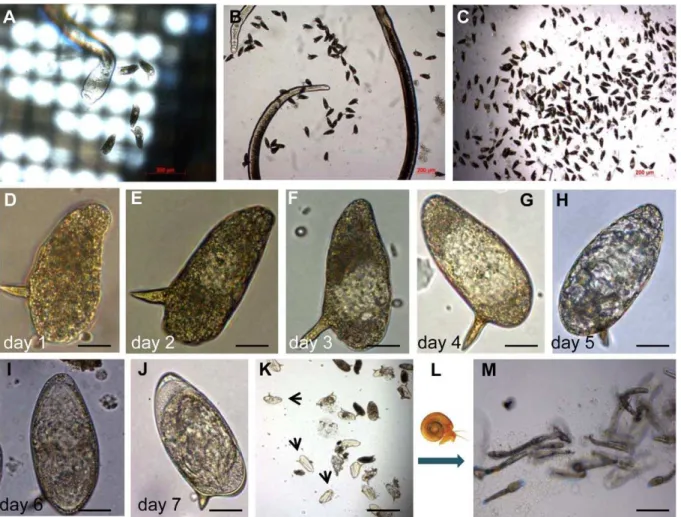
48 hours after perfusion. IVLE and, occa-sionally, adult females fall through the mesh and collect on the bottom of the culture plate (Figure 2A). At 48 hours after perfusion of the worms from mice, we collect IVLE, and concentrate by filtering media containing IVLE through 0.8-mm mesh transwell (BD Biosciences, catalog no. 353097) (Figure 2B, 2C). Thereafter, IVLE are maintained in schistosomule medium where they develop and mature within 7 days (Figure 2D–2J). At that point, we transferred IVLE to sterile water, illuminated the culture with a bright lamp, and observed that many eggs (30% to 40%) hatch within 120 minutes. We observed that miracidia hatched from IVLE infected Biomphalaria glabrata snails from which, in turn, cercariae were released after 6 weeks (Figure 2K–2M). IVLE represents a tractable, developmental stage at which to target transgenes, especially since in developmental stages 0 and 1 (staging system of Jurberg et al. [19]), no cleavage
of the zygote cell has yet taken place. In future studies, we plan to expose IVLE to pseudotyped MLV virions at the time eggs are released from the female schistosome, aiming to introduce transgenes into the schistosome germ line. (The studies with schistosome-infected laboratory mice were undertaken with the approval of the IAC-UC of The George Washington University, Washington, D.C.).
How Can We Increase the Likelihood of Chromosomal Integration?
For transposons, in particular for binary versions of broad host range vectors such aspiggyBacand Sleeping Beauty(e.g., [4,8]), increased efficiency of integration can be accomplished using mRNA encoding the transposase rather than using helper plasmid, and further, optimal ration of transposon and transposase can be titrat-ed. Also, transposase enzyme can be employed instead of mRNA of the gene
Figure 2. Representative pictures of the in vitro laid eggs (IVLE) collection, concentration, and in vitro development.(A) Female of
S. mansonireleasing eggs one day after perfusion. The mesh of the netwell is evident. (B) Female surrounded by IVLE in the bottom of the well. (C) IVLE during the concentration process 48 hours after perfusion. (D–J) Representative images of an individual egg laid in vitro through the developmental process from day 1 (D), 2 (E), 3 (F), 4 (G), 5 (H), and 6 (I) to day 7 (J) after perfusion. (K) IVLE during the hatching process; arrows indicate empty eggshells. (L) Diagram of snail infection. (M) Cercariae released from snails infected with miracidia from IVLE, at 42 days after snail infection. Scale bars: (A–C,K,M), 200mm; (D–J), 20mm.
[3]. For retroviruses, in particular MLV, with which we have some experience, increasing the titer of active virions is a sound way to improve prospects for productive transduction of target germ line cells. Given the progress with trans-genesis of schistosomes with pseudotyped MLV [9–11,20,23,24], comments on viri-on productiviri-on are included below.
Can virion production be optimiz-ed? There are several factors to consider for an optimal virus production, including producer (host) cell strain and culture conditions, cell density and vitality by the
time of the DNA transfection, amount and quality of plasmid DNA used for transfection, and recovery of viral particles. Factors that reduce the retrovirus half-life also should be considered, i.e., storage of virions, freeze-and-thaw cycles, temperature, pH, and presence of serum [25]. The higher the viral titer, the better the prospects are for chromosomal insertion of the proviral transgene [26]. Although protocols to produce retrovirus in vitro optimize to improve titers, contamination of virions with defective particles is a frequent problem. Defective virions include
particles without envelope, without RNA, or with RNA but non-infectious [27]. Using at least two approaches to estimate viral titer is recommended [25,27]. One approach should estimate the titer of infectious particles, i.e., a functional, biological assay where a cell line is infected with serial dilution of the virions and colonies of cells are selected by maintenance in antibiotic for which resistance is conferred by the retrovirus. Second, a quantitative approach to estimate the copy number of particles by qPCR should also be performed in parallel. With findings from the parallel assay, total Figure 3. Correlating RNA titer with infectivity. Schematic diagram of two MLV and HIV representative constructs and corresponding micrographs of the particles (from Higashikawa et al. [25], with permission). To determine infectivity titers, NIH 3T3 or HT1080 cells were infected with qRT-PCR-titrated MLV retrovirus or qRT-PCR-titrated HIV, carrying neomycin (Neo) or blasticidin (Blast) resistance genes, respectively. Cells were selected in G418 or blasticidin for 10 days and resistant colonies were stained and counted.
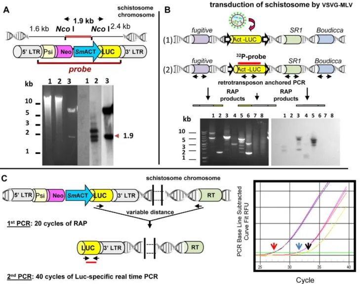
Figure 4. Approaches to identify, clone, and quantify integrated sequences into the schistosome genome.(A) Integration of retroviral provirus into theS. mansonigenome indicated by Southern hybridization analyses of genomic DNA from retrovirus-transduced schistosomes.Top Panel: Schematic representation of retroviral construct pLNHX-SmACT-Luc, showing the position ofNcoI cleavage sites and also the location of the
KpnI fragment employed as the hybridization probe. The retrovirus cassette included the firefly luciferase reporter gene (yellow) driven by the
number of virions particles and of intact, infectious particles can be established [25,27]. We use both approaches to estimate the viral titer and the ratio between copy number of particles and infectious units [24]. Figure 3 presents representative findings from our labo-ratory, for both MLV and HIV pseudotyped virions; in general, titers estimated by qPCR were two to three orders of magnitude greater than those estimated in biological assays. This is not only because qPCR is more sensitive, but also because qPCR detects non infec-tive particles. Accordingly, the lower the titration ratio, the more efficient is the virus production in terms of viable infectious particles.
How Can We Analyze Integration of Transgenes into the Genome?
Integration is a pivotal step in estab-lishing transgenic schistosomes. To con-firm that vectors can integrate into schistosome DNA, methods to deter-mine and extract integration junctions have to be employed. Integration junc-tions cannot be obtained simply by regular PCR procedures because the genomic flanking sequences are un-known. Southern hybridization employ-ing informative restriction enzymes and probes retain a key position in these studies. For example, Figure 4A presents a Southern hybridization analysis to confirm the presence of proviral trans-genes in the genome of schistosomules exposed to pseudotyped MLV virions. However, the definitive proof of integra-tion of the transgenes into the schisto-some chromoschisto-some requires the use of PCR-based approaches directed at clon-ing and sequencing the integration junction (Figure 4B). Given that draft genomes of S. mansoni and S. japonicum
are available, BLAST analysis of cloned sequences flanking the transgenes (e.g., MLV retrovirus) can readily verify that the transgenes have integrated into a schistosome chromosome. A number of PCR-based methods can be employed to recover integration events and unknown host genome sequences flanking the transgene. These include inverse PCR, linker ligation PCR and thermal
asym-metric interlaced (TAIL)-PCR, and Alu -PCR [28,29]. We developed an Alu -PCR-like approach termed retrotranspo-son anchored PCR (RAP) [8,9], which relies on anchoring primers to multi-copy endogenous mobile genetic ele-ments interspersed in the schistosome genome to locate integration junctions of transgenes in the genome of S. mansoni
(Figure 4B). Furthermore, we have adapted RAP for quantitative PCR in order to determine comparatively the number of MLV transgenes within the genome of transduced populations of schistosomes (Figure 4C) [24]. In addi-tion, very high-throughput sequencing using Illumina technology could be utilized, as has been demonstrated for transposon-based insertional mutagene-sis of Salmonella Typhi [3]. This latter approach could determine the exact location of transgenes within the schis-tosome genome.
Outlook
As noted, transgenic schistosomes have been created (e.g., [7–9,23]). However, improvements are needed—and certain to take place—in order to establish transgen-ic schistosomes and protocols. A crucial impediment to date has been the difficulty
of delivering transgenes to chromosomes of the germ line. Targeting integration-competent vectors to IVLE may surmount this limitation. Moreover, there are other points in the developmental cycle where the germ line might be accessed, including the daughter sporocysts where the germ cells are comparatively large (Coustau and Yoshino [30] and references therein). We can look forward to advances in technol-ogies that will drive functional genomics forward quickly, including expansion of in vivo RNAi, high-throughput insertional mutagenesis and, hopefully, gains-of-func-tion approaches involving drug selecgains-of-func-tion of transgenic schistosomes. Advances in S. mansonican be expected to be adapted to the other schistosomes, to the food-borne flukes such as Fasciola and Opisthorchis
species, and to neglected helminth para-sites at large.
Supporting Information
Alternative Language Abstract S1 Translation of Abstract into Thai by Sutas Suttiprapa.
(PDF)
Alternative Language Abstract S2 Translation of Abstract into Spanish by Gabriel Rinaldi.
(PDF)
transgene in schistosome genomic DNAs was calculated by interpolation from a standard curve.Right Panel:Amplification plots observed in MLV-transduced worms in preamplified template usingSR2primer mix (red arrow), in preamplified template using only the luciferase transgene–specific primer as control of ‘‘one-way amplification’’ (blue arrow), and in non-preamplified template (black arrow). Arrows indicate the threshold cycle. RT: endogenous retrotransposon. (Modified from Rinaldi et al. [24], with permission.)
doi:10.1371/journal.pntd.0001230.g004
Key Learning Points
N
Draft genome sequences forAccordingly, there is a pressing need now to develop functional genomics toolsS. mansoniand S. japonicumare now available. for schistosomes to determine the importance of these new genes.N
Functional genomics approaches hold promise to determine the nature andimportance of genes of the human schistosomes.N
Retroviral and transposon-mediated gene manipulation, using integration-competent, vector-based technologies, have been shown to be feasible for schistosomes.N
The retrovirus murine leukemia virus, which is widely used in stem cell andgene therapy research, and thepiggyBactransposon, originally isolated from the genome of a moth, have now been shown to be active in schistosomes, to integrate into schistosome chromosomes, and to provide gains-of-function for the reporter genes firefly luciferase, GFP, and neoR.N
Both MLV andmutagenesis studies, feasible now that draft genome sequences are available.piggyBac both have potential in high-throughput insertionalAcknowledgments
The authors thank Tunika Okatcha, Mary Ayuk, Yousef Alrefaei, and Danielle Skinner
for helpful discussions, and Meredith Brindley for help with the artwork.
References
1. Schistosoma japonicum Genome Sequencing and Functional Analysis Consortium (2009) The Schistosoma japonicum genome reveals features of host-parasite interplay. Nature 460: 345– 351.
2. Berriman M, Haas BJ, LoVerde PT, Wilson RA, Dillon GP, et al. (2009) The genome of the blood fluke Schistosoma mansoni. Nature 460: 352–358. 3. Langridge GC, Phan MD, Turner DJ, Perkins TT, Parts L, et al. (2009) Simultaneous assay of every Salmonella Typhi gene using one million transpo-son mutants. Genome Res 19(12): 2308–2316. 4. Ivics Z, Li MA, Ma´te´s L, Boeke JD, Nagy A, et al.
(2009) Transposon-mediated genome manipula-tion in vertebrates. Nature Methods 6(6): 415–422.
5. Sliva K, Schnierle BS (2010) Selective gene silencing by viral delivery of short hairpin RNA. Virol J 7: 248.
6. Chamberlin HM (2010) C. elegans select. Nat Methods 7: 693–694.
7. Beckmann S, Wippersteg V, El-Bahay A, Hirzmann J, Oliveira G, et al. (2007) Schistosoma mansoni: germ-line transformation approaches and actin-promoter analysis. Exp Parasitol 117(3): 292–303.
8. Morales ME, Mann VH, Kines KJ, Gobert GN, Fraser MJ, Jr., et al. (2007) piggyBac transposon mediated transgenesis of the human blood fluke, Schistosoma mansoni. FASEB J 21: 3479– 3489.
9. Kines KJ, Morales ME, Mann VH, Gobert GN, Brindley PJ (2008) Integration of reporter trans-genes into Schistosoma mansoni chromosomes mediated by pseudotyped murine leukemia virus. FASEB J 22: 2936–2948.
10. Kines KJ, Mann VH, Morales ME, Shelby BD, Kalinna BH (2006) Transduction of Schistosoma mansoni by vesicular stomatitis virus glycopro-tein-pseudotyped Moloney murine leukemia ret-rovirus. Exp Parasitol 112(4): 209–220. 11. Yang S, Brindley PJ, Zeng Q, Li Y, Zhou J, Liu Y,
et al. (2010) Transduction of Schistosoma japo-nicum schistosomules with vesicular stomatitis
virus glycoprotein pseudotyped murine leukemia retrovirus and expression of reporter human telomerase reverse transcriptase in the transgenic schistosomes. Mol Biochem Parasitol 174(2): 109–116.
12. Nkrumah LJ, Muhle RA, Moura PA, Ghosh P, Hatfull GF, et al. (2006) Efficient site-specific integration in Plasmodium falciparum chromo-somes mediated by mycobacteriophage Bxb1 integrase. Nature Methods 3(8): 615–621. 13. O’Neill MT, Phuong T, Healer J, Richard D,
Cowman AF (2011) Gene deletion from Plasmo-dium falciparum using FLP and Cre recombi-nases: implications for applied site-specific recom-bination. Int J Parasitol 41: 117–123. 14. Mann VH, Morales ME, Rinaldi G, Brindley PJ
(2010) Culture for genetic manipulation of developmental stages of Schistosoma mansoni. Parasitology 137: 451–462.
15. Bixler LM, Lerner JP, Ivanchenko M, Mc-Cormick RS, Barnes DW, et al. (2001) Axenic culture of Schistosoma mansoni sporocysts in low O2 environments. J Parasitol 87: 1167–1168. 16. Heyers O, Walduck AK, Brindley PJ, Bleiss W,
Lucius R, et al. (2003) Schistosoma mansoni miracidia transformed by particle bombardment infect Biomphalaria glabrata snails and develop into transgenic sporocysts. Exp Parasitol 105: 174–178.
17. Mann VH, Morales ME, Kines KJ, Brindley PJ (2008) Transgenesis of schistosomes: approaches employing mobile genetic elements. Parasitology 135: 141–153.
18. Castelletto ML, Massey HC, Jr., Lok JB (2009) Morphogenesis of Strongyloides stercoralis infec-tive larvae requires the DAF-16 ortholog FKTF-1. PLoS Pathog 5: e1000370. doi:10.1371/ journal.ppat.1000370.
19. Jurberg AD, Gonc¸alves T, Costa TA, de Mattos AC, Pascarelli BM, et al. (2009) The embryonic development of Schistosoma mansoni eggs: proposal for a new staging system. Dev Genes Evol 219(5): 219–234.
20. Kines KJ, Rinaldi G, Okatcha TI, Morales ME, Mann VH, et al. (2010) Electroporation facilitates introduction of reporter transgenes and virions into schistosome eggs. PLoS Negl Trop Dis 4(2): e593. doi:10.1371/journal.pntd.0000593. 21. Freitas TC, Jung E, Pearce EJ (2007) TGF-beta
signaling controls embryo development in the parasitic flatworm Schistosoma mansoni. PLoS Pathog 3: e52. doi:10.1371/journal.ppat.0030052. 22. Kadandale P, Chatterjee I, Singson A (2009) Germline transformation of Caenorhabditis ele-gans by injection. Methods Mol Biol. 518: 123–133.
23. Tchoubrieva EB, Ong PC, Pike RN, Brindley PJ, Kalinna BH (2010) Vector-based RNA interfer-ence of cathepsin B1 in Schistosoma mansoni. Cell Mol Life Sci 76: 3739–3748.
24. Rinaldi G, Suttiprapa S, Brindley PJ (2011) Quantitative retrotransposon anchored PCR confirms transduction efficiency of transgenes in adult Schistosoma mansoni. Mol Biochem Para-sitol 177: 70–76.
25. Higashikawa F, Chang L (2001) Kinetic analyses of stability of simple and complex retroviral vectors. Virology 280(1): 124–131.
26. Ohishi M, Shioda T, Sakuragi J (2007) Retro-transduction by virus pseudotyped with glycopro-tein of vesicular stomatitis virus. Virology 362: 131–138.
27. Carmo M, et al. (2004) Quantitation of MLV-based retroviral vectors using real-time RT-PCR. J Virol Methods 119(2): 115–119.
28. Liu Y-G, Chen Y (2007) High-efficiency thermal asymmetric interlaced PCR for amplification of unknown flanking sequences. BioTechniques 43: 649–656.
29. O’Doherty U, Swiggard WJ, Jeyakumar D, McGain D, Malim MH (2002) A sensitive, quantitative assay for human immunodeficiency virus type 1 integration. J Virol 76(21): 10942–10950.