SATELLITE CELLS AND THE MUSCLE STEM CELL
NICHE
Hang Yin, Feodor Price, and Michael A. Rudnicki
Regenerative Medicine Program, Ottawa Hospital Research Institute, Ottawa, Ontario, Canada
L
Yin H, Price F, Rudnicki MA.Rev93: 23– 67, 2013; doi:10.1152/physrev.00043.2011.—Adult skeletal muscleSatellite Cells and the Muscle Stem Cell Niche.Physiol in mammals is a stable tissue under normal circumstances but has remarkable ability to repair after injury. Skeletal muscle regeneration is a highly orchestrated process involving the activation of various cellular and molecular responses. As skeletal muscle stem cells, satellite cells play an indispensible role in this process. The self-renewing proliferation of satellite cells not only maintains the stem cell population but also provides numerous myogenic cells, which proliferate, differentiate, fuse, and lead to new myofiber formation and reconstitution of a functional contractile apparatus. The complex behavior of satellite cells during skeletal muscle regeneration is tightly regulated through the dynamic interplay between intrinsic factors within satellite cells and extrinsic factors constituting the muscle stem cell niche/microenvironment. For the last half century, the advance of molecular biology, cell biology, and genetics has greatly improved our understanding of skeletal muscle biology. Here, we review some recent advances, with focuses on functions of satellite cells and their niche during the process of skeletal muscle regeneration.I. INTRODUCTION: SATELLITE CELLS AS... 23
II. FUNCTIONS OF SATELLITE CELLS IN... 30
III. ANATOMIC AND FUNCTIONAL... 41
IV. CONCLUDING REMARKS AND... 53
I. INTRODUCTION: SATELLITE CELLS AS ADULT STEM CELLS IN MUSCLE
Skeletal muscle is a form of striated muscle tissue, account-ing for!40% of adult human body weight. Skeletal muscle is composed of multinucleated contractile muscle cells (also called myofibers). During development, myofibers are formed by fusion of mesoderm progenitors called myo-blasts. In neonatal/juvenile stages, the number of myofibers remains constant, but each myofiber grows in size by fusion of satellite cells, a population of postnatal muscle stem cells. Adult mammalian skeletal muscle is stable under normal conditions, with only sporadic fusion of satellite cells to compensate for muscle turnover caused by daily wear and tear. However, skeletal muscle has a remarkable ability to regenerate after injury. Responding to injury, skeletal mus-cle undergoes a highly orchestrated degeneration and regen-erative process that takes place at the tissue, cellular, and molecular levels. This results in the reformation of inner-vated, vascularized contractile muscle apparatuses. This re-generation process greatly relies on the dynamic interplay between satellite cells and their environment (stem cell niche). During the last half century, advances in molecular biology, cell biology, and genetics has greatly improved our understanding of skeletal muscle regeneration. In particu-lar, extensive research on satellite cells and their niche has
elucidated many cellular and molecular mechanisms that underlie skeletal muscle regeneration. These studies have contributed to the development of therapeutic strategies. These strategies serve to alleviate the physiological and pathological conditions associated with poor muscle regen-eration observed in sarcopenia and muscular dystrophy.
Here, we concentrate on the functions of satellite cells and the regulation of their niche during the process of skeletal muscle regeneration. We first describe the current under-standing of satellite cells with respect to their characteris-tics, heterogeneity, and embryonic origin. We then provide an integrated view of the roles played by satellite cells dur-ing muscle regeneration and normal postnatal muscle growth. We also discuss the contribution of several nonsat-ellite cell populations in muscle regeneration and their lin-eage relationships with satellite cells. Next, we focus on the satellite cell niche with emphasis on the regulatory mecha-nisms associated with each niche component. We further review the links between malfunction of satellite cells and their niche factors during aging. This review focuses on satellite cells and their niche in mammalian models, paying limited attention to the studies of satellite cell biology in other model organisms.
A. A Brief History of Satellite Cells
and intimate association with the plasma membrane of myofibers.
The direct juxtaposition of satellite cells and myofibers im-mediately raised a hypothesis that these cells may be in-volved in skeletal muscle growth and regeneration (329). Indeed, experiments by [3H]thymidine labeling and elec-tron microscopy demonstrated that satellite cells undergo mitosis, assume a cytoplasm-enriched morphology, and contribute to myofiber nuclei (355, 437). Later on, [3
H]thy-midine tracing experiments indicated that satellite cells are mitotically quiescent in adult muscle but can quickly enter the cell cycle following muscle injury (499). The same study also demonstrated that satellite cells give rise to proliferat-ing myoblasts (myogenic progenitors cells), which were pre-viously shown to form multinucleated myotubes in vitro (276, 499, 574). More definitive evidence came from in vitro cultures of individually dissected myofibers, whereby the behaviors of single myofibers and their resident satellite cells during regeneration can be tracked by phase-contrast microscopy (51, 277). It was observed that myofiber necro-sis is accompanied by satellite cell outgrowth, clonal expan-sion, and later fusion to form functional regenerated myo-tubes. These experiments support the notion that it is the satellite cell, rather than the myonuclei, that contribute to postnatal muscle growth and repair.
The pivotal function of the satellite cell in muscle regener-ation stimulated research to determine their regulatory mechanisms. This started with the finding that quiescent satellite cells on single myofibers were activated to enter the cell cycle by a mitogen originating from crushed skeletal muscles (53). In search of this mitogen, it was found that the proliferation and differentiation of satellite cell-derived myoblasts cultured in vitro respond to various growth fac-tors in a dose-dependent manner (10, 11). Numerous studies further revealed that the proliferation and differentiation of satellite cells during muscle regeneration is profoundly influ-enced by innervation, the vasculature, hormones, nutrition, and the extent of tissue injury (121, 224, 250, 332, 360, 410). Interestingly, satellite cells in contact with the plasma mem-brane of intact myofibers were found to have reduced sen-sitivity to mitogen stimulation (50). All of these findings led to an intriguing notion that satellite cells reside in a special microenvironment in vivo, which profoundly affects their behavior.
By definition, stem cells found in adult tissues can both replicate themselves (self-renew) and give rise to functional progeny (differentiate). Although the ability of satellite cells to differentiate into myofibers was clearly proven, their ability to self-renew was once questioned. The first evidence of satellite cell self-renewal came from a single myofiber transplantation assay. It was found that as few as seven satellite cells, when transplanted into irradiated regenera-tion-insufficient mice along with their resident single
myo-fiber, can give rise to hundreds of satellite cells and thou-sands of myonuclei. Most importantly, transplanted satel-lite cells on a single myofiber were able to support subsequent rounds of muscle regeneration (105). These ob-servations demonstrate that satellite cells are bona fide mus-cle stem cells.
Asymmetric division is a common manner of stem cell self-renewal. Close examination of satellite cell divisions on single myofibers revealed that satellite cells can undergo both asymmetric division and symmetric division (280). The fashion of symmetric versus asymmetric division largely depends on the relative position of the daughter cells in relation to the myofiber. This finding strongly argues that satellite cell self-renewal is governed by the structure and signaling present in their immediate niche (280). Further investigation has revealed an important role for noncanoni-cal Wnt signaling in the regulation of satellite cell self-re-newal (293).
The aforementioned observations, together with those from other studies, formed the basis of our current view of satel-lite cells as muscle stem cells whose functions are dictated by their surrounding niche.
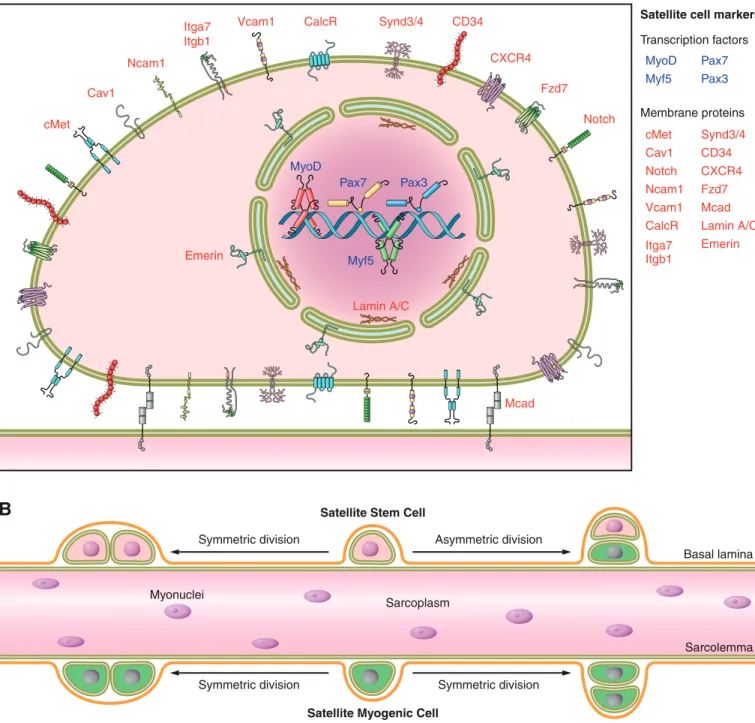
B. Identification and Isolation of Satellite Cells
en-velope proteins lamin A/C and emerin (192). Of these, Pax7 is the canonical biomarker for satellite cells as it is specifi-cally expressed in all quiescent and proliferating satellite cells (478) across multiple species, including human (334),
monkey (334), mouse (478), pig (402), chick (216), sala-mander (353), frog (96), and zebrafish (219). Of note, some of the aforementioned satellite cell markers (e.g., ! 7-in-tergin, CD34) are also expressed on other cell types within
A
B
Synd3/4 CalcR
cMet
Itga7 Itgb1
Ncam1
MyoD
Myf5
Pax7 Pax3
MyoD Myf5
Pax7 Pax3
Emerin
Mcad Lamin A/C
Cav1
cMet Cav1 Notch Ncam1 Vcam1 CalcR Itga7 Itgb1
Synd3/4 CD34 CXCR4 Fzd7 Mcad Lamin A/C Emerin
Vcam1 CD34
CXCR4
Fzd7
Notch
Basal lamina Transcription factors
Membrane proteins
Asymmetric division Satellite Stem Cell
Satellite cell markers
Satellite Myogenic Cell
Sarcolemma Sarcoplasm
Myonuclei
Symmetric division
Symmetric division Symmetric division
skeletal muscle, and thus should not be utilized alone to unequivocally identify satellite cells. Satellite cells can be identified by fluorescence microscopy via combined immu-nofluorescence labeling of laminin and M-cadherin, which respectively label the basal lamina and plasma membrane of satellite cells contacting the myofibers (517). In vivo, satel-lite cell populations can be visualized with the aid of newly developed bioluminescence imaging techniques (454, 558).
Multiple methodologies have been developed to isolate sat-ellite cells from skeletal muscle. The choice of methods largely depends on the isolation scale and the subsequent experiment. In small scale, limited amounts of satellite cells can be released from single myofiber explants by physical trituration (446) or enzymatic digestion (107). In large scale, satellite cells can be isolated from skeletal muscles by fluorescent-activated cell sorting (FACS). In the latter method, single cells are released from muscle tissue chunks by enzymatic digestion, followed by immunofluorescence labeling of satellite cell-specific cell surface markers (posi-tive selection) and defini(posi-tive cell surface markers for non-satellite cell populations (negative selection). As Pax7 is specifically expressed in satellite cells within skeletal mus-cle, satellite cells, in both quiescent and proliferating stages, can be FACS-sorted by fluorescent protein expression in tamoxifen-injected animals carrying both aPax7CreERallele and a fluorescence Cre reporter allele (e.g.,R26R-EYFP). In addition, two transgenic mouse strains, which carry fluo-rescence proteins driven by Nestin- or Pax7-regulatory ele-ments, are also useful for isolating satellite cells by FACS (63, 127).
Protocols have been developed to isolate satellite cells in bulk utilizing their characteristic proliferation and adhesion characteristics under defined culturing conditions (144, 364, 425). It is noteworthy that the satellite cell progeny cultured on regular collagen-coated plastic dishes and un-der activation conditions, also called satellite cell-un-derived primary myoblasts, are molecularly and functionally dis-tinct from the satellite cells freshly isolated from muscles (160, 177, 350). In addition, the gene expression profile of these cultured primary myoblasts differs from that of acti-vated satellite cells in vivo (398). These essential differences are possibly due to the lack of a satellite cell niche in in vitro culturing conditions. One challenge in future studies is to understand the components and role of the satellite cell niche to better control and manipulate their quiescence, activation, proliferation, and differentiation.
C. Heterogeneity of Satellite Cell Population
Satellite cells were considered to be a homogeneous popu-lation of committed muscle progenitors (52). However, re-cent evidence demonstrated that the satellite cell population is heterogeneous and that satellite cells differ in their gene expression signatures, myogenic differentiation propensity,
stemness, and lineage potential to assume nonmyogenic fates.
With regard to gene expression, it was first observed that only a subset of satellite cells expresses Pax3, a close para-log to Pax7 (350, 431). Although Pax3"satellite cells tend
to be associated with skeletal muscle of certain anatomical locations (e.g., diaphragm and truck muscles), the differ-ence of Pax3 expression does not seem to correlate with their embryonic origins, metabolic fiber types, or types of innervating motor neurons. Examination of the expression of satellite cell markers CD34, M-cadherin, and Myf5 by immunofluorescence staining revealed that a subpopulation of satellite cells do not express these markers (37). Similarly, human satellite cells also manifest heterogeneity in their Pax7, neuronal cell adhesion molecule (NCAM), c-Met, and Dlk1 expression (311). By RT-qPCR, two recent stud-ies showed that satellite cells from head or body muscles are distinct in their molecular signatures (225, 456). Notably, the heterogeneity of gene expression in single satellite cells was investigated in a recent study, wherein the expression of Pax7, Pax3, Myf5, and MyoD was interrogated by RT-qPCR for 40 individually FACS-isolated muscle stem cells (CD45#/CD11b#/CD31#/Sca-1#/!7-intergin"/CD34")
(454). All of these muscle stem cells express Pax7, indicat-ing they are satellite cells. In addition to the varied expres-sion of Pax3, it was also found that 25% of investigated satellite cells also express MyoD, a basic helix-loop-helix transcription factor critical for myogenic commitment and differentiation. A recent study discovered a small subpopu-lation of satellite cells, characterized by their surface mark-ers Sca-1"/ABCG2" (as compared with Sca-1#/ABCG2#
for most satellite cells), is able to exclude Hoechst 33342 dye and thus belongs to the skeletal muscle side population (satellite-SP cells) (518). After transplantation into dam-aged muscle, satellite-SP cells can both fuse to regenerating myofibers and return to quiescence in the satellite cell niche. However, by lineage tracing, ABCG2"cells (including
sat-ellite-SP cells) seem to only have minor contribution to myofiber regeneration (146).
vivo revealed two satellite cell populations that are distinct in terms of their mitotic rates (471). It was found that the majority (!80%) of satellite cells readily enter the cell cycle (responsive population), whereas the remaining 20% of sat-ellite cells (reserve population) do this in a much slower manner. It has been proposed that the reserve population maintains in the quiescent state at the beginning of muscle growth/regeneration and only moves into this proliferative state in response to the need for extensive muscle growth/ regeneration (471). In line with this view, a recent study traced satellite cell divisions by a fluorescent dye PKH26, which revealed the minority of PKH26high slow-dividing satellite cells retained long-term self-renewal ability (388). Notably, a recent study thoroughly compared the gene ex-pression profiles and proliferation/differentiation potential of satellite cells isolated from limb and facial muscles of adult and aged mice (387). This study revealed broad satel-lite cell heterogeneity at both the population and single-cell levels. Despite their heterogeneous gene expression profiles, satellite cells isolated from limb (EDL) and facial (masseter) muscles are comparable in their ability to repair a limb (TA) muscle injury after transplantation. This finding suggests that although satellite cells from various muscles have dis-tinct gene expression and behaviors in vitro, their regener-ation potential in vivo might be largely determined by host stem cell niche and microenvironment.
Most importantly, recent studies revealed satellite cell hetero-geneity in terms of their stemness and indicate that only a small percentage of satellite cells are true stem cells (109, 280, 489). By immunofluorescence staining of freshly isolated single myofibers fromMyf5-nLacZmice, our group demonstrated that 13% of quiescent satellite cells on EDL muscles are LacZ#, making them distinct from the LacZ" satellite cells
(280). As Myf5 is a myogenic regulatory factor, the absence of Myf5 expression suggests a less committed cell fate for those LacZ#satellite cells. Moreover, by the Cre-LoxP based lineage
tracing technique, our group further discovered that 10% of satellite cells inMyf5Cre;R26R-YFPmice do not express YFP (Pax7"/YFP#satellite cells), indicating that this small percent
of satellite cells have never expressed Myf5 as did the majority of satellite cells (Pax7"/YFP"). Remarkably, these two distinct
satellite cell subpopulations also differ in terms of their regen-erative potential: the Pax7"/YFP#satellite cells were able to
reconstitute the stem cell niche and repaired muscles in a sus-tainable manner, whereas the Pax7"/YFP"satellite cells
di-rectly underwent myogenic differentiation when transplanted in regenerating muscles ofPax7#/#mice. We further
demon-strated that only Pax7"/YFP# satellite cells could undergo
asymmetric cell divisions, giving rise to a Pax7"/YFP#satellite
stem cell and a Pax7"/YFP" satellite committed progenitor
cell. These findings indicate that a hierarchical lineage progres-sion from the Pax7"/YFP#(satellite stem cell) to the Pax7"/
YFP"(satellite committed progenitors) exists within the total
satellite cell population. Consistent with this notion, multiple studies reported that only a subset of satellite cells undergo
asymmetric division in vivo or in vitro (108, 109, 489). For example, Numb, an inhibitor of Notch signaling and a cell-fate determinant, was found to asymmetrically distribute in some but not all satellite cell divisions (108, 489). By pulse labeling or tandem pulse labeling of growing/regenerat-ing muscles with halogenated thymidine analogs, it was further discovered that all “older” chromosomes (the template chromosome during DNA replication during S phase) cosegregate into the more stemlike daughter cell, whereas “younger” chromosomes are inherited by the more differentiated daughter cell during asymmetric divi-sions of satellite cells both in vivo and in vitro (109, 489). According to the “immortal DNA strand” hypothesis (75), this preferential retention of “older” chromosomes protects stem cells against the accumulation of mutations intro-duced during DNA replications (109, 489). Alternatively, it has been also proposed that the nonrandom segregation of sister chromatids, and hence their different epigenetic states, is essential to the gene expression patterns and cel-lular fates of satellite stem cells and satellite progenitor cells (289). In summary, the aforementioned findings support satellite cell heterogeneity whereby the existence of a hier-archy delineates a small population of true stem cells (sat-ellite stem cells) from a more committed myogenic progen-itor population of satellite cells. The satellite stem cells are less committed to the myogenic lineage and tend to retain older template DNA during division. Through asymmetric division, satellite stem cells self-renew to replenish the stem cell pool and produce more committed myogenic progenitors that par-ticipate in skeletal muscle growth and regeneration.
their differential expression of cell surface markers, CD45 and Sca-1. It was found that the vast majority of myogenic satellite cells are within the CD45#/Sca-1#population and exhibited
no in vitro differentiation potential into fibroblasts or adi-pocytes. In contrast, the minor population of CD45#/Sca-1"
satellite cells can differentiate into both fibroblasts and adi-pocytes in culture, a similarity that is shared with CD45#/
Sca-1"mesenchymal stem cells found in multiple tissues (316,
401, 417, 441, 509, 519). Despite the plasticity of satellite cells in vitro, it is important to note that myogenesis is the predom-inant fate of satellite cells in vivo as fibrosis or adipose infiltra-tion is not normally observed in young healthy muscle. Fur-thermore, recent studies indicate that intramuscular adi-pocytes and fibroblasts can also arise from fibrocyte/adipocyte progenitors (FAPs), which reside in the muscle interstitium (252, 541; and discussed in sect. IIIB1). Indeed, lineage tracing experiments indicate that adipocytes derived from myofibers isolated fromMyoDiCre;R26R-EYFP mice have never tran-scribed MyoD (507). As the vast majority of adult satellite cells were permanently labeled with EYFP in this experiment, this finding argues that most satellite cells from myofiber cultures do not spontaneously differentiate into adipocytes. Further investigation with more definitive lineage tracing (e.g., Pax7CreER;R26R-EYFP) methods would clarify the exact con-tribution of satellite cells to other nonmyogenic lineages both in vitro and in vivo.
As discussed here, multiple lines of evidence demonstrate that satellite cells represent a heterogeneous population. However, our understanding of this heterogeneity is far from complete. First, although several markers can separate the total satellite cell population into functional subpopulations, it is still un-known whether these subpopulations are homogeneous in their function and gene expression. Further studies to identify additional satellite markers will potentially help distinguish the various satellite cell lineages. Moreover, future inves-tigations should attempt to identify the intrinsic differ-ences between satellite cell subpopulations at the molec-ular and functional levels during muscle regeneration. Such findings would elucidate regulatory mechanisms gov-erning the transition between different satellite cell lations and potentially distinct roles of satellite cell subpopu-lations during muscle regeneration. In addition, it would be of great importance to understand the dynamics of satellite cell heterogeneity in response to various environmental cues with regard to research in muscle regeneration and disease.
D. Variance of Satellite Cells Number and Location
In addition to the heterogeneity of satellite cells, the quantity of satellite cells differs between muscles, myofiber types, develop-mental stages, and species. In general, satellite cells account for 30 –35% of the sublaminal nuclei on myofibers in early post-natal murine muscles, and this number declines to 2–7% in adult muscles (9, 227, 450, 469). In adults, the percentage of
satellite cells in soleus muscle is generally two- to fourfold higher than that in tibialis anterior muscle or EDL muscle (190, 466, 500). Within the same muscle, the number of satellite cells found on slow muscle myofibers (type I) is generally higher than those on fast myofibers (type IIa and type IIb) (190, 324, 383). The biological meaning and the potential regulatory mechanisms underlying these phenom-ena are poorly understood. However, it is conceivable that these variances may reflect intrinsic heterogeneity of satel-lite cells on different myofibers and implicate a potential role of myofibers as a niche factor in regulating the homeo-stasis of their resident satellite cells.
Along a myofiber, the distribution of satellite cells is not random. It has been reported that the density of satellite cells is higher at the ends of the myofibers, where the longi-tudinal growth of skeletal muscles happens (14). A higher incidence of satellite cells has been observed at perisynaptic regions compared with that at extrasynaptic regions (190, 269, 567). Moreover, satellite cells have been observed in close proximity to capillaries (100, 466). In fact, 88% of satellite cells in adult human muscles were observed to be located within a 21-#m distance of a capillary (100). This tight association with capillaries seemed to be compromised by denervation (130). These observations indicate that homing of satellite cells is influenced by their niche, both by local motor neurons and blood vessels (discussed in sect. IIIB).
It is noteworthy that some reported variation in satellite cell numbers may be partially due to techniques or statistical analysis employed in satellite cell counting. For example, satellite cell counting based on immunofluorescence label-ing of satellite cell specific markers relies on the comparable expression levels of these markers on all satellite cells under investigation. As such, special caution should be taken into account when interpreting data between independent ex-periments.
E. Origins of Adult Satellite Cells
1. Embryonic origins
By classic techniques of developmental biology, it has long been established that skeletal muscles within both the adult trunk and limbs develop from embryonic somites (462). However, the exact origin of adult satellite cells was ob-scure until recently.
compart-ment was determined using Feulgen staining, which distin-guishes quail-specific interphase nuclei from those of chick. It was found that donor cells from quail somites integrated into the chick limb and contributed to both terminally dif-ferentiated muscle fibers and satellite cells. This finding in-dicated a common somitic origin for all myogenic cell lin-eages, including satellite cells. However, the progenitor cell types at the origin and the developmental route remain unknown.
Advances in mouse genetics, particularly the generation of Pax3 and Pax7 knock-in reporter alleles, allows precise tracing of Pax3/Pax7-expressing myogenic progenitor cells during muscle development in a temporal and spatial man-ner (265, 326, 350, 432, 433). These reporters together with labeling of cells by electroporation (40, 203), retrovi-rus (464), Cre-LoxP based lineage tracing (263, 300, 464), and traditional quail-chick transplantation (203, 464), jointly shed light on the embryonic origins of adult satellite cells. Accumulating evidence indicates that adult satellite cells originate from the dermomyotome (203, 265, 434, 464), an epithelial structure formed on the dorsal part of the somite. The dermomyotome contains multipotent pro-genitor cells, which eventually give rise to multiple adult tissues/cell types including dermal fibroblasts, endothe-lial cells, vascular smooth muscle, brown fat tissue, and all skeletal muscles of the trunk and limbs (71). The cell fate decisions largely depend on the relative position of these multipotent progenitor cells with respect to adja-cent tissues such as the notochord, neural tube, dorsal ectoderm, and myotome (71).
Embryonic muscle development takes place in two succes-sive stages. During the first stage, a group of postmitotic mononucleated myocytes, expressing Myf5 and Mrf4, mi-grate out from the border regions of the dermomyotome and form primitive muscles beneath the dermomyotome (204, 261). These primitive muscles constitute the primary myotome and are the source of fetal and adult trunk mus-cles. During the second stage of muscle growth, the central portion of the dermomyotome undergoes an epithelial-to-mesenchymal transition (EMT). During EMT, tightly packed epithelial cells tease apart and turn in a loose mes-enchymal state prior to assuming different developmental fates: cells in the medial dermomyotome will develop into brown fat, dermis, and trunk muscle, while cells in the lateral dermomyotome will give rise to endothelia and limb muscles. The different cell fates assumed by the same group of progenitor cells are proposed to be due to asymmetric cell division (40, 101). EMT is accompanied by the extensive cell migration (203, 265, 434, 464). With the breakdown of dermomyotome, a group of proliferating progenitor cells, expressing both Pax3 and Pax7, migrate from the central region of the dermomyotome into the previously formed primary myotome. Upon arrival, some progenitor cells con-tinue to proliferate and replenish the progenitor pool. These
cells, which were absent from the primary myotome at ear-lier stages, count for the majority of all proliferating cells in embryonic/fetal trunk muscles (203, 434). Some of the pro-liferating Pax3"/Pax7"progenitor cells persist into late
fe-tal development stages and are enveloped beneath the basal lamina of developing myofibers (203, 434). These cells, which reside in the satellite cell compartment, are presumed to subsequently become the postnatal satellite cells in trunk muscles. Besides proliferation, progenitor cells also exit the cell cycle and begin differentiating into embryonic/fetal truck muscles. Cell cycle withdrawal is concomitant with the expression of the myogenic regulatory factors MyoD and Myf5 (453).
During EMT, another group of proliferating progenitor cells, expressing Pax3 (but not Pax7 in mouse), delaminate from the ventral-lateral border of the dermomyotome and migrate to the limb bud mesenchyme (265, 392, 464). These progenitor cells still maintain their multipotency as they give rise to the limb vascular, lymphatic endothelia, and limb muscles (226, 264). At E11.5 of mouse embryo devel-opment, some progenitor cells start to express Pax7 in the anterior limb buds (433). The expression of Pax7 specifies these cells to the myogenic lineage (242). Similar to the myotome-located progenitors, these Pax3"/Pax7"
progen-itor cells in limb buds undergo proliferation/differentiation while a portion withdraw from cell cycle and become satel-lite cells.
All together, these observations indicate that Pax3"/Pax7"
embryonic progenitor cells are the major source of adult satellite cells in truck and limb muscles; it is, however, note-worthy that the aforementioned observations from lineage tracing and immunofluorescence labeling experiments can-not exclude the possibility that some adult satellite cells may originate from other sources during fetal and postnatal muscle development. For example, embryonic dorsal aorta explants, when cultured and disaggregated in vitro, can efficiently give rise to myogenic precursors (129). These myogenic precursors are similar to satellite cells in their gross morphology and expression of molecular markers. Given that adult satellite cells also express endothelial markers, it was proposed that some adult satellite cells orig-inated from the embryonic dorsal aorta (129). However, recent studies revealed that both skeletal muscles and smooth muscles found in dorsal aorta are derived from the same Pax3" cell population in the paraxial mesoderm
(155). Thus it is also possible that the observed similarities are merely reminiscent of a common embryonic origin be-fore myogenic specification.
from occipital somites. Similar to progenitor cells migrating to limb buds, the progenitor cells for tongue delaminate from the ventral-lateral border of occipital somites. A small fraction of extraocular muscles also arise from the prechordal mesoderm (PM). On the basis of observations from lineage tracing experiments, it has been found that adult satellite cells of the various head muscles originate from their corresponding embryonic muscles and express distinct combinations of signature genes (225). Unlike trunk and limb progenitors, most progenitor cells for head muscles (except for tongue) express MesP1 rather than Pax3.
2. Alternative origins of adult satellite cells
Accumulating evidence indicates that some adult satellite cells may have alternative origins other than dermomyo-tome-derived Pax3"/Pax7"progenitor cells.
First, multiple studies have demonstrated that several types of nonsatellite cells can reconstitute the satellite cell niche and turn into bona fide satellite cells (Pax7-expressing myo-genic cells) after transplantation into regenerating skeletal muscles (for details, see sect. IID). It remains, however, largely unknown to what extent these cells contribute to the adult satellite cell pool and muscle development under physiological conditions. Notably, a recent study utilizing TN-APCreERT2andVE-cadherinCreERT2alleles showed that alkaline phosphatase (ALPL) expressing pericytes, but not VE-cadherin-expressing endothelial cells, can develop into postnatal satellite cells and participate in normal develop-ment of limb muscles (135).
It is noteworthy that some adult satellite cells in mammals may be derived from dedifferentiation of fetal/adult myofi-bers. It has long been established that the dedifferentiation of “terminally” differentiated multinucleated myofibers oc-curs in injured skeletal muscle of urodele amphibians, such as the newt (69). Nevertheless, it remains controversial as to whether mammalian myotubes in vitro or myofibers in vivo can undergo a similar dedifferentiation process. Studies uti-lizing immortalized murine myoblast cell lines (e.g., C2C12 or pmi28 cells) have shown that some myotubes formed by these cells can dedifferentiate into mononucleated myo-genic cells in the presence of protein extract from regener-ating newt muscles (331), bioactive compounds like myos-everin or its derivatives (149, 443), ciliary neurotrophic factor (CNTF) (95), or by genetic manipulations such as overexpression ofMsx1(379),Twist1(232),Barx2(335), or knockdown ofRb1in conjunction with a deficiency for Cdkn2a(p16Ink4a) (395). By a novel fusion-dependent lin-eage tracing technique, two recent studies reported that differentiated myotubes formed by satellite cell-derived pri-mary myoblasts (397) or muscle-derived cells (MDCs) (359) can dedifferentiate into Pax7-expressing mononu-cleated myogenic cells in vitro in response to an inhibitor cocktail (a tyrosine phosphatase inhibitor plus an apoptosis
inhibitor) or within regenerating muscle in vivo. Although still not experimentally tested on myofibers formed during development, these intriguing observations jointly suggest that some adult satellite cells may be “recycled” from multi-nucleated myofibers in vivo during postnatal muscle growth or regeneration. Future studies may employ the fusion-de-pendent lineage tracing technique in chimeric mice to inves-tigate the physiological relevance of this alternative source of satellite cells.
II. FUNCTIONS OF SATELLITE CELLS IN MUSCLE REGENERATION
Skeletal muscles consist of myofibers, neurons, vasculature networks, and connective tissues, of which the structural and functional element of skeletal muscle is the myofiber. Each myofiber is surrounded by the endomysium (also called the basement membrane or basal lamina). Bundles of myofibers are surrounded by the perimysium, while the en-tire muscle is contained within the epimysium. Each myofi-ber is anchored at its extremities to tendons or tendon-like fascia at the myotendinous junctions (MTJs) (531). Myofi-bers are composed of actin and myosin myofibrils repeated as a sarcomere, which is the basic functional unit of skeletal muscle. Responding to the signals from motor neurons, myofibers depolarize and release calcium from the sarco-plasmic reticulum (SR). This drives the movement of actin and myosin filaments relative to one another and leads to sarcomere shortening and muscle contraction.
Based on their physiological properties, skeletal muscle fibers can be grouped into a slow-contracting/fatigue-resistant type and a fast-contracting/fatigue-susceptible type. Myofibers also vary in terms of their myosin heavy chain (MyHC) isoforms (fast or slow) and metabolism types (oxidative or glycolytic). The choice of myosin gene expression is under the dynamic regulation of thyroid hormone and work load (reviewed in Ref. 28). Recent studies demonstrated that the specification of myosin expression is also regulated by intronic microRNAs within MyHC genes (545, 546).
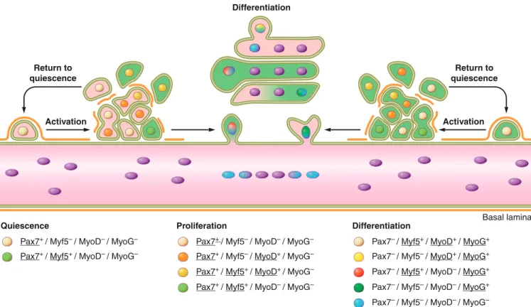
inflamma-tory responses, activation of satellite cells, proliferation, and differentiation of satellite cell-derived myoblasts(FIGURE 2).
This process, starting from myofiber necrosis and ending with new myofiber formation, is called muscle regeneration. It should be stressed that satellite cells play a pivotal role during muscle regeneration under either physiological conditions (e.g., extensive exercise) (400, 457) or pathological conditions (e.g., myotoxin induced injury) (301, 361, 457). This notion is clearly supported by the findings that ablation of the total satellite cell pool (all Pax7" cells) in adulthood completely
abolished muscle regeneration (301, 361, 457). It has been reported that several types of nonsatellite cells can undergo myogenic differentiation and contribute to muscle regenera-tion after transplantaregenera-tion into regenerating muscle (24, 210, 287, 345, 413). Nevertheless, the contribution of these cells to adult muscle regeneration seems to be negligible compared with satellite cells, implying the physiological relevance of nonsatellite cell-based myogenesis might depend on Pax7 ex-pression and/or the existence of considerable numbers of sat-ellite cells.
In this section, we examine the extensive cellular and mo-lecular dynamics during muscle regeneration, with empha-sis on satellite cell. We also review the potential of nonsat-ellite cell lineages on muscle regeneration. At the end of this section, we briefly describe the function of satellite cells in normal postnatal muscle development, and compare and contrast this with muscle regeneration in adulthood.
A. An Introduction to Muscle Regeneration
Muscle regeneration occurs in three sequential but overlap-ping stages:1) the inflammatory response;2) the activation, differentiation, and fusion of satellite cells; and3) the mat-uration and remodeling of newly formed myofibers.
Muscle degeneration begins with necrosis of damaged mus-cle fibers. This event is initiated by dissolution of the myo-fiber sarcolemma, which leads to increased myomyo-fiber per-meability. Disruption of myofiber integrity is reflected by
Pax7+ / Myf5– / MyoD– / MyoG–
Pax7+ / Myf5+ / MyoD– / MyoG–
Basal lamina Differentiation
Return to quiescence
Return to quiescence
Activation Activation
Quiescence
Pax7+ / Myf5– / MyoD– / MyoG–
Pax7+ / Myf5– / MyoD+ / MyoG–
Proliferation
Pax7– / Myf5+ / MyoD+ / MyoG+
Pax7– / Myf5– / MyoD+ / MyoG+
Pax7+ / Myf5+ / MyoD+ / MyoG– Pax7– / Myf5+ / MyoD– / MyoG+
Pax7+ / Myf5+ / MyoD– / MyoG– Pax7– / Myf5– / MyoD– / MyoG+
Pax7– / Myf5– / MyoD– / MyoG–
Differentiation
FIGURE 2. Satellite cell activation, differentiation, and fusion. The myogenic program is orchestrated by key transcription factors that dictate the progression from quiescence, activation, proliferation, and differentia-tion/self-renewal of satellite cells. This results in the transformation of individual satellite cells into a syncytial contractile myofiber. Initially satellite cells are mitotically quiescent (G0phase) and reside in a sublaminar niche.
increased plasma levels of muscle proteins and microRNAs, such as creatine kinase (17) and miR-133a (292), which are usually restricted to the myofiber cytosol. Similarly, the compromised sarcolemmal integrity also allows the uptake of low-molecular-weight dyes, such as Evans blue or pro-cion orange, by the damaged myofiber, which is a reliable indication of sarcolemmal damage associated with exten-sive exercise and muscle degenerative diseases (218, 328, 396). Moreover, myofiber necrosis is accompanied by in-creased calcium influx or calcium release from the SR, which in turn activates calcium-dependent proteolysis and drives tissue degeneration (reviewed in Refs. 7, 19, 39). In this process, calpain, a calcium-activated protease, has been shown to cleave myofibrillar and other cytoskeletal proteins (reviewed in Ref. 145). Myofiber necrosis also activates the complement cascade and induces inflammatory responses (389). Subsequent to inflammatory responses, chemotactic recruitment of circulating leukocytes occurs at local sites of damage (reviewed in Ref. 530). Neutrophils are the first inflammatory cells to infiltrate the damaged muscle, with a significant increase in their number being observed as early as 1– 6 h after myotoxin- or exercise-induced muscle dam-age (168). Following neutrophil infiltration, two distinct subpopulations of macrophages sequentially invade the in-jured muscle and become the predominant inflammatory cells (91). The early invading macrophages, characterized by the surface markers CD68"/CD163#, reach their highest
concentration in damaged muscle at!24 h after the onset of injury and thereafter rapidly decline. These CD68"/
CD163#macrophages secrete proinflammatory cytokines,
such as tumor necrosis factor-! (TNF-!) and interleukin (IL)-1, and are responsible for the phagocytosis of cellular debris. A second population of macrophages, characterized by the surface markers CD68#/CD163", reach their peak
at 2– 4 days after injury. These macrophages secrete anti-inflammatory cytokines, such as IL-10, and persist in dam-aged muscle until the termination of inflammation. Nota-bly, the CD68#/CD163" macrophages also reportedly
fa-cilitate the proliferation and differentiation of satellite cells (80, 81, 303, 341, 503).
A highly orchestrated regeneration process follows mus-cle degeneration. A hallmark of this stage is extensive cell proliferation. Blocking cell proliferation by colchicine treatment (411) or irradiation (423) drastically reduced muscle regenerative capacity. Experiments by [3
H]thymi-dine labeling have clearly demonstrated that the prolifer-ation of satellite cells and their progeny provide a suffi-cient source of new myonuclei for muscle repair (498, 499, 554; reviewed in Ref. 79 and discussed in sect. IIB). It is commonly agreed that following proliferation, myo-genic cells differentiate and fuse to existing damaged fi-bers or fuse with one another to form myofifi-bers de novo. This process, in many but not all respects, recapitulates embryonic myogenesis.
Muscle regeneration can be characterized by a series of morphological characteristics based on histological and im-munofluorescence staining. On muscle cross-sections, newly formed myofibers can be readily distinguished by their small caliber and centrally located myonuclei. These myofibers are often basophilic in the beginning of regener-ation due to protein synthesis and the expression of embry-onic/developmental forms of MyHC (217, 562). On muscle longitudinal sections and on isolated single myofibers, the centrally localized myonuclei were observed in discrete seg-ments of regenerating myofibers or along the entire new myofiber, which suggests that cell fusion during regenera-tion happens in a focal, rather than diffuse, manner (58). Occasionally, concentrated regenerative processes may ap-pear as local protrusions (also called budding) on myofi-bers. Muscle regeneration can often lead to architectural changes of the regenerated myofibers, which are presum-ably due to incomplete fusion of regenerating fibers within the same basal lamina (57, 58, 64, 465). Newly formed myotubes may not fuse to each other, resulting in clusters of small caliber myofibers within the same basal lamina. Al-ternatively, they may fuse only at one end, leading to the formation of forked (also called branching or splitting) myofibers. Myofiber branching was commonly observed in muscles from patients suffering neuromuscular diseases, in hypertrophied muscles, and in aging muscles, suggesting this phenotype may relate to abnormal muscle regenerative capacity (59, 90). Small regenerating myofibers may also form outside the basal lamina in the interstitium, due to migration of satellite cells or other types of myogenic cells. Finally, the reconstitution of myofiber integrity may be pre-vented by scar tissue that separates the two regenerative sites, leading to the formation of a new myotendinous junc-tion.
At the end of muscle regeneration, newly formed myofibers increase in size, and myonuclei move to the periphery of the muscle fiber. Under normal conditions, the regenerated muscles are morphologically and functionally indistin-guishable from undamaged muscles.
B. Satellite Cell Activation and Differentiation
In intact muscle, satellite cells are sublaminal and mitoti-cally quiescent (G0phase). Quiescent satellite cells are
char-acterized by their expression of Pax7 but not MyoD or Myogenin (116). Examination of$-galactosidase activity in Myf5-LacZmice indicated that the Myf5 locus is active in !90% of quiescent satellite cells, which suggests most sat-ellite cells are committed to the myogenic lineage (37).
cells (MPC) or adult myoblasts. Satellite cell activation is governed by multiple niche factors and signaling pathways (discussed in detail in sect. IIIA). Satellite cell activation is not only restricted to the site of muscle damage. In fact, localized damage at one end of a muscle fiber leads to the activation of all satellite cells along the same myofiber and migration of these satellite cells to the regeneration site (473). Satellite cell activation is also accompanied by exten-sive cell mobility/migration. It has been observed that sat-ellite cells can migrate between myofibers and even muscles across barriers of basal lamina and connective tissues dur-ing muscle development, growth, and regeneration (241, 251, 557). Recently, sialomucin CD34, whose expression is high on quiescent satellite cells but dramatically reduced during satellite cell activation, was demonstrated to act as an antiadhesive molecule to facilitate migration and pro-mote the proliferation of satellite cells at very early stages of muscle regeneration (8). In addition, dynamic regulation of Eph receptors and ephrin ligands in activated satellite cells and regenerating myofibers have been shown to direct sat-ellite cell migration (506).
Unlike quiescent satellite cells, myogenic precursor cells are characterized by the rapid expression of myogenic tran-scription factors MyoD (111, 114, 116, 175, 207, 497, 572, 585) and Myf5 (111, 116). Of note, the presence of MyoD, desmin, and Myogenin in satellite cells was observed as early as 12 h after injury, which is before any noticeable sign of satellite cell proliferation (426, 497). This early expres-sion of MyoD is proposed to be associated with a subpop-ulation of committed satellite cells, which are poised to differentiate without proliferation (426). In contrast, the majority of satellite cells express either MyoD or Myf5 by 24 h following injury (111, 116, 585) and subsequently coexpress both factors by 48 h (111, 116). The ability of satellite cells to upregulate either MyoD or Myf5 suggests these two transcription factors may have different functions in adult myogenesis.
First, MyoD#/# mutant mice display markedly reduced
muscle mass (338). This atrophy phenotype is reportedly due to delayed myogenic differentiation (564, 573). Simi-larly, muscle regeneration is also impaired in MyoD#/#
mice, resulting in an increased number of myoblasts within the damaged area (338). TheseMyoD#/#myoblasts persist
for extended periods of time, fail to differentiate, and do not fuse into myotubes. This is consistent with the notion that MyoD#/#myoblasts, when cultured in myogenic
differen-tiation conditions, continue to proliferate and eventually give rise to a decreased number of differentiated mononu-cleated myocytes (114, 452, 573). Intriguingly, trans-plantedMyoD#/#myoblasts have been reported to survive
and engraft intoMyoD"/" regenerating muscles with
im-proved efficacy (compared with wild-type myoblasts). This phenotype is reportedly due to their increased stem cell characteristics and repressed apoptotic potential (22, 231).
After regeneration, these transplantedMyoD#/#myoblasts
not only give rise to myonuclei but also contribute to the satellite cell pool (22). On the other hand, ectopic expres-sion of MyoD in NIH-3T3 and C3H10T1/2 fibroblasts is sufficient to activate the complete myogenic program in these cells (234). Taken together, these observations indi-cate that expression of MyoD is an important determinant of myogenic differentiation, and in the absence of MyoD, activated myoblasts have a propensity for proliferation and self-renewal (452). In contrast to the MyoD#/# mice,
Myf5#/# mutant mice show a myofiber hypertrophy
phe-notype (187), and the proliferation ofMyf5#/#myoblasts is
compromised (187, 542). Together, these results implicate a distinct role for Myf5 in adult myoblast proliferation, while MyoD is essential for differentiation. Notably, the disparate functions of Myf5 and MyoD in adult muscle regeneration parallel the proposed roles for these transcrip-tion factors throughout the development of distinct myo-genic lineages during embryogenesis (188, 213, 256 –259; reviewed in Ref. 260). Together, the aforementioned obser-vations suggest a hypothesis that satellite cells enter differ-ent myogenic programs depending on whether Myf5 or MyoD expression predominates (450). Predominance of MyoD expression would drive the program toward early differentiation, as exemplified by the behavior ofMyf5#/#
myoblasts (349). In contrast, predominance of Myf5 ex-pression would direct the program into enhanced prolifer-ation and delayed differentiprolifer-ation, as shown by the behavior of MyoD#/# myoblasts (452). Finally, myoblasts
coex-pressing both Myf5 and MyoD would exhibit the interme-diate growth and differentiation propensities as shown by most of satellite cell-derived myoblasts. This hypothesis is consistent with the observation that MyoD and Myf5 have different expression profiles throughout the cell cycle. MyoD expression peaks in mid G1, whereas Myf5 expres-sion is maximal at the G0 and G2 phases of the cell cycle
(273). Therefore, disruptions to the MyoD/Myf5 ratio may determine the choice of myogenic programs. This hypothe-sis also explains the spectrum of proliferation and differen-tiation potential observed in different primary myoblast clones cultured in vitro.
Notably, our group recently revealed a pro-proliferation function of MyoD in myoblasts (191). We found that the"
isoform of p38 kinase (p38") phosphorylates MyoD, which negates the transcriptional activation potential of MyoD and leads to a repressive MyoD complex occupying the Myogenin promoter (191). This positive effect of p38"on myoblast proliferation is also supported by the observation that Myogenin is prematurely expressed in p38"-deficient muscle, which displays markedly reduced myoblast prolif-eration (191). These results also support the notion that the functional state of MyoD depends on cofactors present in the MyoD transcriptional complex.
Moreover, multiple studies demonstrated that MyoD ex-pression does not always warrant myogenic commitment. Monitoring satellite cell lineage progression revealed that some Pax7"/MyoD"proliferating myoblasts could retract
back to a Pax7"/MyoD# state and eventually return to
quiescence (127, 216, 584; discussed in sect. IIC). In addi-tion, the reciprocal inhibition of Pax7 with MRFs (MyoD and Myogenin) has been revealed in C3H10T1/2 fibro-blasts and MM14 myofibro-blasts in vitro (385). It was found that Pax7 decreases MyoD transcription activity and stabil-ity, whereas Myogenin represses Pax7 transcription likely via the HMGB1-RAGE axis (385, 438). Based on these observations, it was proposed that the ratio of Pax7 and MyoD activities are critical for satellite cell fate determina-tion (385). A high ratio of Pax7 to MyoD (as seen in quies-cent satellite cells) keeps satellite cells in their quiesquies-cent state. An intermediate ratio of Pax7 to MyoD allows satel-lite cells to proliferate, but not differentiate. Satelsatel-lite cells with a low Pax7-to-MyoD ratio begin to differentiate, and further reduction in Pax7 levels are observed following ac-tivation of Myogenin.
After limited rounds of proliferation, the majority of satel-lite cells enter the myogenic differentiation program and begin to fuse to damaged myofibers or fuse to each other to form new myofibers. The initiation of terminal differentia-tion starts with the expression of Myogenin and Myf6 (also called Mrf4) (114, 116, 207, 497, 572). The induction of Myogenin expression primarily depends on MyoD and is proposed to enhance expression of a subset of genes previ-ously initiated by MyoD (83). Target genes of MyoD and Myogenin have been revealed by candidate approaches (405), ChIP-on-chip experiments (44, 56, 83), and more recently by ChIP-Seq analysis (84). These investigations jointly reveal a convoluted hierarchical gene expression cir-cuitry centered on MyoD and its immediate downstream targets: Myogenin and Mef2 transcription factors (Mef2s). Based on the temporal expression pattern of MyoD, Myo-genin and Mef2s, a feed-forward regulatory circuit is pro-posed. In this hypothesis, myogenic differentiation is an irreversible procedure and is driven by the sequential ex-pression of key transcription factors (master regulators), which are destined to transduce gene expression signals to
their target genes (45, 405). A large portion of target genes induced by MyoD, Myogenin, and Mef2s are muscle-spe-cific structural and contractile genes, such as those encoding actins, myosins, and troponins. The expression of these genes is essential for the proper formation, morphology, and function of skeletal muscle and thus they are regulated by multiple mechanisms (41).
First, the transcriptional activities of MyoD, Myogenin, and Mef2s are regulated by posttranscriptional modifica-tions (reviewed in Ref. 420). The!and$isoforms of p38 kinase (p38-!/$) have an important role in the expression of muscle-specific genes (405) and in muscle terminal differen-tiation (571). The function of p38-!/$is at least partially responsible for the phosphorylation of Mef2s as inhibition of p38-!/$disrupts the transcriptional activities of Mef2s. In contrast, the expression of constitutively active forms of p38-!/$ promotes myogenesis (117, 221, 390, 420, 571, 589). p38-!/$activity stimulates the binding of MyoD and Mef2s to the promoters of muscle-specific genes, leading to the recruitment of chromatin remodeling complexes, and ultimately the RNA polymerase II holoenzyme (405, 424, 491). Similarly, the transcriptional activity of Myogenin is regulated by protein kinase A (PKA) (308) and protein ki-nase C (PKC)(309). Protein inhibitors of MRFs also control myogenic gene expression. The transcriptional activity of MRFs relies on heterodimerization with E proteins (ITF1, ITF2, E12, E47) (291, 362, 363). This heterodimerization is negatively regulated by a group of inhibition of DNA bind-ing proteins (Ids: Id1, Id2, Id3, and Id4), which are also helix-loop-helix proteins but lack the basic DNA-binding domain (42, 43). Id proteins heterodimerize with E proteins and prevent their association with MRFs, thus abrogating myogenic gene expression (43). Similar results occur when Mist1 directly interacts with MyoD and prevents MyoD from binding E-boxes (298). MyoD activity can be further inhibited by the sequestration of E proteins, by Twist (504). Finally, the transcriptional activity of MyoD is also deter-mined by specific cofactors present on the promoters of myogenic genes (reviewed in Ref. 450). In vitro, MyoD associates with histone acetyltransferases (HATs) p300 and p300/CBP (CREB-binding protein)-associated factor (PCAF) on E-box motifs of its target genes (419). This as-sociation is presumed to induce histone acetylation and transcriptional activation (45). MyoD also interacts with histone deactylases (HDACs), which negatively regulate the transcriptional activity of MyoD either directly (325) or in a Mef2-dependent manner (319).
miR-206 together called MyomiRs). These myogenic microRNAs, in conjunction with other microRNAs, mod-ulate the expression levels of key myogenic transcription factors and regulators, such as Pax3 (119, 196, 231), Pax7 (94, 139), SRF (93), c-Met (526, 577), and Dek (98) during satellite cell activation/proliferation and differentiation (also reviewed in Refs. 374, 565). Furthermore, recent stud-ies have demonstrated the requirement for caspase-3 and its activation of CAD (caspase-activated DNase) in initiating myoblasts differentiation in vitro (164, 290). Activation of CAD by caspase-3 induces double-stranded DNA breaks (DSBs) in the genome and the association of CAD with various target promoters. One such promoter is the cyclin-dependent kinase (CDK) inhibitor p21(Waf1/Cip1) which following binding by CAD is activated (290). MyoD also induces the expression of p21(Waf1/Cip1) (209, 215) and subsequent permanent cell cycle arrest. p21 is known to dephosphorylate the retinoblastoma protein (Rb) and cause the inactivation of the Rb-associated E2F family of tran-scription factors known to activate S-phase genes (reviewed in Ref. 559). Thus, through this pathway, MyoD induces permanent cell cycle withdrawal in myoblasts. Consis-tently, myoblasts inp21#/#mice are defective in cell cycle
arrest and myotube formation, leading to increased apopto-sis (555, 588). Similarly, Rb-deficient myoblasts cannot complete cell cycle withdrawal and arrest during both S and G2phases of the cell cycle (378).
After exiting the cell cycle, myogenic cells undergo cell-to-cell fusion to repair damaged myofibers or form nascent multinucleated myofibers. The cellular events in this com-plex process have been extensively studied (274, 312, 418, 428, 553). Akin to embryonic myogenesis, de novo forma-tion of myofibers during muscle regeneraforma-tion happens in two stages. In the first stage, individual differentiated myo-blasts fuse to one another and generate nascent myotubes with few nuclei. In the second phase, additional myoblasts incorporate into the nascent myotubes, forming a mature myofiber with increased size and expression of contractile proteins. In recent years, studies in myoblast fusion in vitro have identified a cadre of cell surface, extracellular, and intracellular molecules, which are important for these two stages of myogenic cell fusion (reviewed in Ref. 238). For example, cell membrane proteins$1-integrin (474), VLA-4 integrin (445), integrin receptor V-CAM (445), caveo-lin-3 (180), and transcription factor FKHR (Forkhead in human rhabdomyosarcoma, also called FOXO1a) have been shown to act in myoblast-to-myoblast fusion. Whereas the cytokine IL-4 (238) and calcium and cal-modulin activated NFATC2 (nuclear factor of activated T cell isoforms C2) pathway (237) is critical for the fu-sion of myoblasts with nascent myotubes.
As discussed here, activation of satellite cells following mus-cle injury results in the expansion of the myogenic cell pool and leads to the initiation of the myogenic program. This
program, orchestrated by key transcription factors, dictates the balance between proliferation and differentiation and drives the functional transformation from individual prolif-erating myogenic cells to a syncytial contractile myofiber. Although numerous studies have shed light on this complex process, there are still many interesting questions that re-main to be answered. For example, what are the intrinsic and extrinsic mechanisms that determine the scale of satel-lite cell proliferation in vivo, which essentially generate suf-ficient but not excessive numbers of myogenic cells for mus-cle regeneration? Similarly, what mechanisms regulate the magnitude of muscle regeneration in response to variable levels of damage and eventually prevent muscle atrophy or hypertrophy? With the recent advances in high-throughput sequencing and system biology, is it possible to elucidate a core regulatory network of myogenic gene expression and employ such knowledge on directing myogenic determina-tion and differentiadetermina-tion from multipotent cells? The an-swers to these questions will improve our understanding of muscle regeneration and facilitate future development of therapeutic approaches to cure muscle diseases.
C. Satellite Cell Self-Renewal
A hallmark of stem cells is the ability to self-renew. Stem cells can divide and self-renew in two fashions: ric cell division and symmetric cell division. In asymmet-ric cell division, one parental stem cell gives rise to two functionally different daughter cells: one daughter stem cell and another daughter cell destined for differentia-tion. In symmetric cell division, one parental stem cell di-vides into two daughter stem cells of equal stemness. In either fashion, the number of stem cells is maintained at a constant level. Stem cell self-renewal by asymmetric cell division is exemplified by neuronal stem cells (590). Stem cell self-renewal by symmetric cell division is frequently observed in hematopoietic stem cells and mammalian male germline stem cells (spermatogonia) (570).
The self-renewing capability of satellite cells is clearly dem-onstrated by their remarkable ability to sustain the capacity of muscle to regenerate. For example, in a single myofiber transplantation experiment (105), 7–22 satellite cells to-gether with their intact myofibers were transplanted into irradiated muscles of immunodeficient dystrophic ( scid-mdx) mice. It was found that one grafted myofiber can give rise to over 100 new myofibers, which contain!25,000 – 30,000 differentiated myonuclei. In addition, the grafted satellite cells can undergo a 10-fold expansion via self-re-newal. The expanded satellite cells are functional as they can be activated and support further rounds of muscle re-generation (105). Similarly, the self-renewing capability of satellite cells was further proven by single satellite cell trans-plantation experiments (454). In this study, single Lin#/!
7-integrin"/CD34"cells freshly isolated by FACS were
observed that progeny from these single cells not only gen-erated myofibers, but also migrated to the satellite cell niche and persisted in host muscles (454).
An intriguing question is how satellite cells renew them-selves. By using the Cre-LoxP based permanent lineage trac-ing technique (Myf5Cre;R26R-loxP-stop-loxP-YFP), our group first demonstrated that satellite cells can undergo both asymmetric and symmetric divisions within their nat-ural niche environment in mildly damaged EDL muscle (280). The choice of asymmetric versus symmetric division is largely correlated to the mitotic spindle orientation rela-tive to the longitude axis of the myofiber. Asymmetric divi-sions are only observed for Pax7"/Myf5# satellite cells,
which give rise to one satellite stem cell (Pax7"/Myf5#) and
one satellite myogenic (Pax7"/Myf5") cell. Asymmetric
di-vision predominantly happens when the mitotic spindle is perpendicular to the myofiber axis (apical-basal division) with the satellite stem cell (Pax7"/Myf5#) in close contact
with the basal lamina (basal) and the Pax7"/Myf5"satellite
myogenic cell adjacent to the myofiber plasma membrane (apical). Occasionally, it was observed that Pax7 expression is dampened in the apical satellite cell, suggesting a progres-sion towards terminal differentiation. Furthermore, the asymmetric nature of this kind of division is also under-scored by the observation that the apical Pax7"/Myf5"
cells express higher levels of the Notch ligand Delta-1, whereas the basal Pax7"/Myf5# satellite cells express
higher levels of the Notch-3 receptor. Symmetric divisions were observed for both Pax7"/Myf5# satellite cells and
Pax7"/Myf5" satellite cells and in either case the mitotic
spindle was frequently parallel to the myofiber axis (planar division) and both daughter cells were in contact with the myofiber plasma membrane and the basal lamina.
When both satellite stem cells and satellite myogenic cells are freshly sorted and transplanted into Pax7#/# muscles
lacking any functional endogenous satellite cell, both cell types can colonize the host muscle. However, only Pax7"/
Myf5#satellite cells can efficiently reconstitute the satellite
cell compartment (!7-fold more efficient than Pax7"/
Myf5"). These observations indicate that Pax7"/Myf5#
satellite cells represent a bona fide stem cell population, which can undergo self-renewal by both asymmetric divi-sion and symmetric dividivi-sion (280).
Although asymmetric self-renewal may be sufficient under physiological conditions, satellite stem cells may favor sym-metric self-renewal divisions to replenish and expand their population in response to acute need for a large number of satellite cells after injury or disease state (354). Indeed, it was observed that Pax7"/Myf5# satellite stem cells
repre-sent!10% of total satellite cells in intact muscles, whereas this percentage increases to !30% at 3 wk after injury (280). In search of intrinsic and extrinsic cues regulating Pax7"/Myf5#satellite stem cell self-renewal, our group
fur-ther revealed that symmetric self-renewal is promoted by a niche factor, Wnt7a, during muscle regeneration (293; dis-cussed in sect. IIIA1A). Therefore, the mode of satellite cell self-renewal is governed by the satellite cell niche.
With regards to activated satellite cells returning to quies-cence, observations on the dynamic expression of Pax7, MyoD, and Myogenin in ex vivo cultured single myofibers suggest it is possible (127, 216, 584). After myofiber isola-tion (the isolaisola-tion procedure per se is a form of injury to initiate muscle regeneration), satellite cells are activated and rapidly start to express MyoD (572, 584). It has been observed that almost all myoblasts express MyoD after !24 h of in vitro culture (584). While most of these proliferating Pax7"/MyoD" myoblasts differentiate into
Pax7#/MyoD"/Myogenin"cells after 72 h of culture, some
Pax7"/MyoD" myoblasts (also called reserve cells)
none-theless have been observed to repress MyoD expression and maintain Pax7 expression and eventually withdraw from the cell cycle (216, 584). These Pax7"/MyoD#satellite cells
exclu-sive but rather represent two different perspectives of the same regeneration process. Future studies are needed to identify the essential differences between Sprouty-1-de-pendent and Sprouty-1-indeSprouty-1-de-pendent populations and un-derstand whether these differences reflect any other func-tional distinction. In addition, it would be interesting to investigate the regulatory mechanisms governing Sprouty-1 expression, for example, whether the reappearance of Sprouty-1 is regulated by Pax7 or Wnt signaling.
D. Contributions and Therapeutic Potential of Nonsatellite Cells in Trauma-Induced Muscle Regeneration
1. Bone marrow stem cells
The myogenic potential of bone marrow cells was demon-strated by either intravenous injection of bone marrow cells into subjects following muscle injury or directly via intra-muscular injection of whole bone marrow into injured mus-cles. Both of these methods led to the incorporation of bone marrow-derived cells into newly formed myofibers within regenerating muscles (54, 166, 210). Because of the ex-tremely low efficacy of incorporation, doubts persist re-garding the ability of bone marrow-derived cells to recon-stitute the satellite cell niche (210). Nevertheless, lineage tracing experiments demonstrated that bone marrow cells, when systematically injected into irradiated mice, were able to reconstitute the satellite cell niche and expressed satellite cell markers, such as Myf5, c-Met, and!7-integrin (287). These bone marrow-derived satellite cells can undergo myogenic differentiation in vitro and give rise to myofibers when injected into damaged muscles (287). Adult bone marrow contains hematopoietic stem cells (HSC) and stro-mal cells (also called mesenchystro-mal stem cells). Due to a lack of a standardized definition, conflicting results exist regard-ing the exact bone marrow-derived progenitor cell type(s) that contribute to muscle regeneration. Bone marrow-resid-ing hematopoietic stem cells, isolated by cell surface mark-ers c-Kit"/Sca-1" or CD45"/Sca-1", or CD45"/c-Kit"/
Sca-1" are able to incorporate into newly forming
myofi-bers (1, 78, 112, 581), although it was unclear whether the progeny of these cells were able to occupy the satellite cell niche. It was also demonstrated that Mac-1lowbone
mar-row SP cells, particularly those c-Kit"/CD11b# immature
cells, were able to generate myofibers (147, 382). On the other hand, mesenchymal stem cells were able to incorpo-rate into myofibers in injured muscles (140, 488) and gave rise to Pax7" satellite cells, which can support multiple
rounds of muscle regeneration (140). Interestingly, the high myofiber turnover rate induced by muscle stress and injury seems to facilitate bone marrow cell engraftment into regen-erating muscles (1, 68, 287, 460, 487). These results further emphasize the importance of the microenvironment on sat-ellite cell specification. In accordance with this, stromal cell-derived factor-1 (SDF-1)/chemokine receptor 4 (CXCR4)
signaling and chemokine receptor 2 (CCR2) have been im-plicated in guiding bone marrow cells towards muscle in-jury sites (429, 510). However, it has also been reported that SDF-1/CXCR4 signaling is dispensable for the homing of CD45" hematopoietic lineage cells to injured muscle,
which are instead responsive to HGF/c-Met signaling (447). Overall, it is noteworthy that bone marrow transplantation has not been reported to robustly or therapeutically ame-liorate any muscle disease. Future studies are required to define the subpopulation(s) of bone marrow cells, which have long-term myogenic potential in vivo especially in the context of muscle diseases. Also, it would be interesting to investigate whether bone marrow-derived cells are involved in postnatal muscle growth since satellite cells with similar markers have been identified in adult muscles.
2. Muscle side population cells
Similar to bone marrow, skeletal muscle contains a unique cell population termed muscle side population (SP) cells, which can be isolated based on their ability to efflux Hoechst 33342 dye (210, 245). The majority of muscle SP cells are characterized by CD45#/c-Kit#/Sca-1"/ABCG2"/
Pax7#/Myf5#/Desmin# and reside in the skeletal muscle
interstitium juxtaposed to blood vessels, which make them distinct from satellite cells and bone marrow-derived SP cells (24, 146, 210, 439, 464). Multiple lines of evidence indicate that muscle SP cells are heterogenic (24, 464). Ini-tially, a minor population of CD45" muscle SP cells was
identified (24, 464, 478). These CD45" muscle SP cells
persist in Pax7#/# germline null mice and can efficiently
differentiate into hematopoietic cells in culture (24, 478). In addition, CD45"muscle SP cells also have myogenic
poten-tial when isolated from both wild-type andPax7#/#mice.
They can be induced to differentiate into myogenic cells following coculture with primary myoblasts or through the forced expression of Pax7 or MyoD (24, 477). In addition, lineage tracing experiments have revealed that only the main population of CD45#muscle SP cells arise from
em-bryonic Pax3"hypaxial somitic cells, suggesting the minor
CD45"SP cells might have distinct origins, possibly from
endothelial, hematopoietic stem, or bone marrow cells (464). This is consistent with results from transcriptome analysis of muscle SP cells in resting and regenerating mus-cles, which indicate the increased expression of c-Kit and CD45 in the muscle SP population 5 days after injury (337). Furthermore, it was also demonstrated that somite-derived CD45#SP cells have more myogenic potential than CD45"
SP cells when cultured in vitro, suggesting that the develop-mental origin of SP cells affect their intrinsic myogenic ca-pacity (464). Lastly, a recent study revealed that a subpop-ulation of satellite cells also have an SP phenotype (518). These SP cells that reside in the satellite cell compartment, termed satellite-SP cells, are characterized by CD45#
/Sca-1"/ABCG2"/Pax7"/Syndecan-4", which makes them
dis-tinct from the majority of CD45#SP cells in muscle.