Dopamine Receptor DopR in the Central Complex to
Promote Ethanol-Stimulated Locomotion in
Drosophila
Eric C. Kong1, Katherine Woo2, Haiyan Li3, Tim Lebestky4, Nasima Mayer5, Melissa R. Sniffen2, Ulrike Heberlein1,2, Roland J. Bainton5, Jay Hirsh3, Fred W. Wolf1*
1Ernest Gallo Clinic and Research Center, Emeryville, California, United States of America,2Department of Anatomy, University of California San Francisco, San Francisco, California, United States of America,3Department of Biology, University of Virginia, Charlottesville, Virginia, United States of America,4Division of Biology, California Institute of Technology, Pasadena, California, United States of America,5Department of Anesthesia, University of California San Francisco, San Francisco, California, United States of America
Abstract
Dopamine is a mediator of the stimulant properties of drugs of abuse, including ethanol, in mammals and in the fruit fly
Drosophila. The neural substrates for the stimulant actions of ethanol in flies are not known. We show that a subset of dopamine neurons and their targets, through the action of the D1-like dopamine receptor DopR, promote locomotor activation in response to acute ethanol exposure. A bilateral pair of dopaminergic neurons in the fly brain mediates the enhanced locomotor activity induced by ethanol exposure, and promotes locomotion when directly activated. These neurons project to the central complex ellipsoid body, a structure implicated in regulating motor behaviors. Ellipsoid body neurons are required for ethanol-induced locomotor activity and they express DopR. Elimination of DopR blunts the locomotor activating effects of ethanol, and this behavior can be restored by selective expression ofDopRin the ellipsoid body. These data tie the activity of defined dopamine neurons to D1-like DopR-expressing neurons to form a neural circuit that governs acute responding to ethanol.
Citation:Kong EC, Woo K, Li H, Lebestky T, Mayer N, et al. (2010) A Pair of Dopamine Neurons Target the D1-Like Dopamine Receptor DopR in the Central Complex to Promote Ethanol-Stimulated Locomotion inDrosophila. PLoS ONE 5(4): e9954. doi:10.1371/journal.pone.0009954
Editor:Mark A. Frye, UCLA, United States of America
ReceivedJanuary 27, 2010;AcceptedMarch 11, 2010;PublishedApril 1, 2010
Copyright:ß2010 Kong et al. This is an open-access article distributed under the terms of the Creative Commons Attribution License, which permits unrestricted use, distribution, and reproduction in any medium, provided the original author and source are credited.
Funding:Funding for this research was provided by United States National Institutes of Health grant AA014594-04 (FWW), GM27318 (JH), DA19573 (UH), and GM081863-03 (RJB). The funders had no role in study design, data collection and analysis, decision to publish, or preparation of the manuscript.
Competing Interests:The authors have declared that no competing interests exist.
* E-mail: frederick.wolf@ucsf.edu
Introduction
Alcoholism is a major societal problem with great medical, financial and social costs. Environmental and genetic factors contribute to the susceptibility to alcoholism, and it is well established that the initial sensitivity of an individual to ethanol correlates with the likelihood of developing alcohol use disorders [1]. Similarly, breeding studies in rodents have identified posi-tive correlations between the locomotor stimulant properties of acute ethanol exposure, a measure of ethanol sensitivity, and its reinforcing properties, suggesting that a genetic link for these traits may be evolutionarily conserved [2,3]. Acute and repeated exposure to ethanol as well as to most other abused drugs increases dopamine (DA) levels in the mesolimbic region of the brain [4]; this brain region mediates many aspects of drug responding and reinforcement in a wide variety of behavioral paradigms [5]. For ethanol, mesolimbic DA is critically important for the regulation of drug sensitivity, consumption and preference [6–8]. Definition of the molecular, cellular, and neural circuit effects of acute ethanol exposure can provide a mechanistic understanding of how ethanol co-opts normal brain functions, and a foundation for understanding the complex shifts in behavior and physiology that underlie the development of addiction.
In the fruit fly Drosophila melanogaster, acute, repeated, and chronic exposure to ethanol, cocaine, or nicotine affects behavior in ways that appear similar to mammals [9]. Acute ethanol exposure stimulates locomotor activity at low doses, and promotes motor incoordination and sedation at higher doses [10,11]. Repeated ethanol exposure results in sensitization to its locomotor activating effects and tolerance to its incoordinating and sedating effects [12–14], behavioral changes that can lead to increased ethanol intake in higher organisms. Moreover, flies show a preference for ethanol intake [15]. Importantly, DA signaling is critical for the actions of ethanol and cocaine in flies: disruption of DA signaling blunts the locomotor activating effects of ethanol, alters cocaine sensitivity, and decreases cocaine sensitization [16–18]. As in mammals, DA is also a pleiotropic modulator of behavior in flies, regulating arousal state, associative learning, and courtship behaviors [16,19–24]. Understanding DA function at the molecular and circuit levels will help to define the relationships between these apparently distinct behaviors [25–28], and may provide insight into how drugs of abuse impinge upon natural behaviors.
The neuronal substrates for the effects of ethanol on behavior in
approximately 140 DA neurons group into seven clusters in each hemisphere (Fig. 1D), and each DA neuron elaborates a complex projection that is stereotyped within a cluster [29,30]. The connectivity and functional properties of the DA neurons are just beginning to be explored. The goal of this study was to determine if specific DA neurons and their targets in flies function in behavioral responses to acute ethanol exposure. Using genetic tools to inactivate or activate neurons, we identified a small set of DA neurons and their targets that promote locomotor stimulation by acute ethanol exposure. Additionally, we showed that the D1-like DA receptor DopR functions in the target neurons in the central complex, a brain region implicated in motor control. Our identification of specific DA and DA target neurons will allow detailed analysis, by genetic and other means, of circuit function in drug-induced and other behaviors, including those with reward-ing properties. Moreover, circuit definition will help to place into context genes whose functions in drug abuse are less well understood.
Results
Dopaminergic neurons promote ethanol-induced locomotor activity
Flies placed in a behavioral testing chamber acclimate within 10 min and subsequently maintain low levels of activity (see DopR mutant characterization results below for details) [11]. Upon exposure to a continuous stream of ethanol vapor, acclimated flies immediately enter into a transient olfactory-mediated startle response (Fig. 1A–B). Following a brief period of quiescence, flies then exhibit a more sustained period of enhanced locomotor activity, the hyperactive phase, achieving peak speeds at 10–12 min. As flies continue to accumulate etha-nol internally, they become progressively more uncoordinated, decrease overall locomotion, and eventually become sedated. The peak of the hyperactive phase corresponds to an inebriating 15–30 mM internal ethanol concentration [11]. Sedated flies recover following the termination of ethanol exposure [12]. The
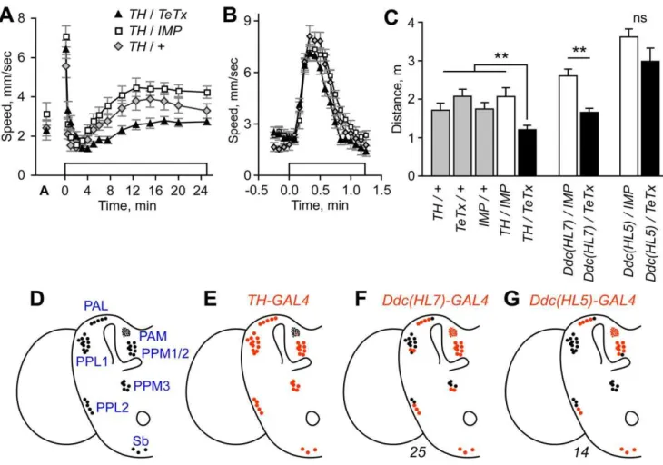
Figure 1. Blockade of evoked release from dopaminergic neurons reduced ethanol-induced hyperactivity. A. Expression of active tetanus toxin (TeTx) or an inactive form (IMP) as a control in tyrosine hydroxylase (TH) neurons (full genotype:TH-GAL4/+;UAS-TeTx/+). Open box
indicates time of ethanol vapor exposure (47% ethanol vapor concentration). ‘A’ indicates locomotor speed of acclimated flies in humidified air just prior to ethanol exposure.B. Higher resolution analysis of the olfactory startle response. Peak speed achieved between 0–1 min ethanol exposure did not differ between genotypes (P = 0.2722, 1 way ANOVA, n = 10).C. Distance traveled for hyperactivity onset.TH/TeTxwas different from indicated control genotypes (**P,0.01, 1-way ANOVA, Dunnett’s multiple comparison test, n = 10). Expression ofTeTxwithDdc(HL7)but notDdc(HL5)resulted in reduced ethanol-induced hyperactivity (Ddc(HL7): **P = 0.0006,Ddc(HL5): P = 0.139, 2 sample t-test, n = 9–10). All transgenes were heterozygous in animals tested for behavior.D. Dopaminergic neuron cell body positions in one hemisphere of the adult brain. Drawing was adapted from Friggi-Grelin et al. [20]. DA neuron nomenclature describes the location of the cell bodies in the adult brain (for example the PPM3s are one of three clusters of protocerebral posterior medial DA cells) [30].E–G. The average number of TH-positive cells that expressed the indicated GAL4 transgenes (red). Only PAM cell number is underrepresented. Number of hemispheres counted is indicated below the diagrams. TheTH-GAL4expression pattern has been reported previously [20,29].
magnitude of the hyperactive phase is dose-dependent, with lower concentrations of ethanol vapor resulting in higher levels of hyperactivity, due in part to reduced sedation [11].
We asked if blockade of evoked neurotransmitter release from DA neurons in the CNS affected ethanol-induced hyperactivity. To do this, we expressed in DA neurons the tetanus toxin light chain (TeTx) that cleaves and inactivates the neuronal-specific isoform of synaptobrevin, blocking synaptic transmission [31]. This was accomplished by driving expression of the UAS-TeTx
transgene with TH-GAL4 that places the yeast transcriptional activator GAL4 under the control of the tyrosine hydroxylase (TH) locus [20,31]. InTH/TeTxflies, ethanol-induced hyperactivity was reduced (Fig. 1A). Expression of catalytically inactive TeTx (IMP) had no effect, indicating that protein overexpression in TH neurons did not impinge upon neuron function. Determining the distance traveled during the onset of the hyperactive phase (2– 12.5 min exposure) revealed a significant reduction in locomotor activity specifically in TH/TeTx flies (Fig. 1C). Similarly, locomotor speed achieved at 7.5 min of ethanol exposure was reduced in TH/TeTx flies (Table S1). However, using a higher resolution data analysis, we detected no change in the magnitude of the olfactory-mediated locomotor startle response of TH/TeTx flies (Fig. 1B). Additionally, locomotor coordi-nation appeared normal inTH/TeTxflies, both by direct obser-vation of fly movement, and in a negative geotaxis climbing assay, indicating that motor behaviors were grossly normal, and that at least one sensorimotor circuit was fully functional. Ethanol absorption was also unaffected (TH/TeTx: 38.3 mM,
TH/IMP: 39.1 mM, 30 min at 47% ethanol vapor, P = 0.69, t-test, n = 3 groups of 25 flies each). Thus, blockade of evoked release from DA-producing neurons resulted in a speci-fic decrease in the ethanol-induced stimulation of locomotor activity.
To confirm that disruption of DA neurotransmission was the basis for the observed ethanol-induced hyperactivity defect, we performed two additional experiments. First, the Drosophila DA transporter DAT is expressed in DA neurons, and flies mutant for DAT (DATfmn) have been characterized for their altered sleep-like behavior [22,32]. Flies homozygous forDATfmnwere viable, fertile, grossly normal for motor behaviors, and exhibited a normal olfactory startle response. Ethanol-induced hyperactivity, however, was markedly reduced inDATfmnflies (Fig. S1A), indicating that disruption of DA reuptake and of evoked release from DA neurons had similar effects. Second, we confirmed that DA depletion by pharmacological means in adult flies reduced ethanol-induced hyperactivity (Fig. S1B) [16]. Thus, the effects of genetic and pharmacological manipulation of DA levels are consistent with the promotion of ethanol-induced hyperactivity by DA signaling in adult animals.!
Role of PPM3 neurons in promoting ethanol-induced locomotor activity
We next asked whether DA signaling from many or a few neurons was required to promote ethanol-induced hyperactivity. As a first step, we built Dopa decarboxylase-GAL4 (Ddc-GAL4) transgenes that express GAL4 in subsets of TH- and 5-HT-positive neurons (Fig. S1C); Ddc executes the final step in DA synthesis and the locus is well characterized [33]. We then determined the behavioral effects of blocking evoked release in the patterns specified by the Ddc transgenes, and correlated the behavioral effects with expression in TH-positive neurons. Expression of TeTx withDdc(HL7)-GAL4resulted in decreased ethanol-induced hyperactivity, whereas expression with Ddc(HL5)-GAL4 did not (Fig. 1C, Fig. S1E,G). Previous studies demonstrated (and we
confirmed) thatTH-GAL4fully labeled all TH-positive clusters in the adult brain, except for the PAMs where approximately 12 of 100 cells showed expression (Fig. 1D,E) [20,29]. With Ddc(HL7)-GAL4, fewer TH-positive cells were labeled in the PAL, PPL1, PPL2, and PPM3 clusters (Fig. 1F, Fig. S1D).Ddc(HL5)-GAL4
expression was further restricted, with no expression detected in the PPL1 and PPM3 clusters (Fig. 1G, Fig. S1F). We also detected 1–2 fewer labeled cells in the PPL2 and PPM1/2 clusters in Ddc(HL5)-GAL4. In the thoracic ganglion, nearly all TH-positive neurons were labeled by Ddc(HL5)-GAL4 (Fig. S2A), indicating that thoracic DA neurons were not necessary to promote ethanol-induced hyperactivity. These data suggested that DA promotion of ethanol-induced hyperactivity was localized to specific subsets of DA neurons, or, alternatively, that activity in a substantial fraction of DA neurons was required for normal ethanol responses.
To test the possibility that discrete DA neurons regulated ethanol-induced hyperactivity, we searched for GAL4 transgenic lines that expressed specifically in the DA neurons present in
Ddc(HL7)-GAL4and absent inDdc(HL5)-GAL4. One GAL4 line,
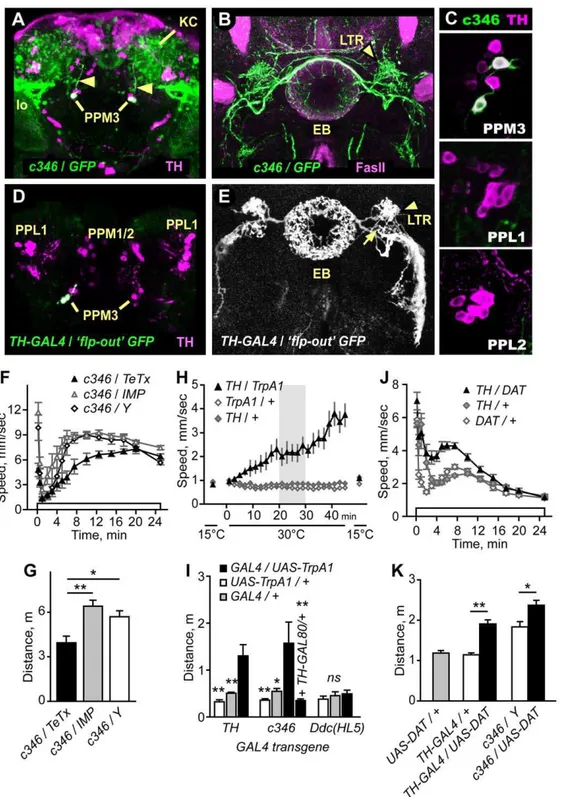
c346, was expressed in two of the 6–8 TH-positive PPM3 neurons and in no other TH-positive neurons (Fig. 2A–C).c346sparsely labeled other neurons in the adult CNS, including a subset of the mushroom body Kenyon cells, a group of cells located in the lateral protocerebrum that projected to the lobula of the optic lobe, and approximately 15–20 cells in the thoracic ganglion (Fig. S2B). The two TH-positive c346-expressing PPM3 cell bodies, located at the posterior surface of the brain, extended a fasciculated process anteriorly towards the central complex (Fig. 2B, Movie S1). The central complex includes four interconnected neuropils: the ellipsoid body, fan-shaped body, noduli, and the protocerebral bridge. Specific regions of the central complex are heavily innervated by dopaminergic processes [29,30,34]. The c346-labeled PPM3 processes branched at the central complex, and elaborated dense networks of processes in three regions: the central complex ellipsoid body ring and lateral triangles, and the ventral body region located ventral and lateral to the central complex.
To confirm that PPM3 neurons innervated the ellipsoid body, we used the ‘flp-out’ technique to randomly label subsets of neurons in theTH-GAL4expression pattern. All PPM3 neurons detected by this method innervated the central complex. Three distinct patterns of innervation were found: dense arborization in a discrete layer of the fan-shaped body either with or without additional arborization in the noduli (not shown), and dense arborization in the ellipsoid body ring and lateral triangles (Fig. 2D,E). This latter innervation pattern appeared identical to that observed inc346. These innervation patterns, also observed in a recent survey of DA neuron projection patterns [29], indicate that the PPM3 DA neurons make extensive connections in the central complex, and thatc346specifically labels PPM3 neurons that project to the ellipsoid body. We detected no innervation of the central complex by other TH-positive neurons. However, one PPL1 neuron has been reported to make an arborization in the fan-shaped body [29].Ddc(HL7) but notDdc(HL5) also showed expression in neurons that innervated the ellipsoid body, suggesting that Ddc(HL7) and c346 labeled the same subset of PPM3 neurons (Fig. S3).
Importantly, expression of TeTx but not IMP in the c346
pattern resulted in reduced ethanol-induced hyperactivity (Fig. 2F,G), indicating that evoked release from neurons in the
Figure 2. TH-positive PPM3 neurons that project to the ellipsoid body promote ethanol-induced hyperactivity. A.c346(detected by UAS-GFP, green) was expressed in two TH-positive (magenta) PPM3 neurons per hemisphere. The c346PPM3 processes projected anteriorly (arrowheads) towards the central complex.c346was also detected in the mushroom body Kenyon cells (KC) and in neurons that projected to the lobula (lo).B.c346(green) processes project to the lateral triangles (LTR) and ring of the ellipsoid body (EB), counterstained with FasII (magenta).C. c346expression in TH-positive cells is limited to the PPM3s.D,E. Labeling of individualTH-GAL4cells using the flp-out technique. Two GFP-labeled TH cells in the PPM3 cluster projected anteriorly to the LTR region (entry point is marked with an arrow in E). The process branched to innervate the EB ring and LTR, and the ventral body region.F,G. Distance traveled was reduced inc346/TeTx(*P,0.05, **P,0.01, 1-way ANOVA, Tukey’s multiple comparison test) whereas the startle response was not (P = 0.0925, 1-way ANOVA, n$6).H. Transient activation of TH neurons increased locomotor activity. Expression of the heat-activated TrpA1 ion channel in TH neurons at 15uC (off) and 30uC (on).I. Distance traveled from 20–30 min at 30uC for TH/TrpA1(**P,0.01, 1-way ANOVA, Dunnett’s multiple comparison test, n$5).c346/TrpA1also increased locomotor activity. Flies of the genotype c346/Y;TH-GAL80/+;UAS-TrpA1/+, where GAL4 activity was blocked by GAL80 solely inc346PPM3 neurons, showed no increase in locomotor activity
(*P,0.05, **P,0.01, 1-way ANOVA, Dunnett’s multiple comparison test toc346/Y;UAS-TrpA1/+, n$6). Activation of TrpA1 in theDdc(HL5)pattern had no effect (P = 0.6243, 1-way ANOVA, n$5). J,K. Overexpression of DAT inTH-GAL4 or c346 neurons increased ethanol-induced hyperactivity. (*P = 0.0123, **P = 0.0005, 2 sample t-test, n$10).
PPM3 neurons regulate locomotor activity
If DA release in a subset of DA neurons promotes ethanol-induced hyperactivity, then directly activating these neurons may also increase locomotor activity. To address this possibility, we expressed the heat-activated TrpA1 ion channel (UAS-TrpA1) in
TH-GAL4orc346neurons, and assessed the behavioral effects of transient neuronal activation [35]. At 15uC, when TrpA1 is inactive, TH/TrpA1flies showed low levels of locomotor activity (Fig. 2H). When the temperature was raised to 30uC, above the temperature required for TrpA1 activation, TH/TrpA1 flies increased locomotor activity levels, whereas control flies did not (Fig. 2H,I). Interestingly, returning TH/TrpA1 flies to 15uC almost immediately resulted in a cessation of locomotion. Moreover, repeated temperature cycling caused repeated cycling between states of inactivity and activity (not shown), suggesting that locomotor activity may be specified by continued signaling by DA neurons. These data indicated that acute activation of DA neurons promotes locomotor activity.
When TrpA1 was activated in c346-expressing neurons, flies also increased locomotor activity (Fig. 2I). Importantly, activation of neurons in the Ddc(HL5) pattern had no effect. Thus, the stimulation of locomotor activity by activation of eitherTH-GAL4
orc346neurons was specific, and the subset of DA neurons labeled by theDdc(HL5)transgene (PAL, PAM, PPL2, PPM1/2, Sb) could not be the sole source of a DA-dependent locomotor activation signal. To determine whether the stimulation of locomotor activity byc346neurons was due to activation of the PPM3s, we included
TH-GAL80 to block c346 GAL4 activity specifically in TH-expressing neurons. Inc346;TH-GAL80/+;UAS-TrpA1/+ flies, no stimulation of locomotor activity was detected (Fig. 2I). These data indicate that the dopaminergic PPM3 neurons that project to the ellipsoid body ring and lateral triangle promote locomotor activity when stimulated.
Lastly, we asked whether increasing DAT levels in DA neurons affected locomotor activity. Overexpression of DAT in either the
TH-GAL4 or the c346 pattern resulted in increased ethanol-induced hyperactivity (Fig. 2J,K), the opposite behavioral response as compared to the loss-of-function DATfmn mutants (Fig. S1A). Taken together, these data suggest that ethanol-induced stimulation of locomotor activity was due at least in part to DA signaling from PPM3 neurons that project to the ellipsoid body ring and lateral triangle region of the brain.
Central complex ellipsoid body neurons promote ethanol-induced locomotor activity
The central complex, the putative downstream target of the PPM3 DA neurons, plays multifaceted roles in the regulation of movement behaviors, including locomotion and flight (for review, see [36] and it has been implicated in the development of ethanol tolerance [14,37]. To determine if the central complex contributes to ethanol-induced hyperactivity, we expressed TeTx in neuronal patterns dictated by a collection of 24 GAL4 driver lines that showed expression in discrete subsets of central complex neurons (CC-GAL4) (Table 1). The phenotypes ofCC-GAL4/TeTxflies fell into four classes: non-viable, non-responsive (no startle or hyperactive phase), defective in ethanol-induced hyperactivity, and unaffected. There was no obvious correlation between TeTx-induced lethality and central complex expression pattern, suggesting that evoked release from the central complex is non-essential. Six of the seven non-responsive class CC-GAL4 lines expressed GAL4 either in the fan-shaped body or in small field neurons that connect different regions of the central complex. This finding is consistent with previous evidence for a role of the central complex in locomotion [38].
FourCC-GAL4lines (4.67,5.30,11.148, andc819/c547) resulted in reduced ethanol-induced hyperactivity when crossed to TeTx (Fig. 3A, Fig. S4A–C). All four lines showed expression in the R2/R4 subset of ellipsoid body neurons (Fig. 3B–D, Fig. S4G,H). These lines also showed expression in other regions of the brain and in the thoracic ganglion (Fig. S2). However, we detected no clearly discernible overlap of expression outside of the ellipsoid body (see below). One line,4.67, was also expressed in a subset of TH-positive neurons. Finally, eight CC-GAL4 lines showed no apparent behavioral defect when crossed toUAS-TeTx
(Fig. 3A). Three of these eight lines (189Y, c232, c561) were expressed in ellipsoid body neurons belonging to the R1, R3, and R4d classes (Fig. 3E–G, Figure S4D–F). Taken together, these data demonstrate a strong correlation between blocking evoked release in ellipsoid body neurons of the R2/R4 class and reduced ethanol-induced hyperactivity.
Other brain regions implicated in locomotor control or ethanol responses appear to be dispensable for the promotion of ethanol-induced hyperactivity. First, the mushroom bodies regulate overall locomotor activity levels, but not sensitivity to the incoordinating and sedating effects of ethanol [39,40]. Similarly, we found that ethanol-induced hyperactivity was unaffected following chemical ablation of the mushroom bodies (Fig. S5). Furthermore, the GAL4 line 189Y was expressed in the mushroom bodies and showed no phenotype when crossed to TeTx (Fig. 3E) [41]. Finally, we were unable to rescue the ethanol hyperactivity Table 1.Expression of TeTx in GAL4 patterns that include the central complex.
GAL4 Line GAL4/TeTx Class CC Expression
4.67 Hyperactivity defective EB R2/R4
5.30 Hyperactivity defective EB R2/R4
11.148 Hyperactivity defective EB R2/R4
c819/c547 Hyperactivity defective EB R2/R4m
3.16 Unaffected FSB
4.13 Unaffected FSB
4.69 Unaffected EB R neurons
11.17 Unaffected EB R1/R3
189Y Unaffected EB R3
c232 Unaffected EB R3/R4d
c561 Unaffected EB R1
11.33 Non-responsive FSB
11.243 Non-responsive FSB
12.30 Non-responsive FSB
78Y Non-responsive Small-field
c42 Non-responsive EB R2/R4m, FSB
c107 Non-responsive Small field
c161 Non-responsive Small field
2.70 Non-viable FSB
5.21 Non-viable EB R4
5.36 Non-viable EB R1 or R3
6.61 Non-viable EB R2/R4
11.32 Non-viable EB R neurons
11.113 Non-viable EB R Neurons
phenotype of DopR mutants utilizing mushroom body GAL4 drivers (see below). These data suggest that the mushroom bodies are not involved or play a redundant role in ethanol-induced hyperactivity. Second, the thoracic ganglion contains sufficient circuitry for some motor programs, including coordinated locomotion [42,43]. We exposed freshly decapitated flies to ethanol vapor to ask if ethanol directly stimulates thoracic
ganglion locomotor circuits. Decapitated flies maintained the righting response and the ability to groom, and walked a few steps when prodded, indicating that thoracic ganglion circuitry was functional. However, ethanol exposure elicited no increase in locomotor activity (Fig. S2E). Finally, neurosecretory cells of the pars intercerebralis regulate ethanol sensitivity [44]. However, pars intercerebralis expression in the CC-GAL4 lines did not correlate with TeTx behavioral phenotypes in our experiments (Fig. 3D–F). Collectively, our findings are consistent with a model whereby evoked neuronal activity in specific regions of the central complex, most likely the R2 and/or R4 neurons of the ellipsoid body, promotes ethanol-induced hyperactivity.
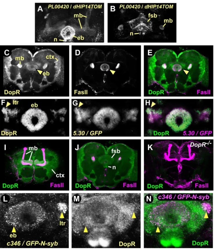
D1-like DopR receptor is expressed in the ellipsoid body The D1-like DA receptor DopR (DmDop1, dDA1) is present in the central complex and the mushroom bodies [27,45]. To ask if DopR is present in the ellipsoid body, we first determined the expression pattern of a GAL4 enhancer trap, PL00420, that resides in the first intron of theDopR locus (see below). Brains labeled withPL00420/UAS-dHIP14tdTOM(preferentially labeling presynaptic sites of neurons [46]) showed robust expression in the central complex ellipsoid body ring, noduli, and fan shaped body (Fig. 4A,B). We also observed labeling of the mushroom bodies, subsets of antennal lobe glomeruli and other less well-defined regions of the protocerebrum. Furthermore, we stained brains of our genetic background control strain with antiserum raised against a peptide derived from the third intracellular loop of DopR (Fig. 4C–J). DopR protein was expressed in the ellipsoid body ring (Fig. 4C–E, arrowhead), fan shaped body, noduli (Fig. 4J), and mushroom bodies (Fig. 4I). These patterns were specific, as all immunostaining was dramatically reduced inDopRmutant flies that expressed very low levels of DopR transcript (see below) (Fig. 4K). Furthermore, DopR was present in both the ring and lateral triangles of the ellipsoid body, where DopR staining overlapped with neuronal processes of R2/R4 neurons labeled by
5.30-GAL4(Fig. 4F–H). Finally, presynaptic terminals of thec346
PPM3 neurons required for ethanol-induced hyperactivity over-lapped with or were closely apposed to DopR in the ellipsoid body and the lateral triangles (Fig. 4L–N). Thus, the D1-like DopR receptor is expressed in many structures throughout the adult brain, including the R2/R4 neurons of the ellipsoid body, and these neurons may be in synaptic contact with the PPM3 neurons.
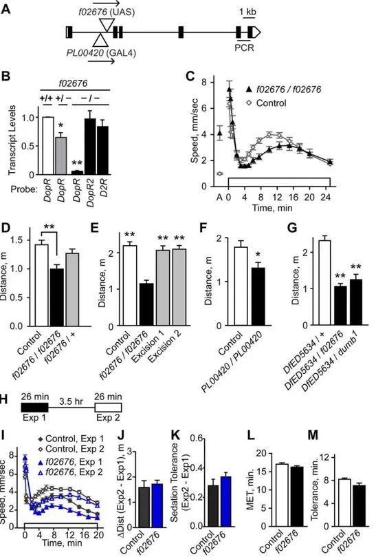
DopR is required for ethanol-induced hyperactivity To ask if DopR contributes to ethanol-induced hyperactivity, we tested the effects reducing the levels ofDopR. DopRf02676is a transposon insertion allele tha thet dramatically reduces DopR protein levels [21] (Fig. 4K, 5A). In agreement with this, we found that DopR transcript was reduced by 95% in the heads of flies homozygous forDopRf02676(Fig. 5B). No compensatory changes were detected in transcript levels for the DA receptorsDopR2and
D2R. Similar to the effects of blocking evoked neurotransmis-sion from dopaminergic and ellipsoid body neurons,DopRf02676
mutants showed reduced ethanol-induced hyperactivity (Fig. 5C,D), and no change in ethanol sedation sensitivity (Fig. S6A) or ethanol absorption (DopRf02676: 35.2 mM, control: 36.3 mM, 30 min at 47% ethanol vapor, P = 0.66, t-test, n = 6). Precise excision of f02676 restored both DopR transcript levels and ethanol-induced hyperactivity levels to those of controls, indicating that the f02676 transposon insertion was responsible for the molecular and behavioral phenotypes (Fig. 5E, Fig. S6C). Flies homozygous for thePL00420 transposon insertion showed 30% reduced DopR transcript levels and reduced ethanol-induced hyperactivity (Fig. 5F, Fig. S6E). Moreover, either f02676 or Figure 3. Functional mapping of ellipsoid body neurons in
ethanol-induced hyperactivity. Blockade of evoked release in patterns that included R2/R4 neurons of the ellipsoid body (B–D, GAL4 transgenic lines 5.30, 11.148, and c819) resulted in reduced ethanol-induced hyperactivity (A). Ethanol-induced hyperactivity was not reduced by TeTx expression in patterns that included R1 (c561), R3 (189Y,c232), or R4d (c232) neurons of the ellipsoid body (E–G). *P,0.05, **P,0.01, 1-way ANOVA for a given GAL4 transgene, Tukey’s multiple comparison test, n$5. For c819/TeTx vs. c819/IMP, **P = 0.0041, 2 sample t-test, n = 8. B–G. Expression of UAS-GFP (green) by the indicated GAL4 transgene, counterstained with FasII antibodies (magenta). Arrowheads indicate innervation of the ellipsoid body ring. PI: pars intercerebralis, MB: mushroom bodies.
Figure 4. The D1-like DA receptor DopR expression pattern in the adult brain includes the R neurons of the central complex ellipsoid body. A–B. Presynaptic regions ofDopR-expressing neurons. GAL4 enhancer trapPL00420in theDopR locus driving expression of tdTomato-GFP tagged with presynaptic protein HIP14.A. Expression in the mushroom body lobes (mb), ellipsoid body neurons (eb), and central complex noduli (n).B. Expression in the central complex fan shaped body (fsb) and noduli, and the mushroom body peduncles.C–E. Genetic background control brains stained with DopR (green) and FasII (magenta) antisera. Each panel is a 4.3mm confocal section of a whole mount brain.
Ellipsoid body (arrowhead), mushroom body peduncles, and cellular cortex (ctx) labeling with DopR antisera.F–H. Ellipsoid body lateral triangles (arrowhead) and ring showed elevated DopR levels that were coincident with the5.30GAL4 enhancer trap expression pattern in 10mm thin sections. I. Mushroom bodies and cellular cortex staining in controls.J. Fan-shaped body and noduli DopR staining in controls.K.DopRf02676mutant brain (DopR2/2) stained with DopR (green) and FasII (magenta) antisera. 100
mm confocal projection shows nearly absent DopR staining throughout the
Figure 5. Molecular and behavioral characterization ofDopRmutants. A.DopRgene structure. Positions of the transposonsf02676and PL00420are indicated in theDopRlocus. Arrows indicate orientation ofUASsites forf02676andGAL4forPL00420. Black rectangles indicate the single predicted open reading frame.B. Relative to genetic background controls,DopRtranscript levels were nearly eliminated inDopRf02676
homozygotes (2/2), whereas transcript levels of the DA receptorsDopR2andD2Rwere unchanged, as determined by quantitative RT-PCR (*P,0.05, **P,0.01, 1 way ANOVA, Dunnet’s multiple comparison test to control, n = 3 replicates).DopRPCR probeset location is indicated in A.C,D. Ethanol-induced locomotor activity was reduced inDopRf02676
homozygotes (**P,0.01, 1 way ANOVA, Tukey’s multiple comparison test, n$11).E. Precise excision of f02676reverted the ethanol-induced hyperactivity phenotype (**P,0.01, 1 way ANOVA, Dunnett’s multiple comparison test, n = 12).F. Reduced ethanol-induced hyperactivity inDopRPL00420
thedumb1inversion breakpoint allele ofDopRplaced intransto a deficiency that deleted theDopR locus showed reduced ethanol-induced hyperactivity (Fig. 5G, Fig. S6G). Taken together, these data demonstrate that decreased levels of the DopR receptor resulted in reduced ethanol-induced hyperactivity. Because the effects of the DopR mutations on ethanol-induced locomotor activation were partial, other DA receptors may also play a role in this behavior.
DopRf02676 mutants also showed increased activity prior to ethanol exposure (‘A’ in Fig. 5C). While opposite in sign, increased pre-exposure activity could influence the magnitude of the decrease in ethanol-induced hyperactivity in DopRf02676. To quantify this behavior, we recorded the locomotion of flies without an added stimulus (‘unstimulated activity’) (Fig. S6B). Immedi-ately after being placed in the observation chamber, control flies showed moderate levels of activity (4.7 mm/sec), and they quickly acclimated to their environment, maintaining low levels of activity for at least 1 hr (1.2–2.5 mm/sec). DopRf02676 mutants showed higher initial levels of activity (8.6 mm/sec), and whileDopRf02676
flies also acclimated, they maintained two-fold higher activity levels (4.9–5.3 mm/sec) (Fig. S6B). This phenotype was due to thef02676insertion (Fig. S6D). However, unstimulated activity was unaffected inDopRPL00420homozygotes (Fig. S6F). Moreover, we previously demonstrated a lack of correlation between pre-exposure locomotor activity and ethanol-induced hyperactivity levels [11]. Thus, flies with strongly reducedDopRlevels showed increased unstimulated activity and this behavior could be dissociated from the role ofDopRin ethanol-induced hyperactivity. Ethanol tolerance is a form of acquired resistance that facilitates increased drug intake, a major risk factor for the development of alcohol use disorders [47]. The development of ethanol tolerance in flies involves central complex neurons and the ellipsoid body [14,37]. We found that DopRf02676 mutants performed indistin-guishably from genetic background controls in a rapid tolerance paradigm, showing both increased ethanol-induced hyperactivity and increased resistance to the sedating effects of ethanol during a second exposure to ethanol vapor (Fig. 5H–M). Thus,DopRis not required for rapid ethanol tolerance.
DopR functions in the ellipsoid body for ethanol-induced locomotor activity
Our data suggested that DopR may function in the ellipsoid body to promote ethanol-induced hyperactivity. To test this, we carried out genetic rescue experiments, supplying functional DopR to specific brain regions in animals otherwise lackingDopR. We took advantage of GAL4-binding sites (UASelements) that are pre-sent in thef02676transposon. Flies of the genotypeTub-GAL4/+;
DopRf02676/+ that express GAL4 ubiquitously from the tubulin promoter showed an almost six-fold increase inDopR transcript levels. While theDopRtranscript produced in these flies is expected to lack sequences encoded by the first exon, resulting in the loss of the predicted translation initiation site (Fig. 5A), in-frame methionines encoded in the second exon are likely to initiate translation [21]. Moreover, DopR lacking the long N-terminal extracellular region remains functional in cultured cells [48]. Finally, flies of the genotype5.30-GAL4/+;DopRf02676/DopRf02676
showed selectively restored expression of DopR protein in the ellipsoid body ring and lateral triangles (Fig. 6B) indicating that
DopR protein was generated and it localized to regions of the ellipsoid body where synaptic connections are likely to be made.
Selectively restoring expression of DopR inDopRf02676mutants in patterns that included the R2/R4 class ellipsoid body neurons resulted in rescue of ethanol-induced hyperactivity (Fig. 6A, Fig. S7A,B), indicating that DopR expression in these neurons was sufficient for promotion of hyperactivity by ethanol. Restoring DopR expression in the R3 neurons of the ellipsoid body resulted in a modest but statistically insignificant increase in ethanol-induced hyperactivity with one driver (189Y,Fig. S6C) but not another (c232,Fig. S7D). As DopR is expressed broadly in the
Exposure 2 minus Exposure 1 (DDist) (P = 0.6691, paired t-test, n = 15).K. Sedation tolerance, the fraction awake exposure 2 minus exposure 1, was unaffected (P = 0.27, 2 sample t-test, n = 15).L. Ethanol sensitivity as measured in the inebriometer was unaffected (P = 0.16, 2 sample t-test, n = 5).M. Rapid tolerance, as measured in the inebriometer, was unaffected (P = 0.09, 2 sample t-test, n = 5).
doi:10.1371/journal.pone.0009954.g005
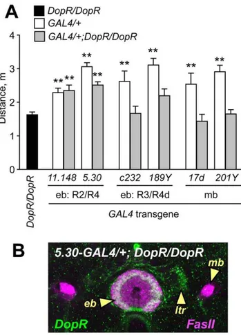
Figure 6. Genetic rescue of DopR mutant ethanol-induced hyperactivity by restricted expression ofDopRin the ellipsoid body. A. Distance traveled for animals homozygous forf02676(DopR/ DopR), and heterozygous for GAL4 transgenes to drive expression of DopR in patterns that include the ellipsoid body R2/R4 neurons (11.148, 5.30), R3/R4d neurons (c232,189Y), or the mushroom bodies (17d,201Y). Restored expression of DopR in the R2/R4 neuron patterns but not others resulted in increased ethanol-induced hyperactivity as compared toDopR/DopR(**P,0.01, 1 way ANOVA, Dunnett’s multiple comparison test, n$10).B. Rescuing strategy results in selectively localized DopR protein expression. Confocal section of an animal homozygous for f02676and carrying the GAL4 enhancer trap5.30, stained for DopR protein (green) and FasII (magenta). 5.30-GAL4 drives expression of DopR protein in the ring of the ellipsoid body (eb) and the lateral triangles (ltr), but not in the mushroom bodies (mb). DopR expression in the R2 neurons is indicated by the arrowhead.
ellipsoid body (Fig. 4A,C), there may be an undetected overlap in expression patterns of the ellipsoid body GAL4 drivers, or there may be differences between the effects of blocking evoked neuronal output and restoring neuronal input. Importantly, restored expression of DopR in the mushroom bodies, where DopR is also normally expressed, did not rescue DopRf02676
ethanol-induced hyperactivity defects (Fig. 6A, Fig. S7E–F), demonstrating that DopRbehavioral rescue in the ellipsoid body was specific. Overexpression of DopR did not increase ethanol-induced hyperactivity (Fig. S7G,H), indicating that the genetic rescue was due to restoration of normal DopR function. Thus,
DopR expression in the ellipsoid body was sufficient for the promotion of ethanol-induced hyperactivity.
Discussion
DA regulates specific behavioral responses elicited by acute exposure to drugs of abuse in mammals and in flies. For low to moderate doses of ethanol, DA signaling is associated with euphoria (the subjective high), locomotor activation, and drug seeking. Our studies extend the understanding of the role of DA in ethanol responses in flies in three main ways. First, we identified specific DA (PPM3) and DA target (central complex ellipsoid body) neurons that promote locomotor activation by acute ethanol exposure. Second, we showed that the D1-like receptor DopR is required for promotion of locomotor activity elicited by acute ethanol exposure, and that its function for this role localized to the ellipsoid body. Importantly, DA signaling through DA D1 receptors also promotes locomotor activation by ethanol in mice [6,49]. Finally, these DA and DA target neurons were dispensable for the olfactory startle response and negative geotaxis, indicating that the circuit regulating locomotor activation by ethanol is separable from at least some other locomotor activation circuits. Our findings allow us to propose a simple DA circuit: ethanol may act either directly or indirectly on PPM3 DA neurons, eliciting a DA locomotor activation signal that is received by DopR in the ellipsoid body, where presumably it is parsed to invoke an appropriate motor response.
Definition of specific behavioral roles for DA neurons Silencing of nearly all DA neurons (TH-GAL4) or discrete subsets (Ddc(HL7)-GAL4 and c346) resulted in reduced ethanol-induced hyperactivity, whereas silencing of further restricted DA neuron subsets (Ddc(HL5)-GAL4) did not. Comparison of expres-sion patterns for TH, Ddc(HL7) and Ddc(HL5) revealed a correlation between effects of TeTx and expression in the PPL1 and PPM3 DA neuron clusters, and a lack of correlation with other DA neuron clusters (Fig. 1). Assignment of promotion of ethanol-induced hyperactivity to the PPM3 neurons is based on the phenotypic effects of TeTx silencing of neurons in thec346
expression pattern, stimulation of locomotion by direct activation of DA neurons in thec346pattern, and potentiation of ethanol-induced hyperactivity by overexpression of DAT in either TH or
c346neurons (Fig. 2). Neurons in the PPM3 cluster project to distinct yet highly stereotyped locations in the central complex and are the only DA neurons to target the ellipsoid body and lateral triangles. Thec346PPM3s project to the ellipsoid body and lateral triangles while other PPM3s project to the fan-shaped body and noduli (Fig. 2) [29].Ddc(HL7), likec346, is expressed in a subset of PPM3 neurons (Fig. S1), that project to the ellipsoid body (Fig. S3, Movie S1). c346-negative PPM3 and the PPL1 neurons may also contribute to ethanol-induced hyperactivity and DA-depen-dent locomotor activation.
Specific DA-dependent behaviors may map to discrete sets of DA neurons. First, the projection patterns of DA neurons from different clusters are stereotyped and largely non-overlapping [29]. Second, associative olfactory learning is DA-dependent [21,24], and requires a subset of DA neurons in the PPL1 cluster that project to the mushroom bodies [26]. We did not uncover a DA-dependent role for the mushroom bodies in ethanol-induced hyperactivity (Fig. 3,6, Fig. S5), suggesting there exists a separation between DA-dependent locomotor activation and olfactory conditioning. Other DA-influenced behaviors in flies include courtship, arousal state, and a neural correlate of a visual perceptual response [19,27,50]. Identifying the DA-dependent neural circuitry for each behavior is likely to help define shared and distinct underlying mechanisms in behavioral control, and will be important for the accurate assessment of gene function in behavior.
What are the inputs and outputs for the PPM3 DA neurons that carry the ethanol locomotor activation signal? Presynaptically localized GFP in the PPM3s labeled the ellipsoid body lateral triangles and ring, suggesting that these are sites of DA release (Fig. 4L–N). Morphological analysis of ellipsoid body lateral triangle neurons suggests that this region is dense with dendrites [51], and functional localization ofDopRto the ellipsoid body for ethanol-induced hyperactivity suggests that this region may be a synaptic target of the PPM3s. The neurons upstream of the PPM3s are not defined, but may include neurons in the central complex or in contact with the lateral processes that extend into the ventral body region (Fig. 2B,D). Consistent with this latter possibility, we found that postsynaptically targeted GFP expressed in the PPM3s localized primarily to these lateral processes (not shown). Neurons upstream of the PPM3s could encode information from sensory cues that elicit a locomotor response, including for example the chemical or visual perception of other flies, odors, or food. Alternatively, the PPM3s may function downstream of higher order processing circuits that encode previous experience or interoceptive cues such as satiation state [52]. Descriptions of synaptic connectivity outside of the well defined olfactory and visual circuits will be needed to better understand the dopami-nergic control of locomotor activity.
Effects of DAT manipulation on ethanol behavioral responses
activation of TH neurons is consistent with this interpretation (Fig. 2H).
Role of the ellipsoid body in control of motor behavior and ethanol responses
The ellipsoid body is a prominent central neuropil that has been implicated in regulating unstimulated locomotor activity levels [55], the visual control of walking and flight [56–58], ethanol tolerance [37], and a persistent state of arousal induced by repeated mechanical stimulation [27]. Current findings suggest that specific motor behaviors may map to distinct neuronal classes within the ellipsoid body; for example, orientation during visually guided walking maps to the R1 or R3 neurons [57,59], whereas ethanol-induced hyperactivity maps to the R2/R4 neurons (Fig. 3, 6). While the specific inputs and outputs of the ellipsoid body are not yet known (with the exception of the DA input described here), the above findings, taken together with earlier structural and lesioning studies, have led to the hypothesis that the ellipsoid body may be involved in parsing information from various sensory modalities to engage the appropriate motor output [36]. Interestingly, DopR in the R2/R4 neurons also regulates arousal state [27]. However, while ellipsoid body-expressed DopR promotes locomotor activity induced by ethanol, it suppresses locomotor activity induced by repeated mechanical stimulation. Although alternative interpretations exist, these apparently opposite roles for DopR may suggest the integration of other sources of information to set the sign of dopamine signaling in the ellipsoid body with respect to motor output. Conceptually consistent with this idea, a previous study showed that DA neuron stimulation in low activity flies increased activity, whereas high activity flies showed the opposite response [60]. Additionally, the contribution to fly locomotor behaviors of other D1-class (DopR2/
DAMB/DopR99B, DmDopEcR) and D2-class (D2R) dopamine receptors is largely unknown [61–64]. Pharmacological and gene knockout studies in rodents indicate that both D1-class and D2-class DA receptors regulate the locomotor stimulant effects of ethanol [65,66].
The R2/R4 neurons of the ellipsoid body also regulate ethanol sedation sensitivity and sedation tolerance via a Homer protein-dependent mechanism [37]. Therefore, the ellipsoid body R2/R4 neurons regulate ethanol-induced locomotor activity in a DA-dependent fashion, and ethanol sedation sensitivity and tolerance in a Homer-dependent fashion. In mammals, DA and glutamate are thought to play distinct a roles in the nucleus accumbens for the behavioral effects of ethanol [67]. Dopaminergic neurons from the ventral tegmental area and glutamatergic neurons from cortical and other brain regions make synapses onto GABAergic medium spiny neurons in the nucleus accumbens where they regulate ethanol-induced locomotor activity (DA) and ethanol consumption and withdrawal (glutamate) [68]. It is of note that there are both GABAergic and glutamatergic neurons in the ellipsoid body [69,70], and that a subset of R2/R4 neurons in the
5.30 and 11.148GAL4 expression patterns are GABAergic (not shown). Thus, Drosophila may serve as a model to define some fundamental properties of neural circuit function as well as gene action that may be relevant to the actions of drugs of abuse in mammals.
Locomotor activation and reward
Selective breeding studies in rodents uncovered a positive correlation between the locomotor stimulant and reinforcing properties of ethanol [2,3], and both properties of ethanol are mediated at least in part through the mesolimbic DA pathway [71,72]. Moreover, human studies demonstrated a correlation
between ethanol sensitivity and the propensity to develop alcohol use disorders [1]. The mesolimbic DA-dependent acute locomotor and reinforcing effects of cocaine, however, are genetically separable [73,74], suggesting that DA can encode the acute and reward learning effects of at least some drugs of abuse in a complex manner. Our current findings show that there exists a DA circuit that mediates the locomotor stimulant properties of ethanol in flies. Application of learned reward-like behavior models for drugs of abuse and other stimuli to DA circuit analysis in flies will aid in understanding how this organism codes for complex DA-dependent behaviors.
Because DopR regulates arousal levels in a set of ellipsoid body neurons that overlap with those required for ethanol-induced hyperactivity [27], it is possible that ethanol-stimulated locomotion represents a form of arousal in flies. A proposed function for mesolimbic DA in mammals is to impart salience to increase the motivational state with regard to cues in the environment [4]. It will be important to determine whether locomotor activating stimuli other than ethanol also utilize this circuit, and to determine how ethanol interacts with other locomotor-activating cues that elicit approach and avoidance responses.
Materials and Methods
Genetics and Molecular Biology
All strains were outcrossed to the wild-type Berlin strain carrying thewhite(w1118) mutation (control) for 5–10 generations.
DATfmnwas followed molecularly during outcross by PCR with the upstream primer 59-GCTGCTGGCCTATGCATCC and the downstream primers 59-GCAATGCACCCATGTCGCC in the
fmnlesion and 59-ACTGGTTAACAAAGCATCC downstream of thefmnlesion. DopR locus lesions includedf02676 (PBac{WH}),
PL00420 (PBac{GAL4D,EYFP}), dumb1 (In(3LR)234) [21], and
Df(3R)ED5634. Transposon marker genes aremini-w+forf02676 andEYFPforPL00420. Some strains (2.70,3.16,4.13,4.67,4.69,
5.21,5.30,5.36,6.61,11.17,11.32,11.33,11.113,11.148,12.30) were generated by mobilization of the GAL4-containing enhancer trap transposon P{GawB} to random integration sites in the genome.UAS-CD2-mCherrywas a gift from Bing Ye, UAS-tdTomato-GFP-HIP14was from Steve Stowers, andUAS-dTrpA1 was from Paul Garrity. TH-GAL4, UAS-TeTx (UAS-TeTxLC.tnt), UAS-IMP
(UAS-TeTxLC.IMPTNT), andUAS-n-syb-eGFPwere obtained from the Bloomington stock center. TheP{GawB} strains78Y,189Y,
c42,c107,c161,c232,c346,c561, andc819were obtained from the Fly-trap project (www.fly-trap.org). To visualize individual neu-rons in theTH-GAL4pattern, flies heterozygous forTH-GAL4, hs-Flp, andUAS.CD2,y+.CD8-GFPwere heat shocked at 37uC for 10–30 min during the first larval instar. Over 500 brains were analyzed. The transposonf02676 was excised utilizing standard genetic methods and was backcrossed for ten generations to
f02676. DopR antisera was raised in rabbits against the peptide sequence CVAEKQRYKSIRRPKNQPKKFK derived from the intercellular loop region between transmembrane regions five and six, and affinity purified using the peptide (Covance, Denver, PA). Antisera specificity was verified by ELISA and immunostaining of
DopRf02676brains. TheDopRopen reading frame was isolated by PCR from the Berlin strain with the primers 59 -ATGTACA-CACCACACCCA and 59-TCAAATCGCAGACACCTGC and cloned into pUAST to generateUAS-DopR.
mutant strains to the genetic background control. Taqman probesets (Applied Biosystems) used in this study were D2R: Dm01845573_g1,DopR: Dm02134814_m1,DopR2: Dm02151743_ g1, andRpL32: Dm02151827_g1.
Construction of Ddc/GAL4 fusion genes
Numbering is relative to theDdctranscription start site [75], and underlined letters representDdcexons. SeeFig. S1Cfor diagrams of the constructs.
HL5-[DdcA/GAL4]. A fragment of Ddc from Bgl II at 22702 through a PCR-added BamHI site at +160 in exon A, was cloned upstream of GAL4 at a BamHI site of the pGaTB vector [76] to generate construct HL5 [DdcA/Gal4]. The 190 bp portion (224 to +160) downstream of this Ddc fragment was a PCR product and isolated by digestion with NcoI and BamHI. The PCR fragment was generated using oligonucleotides: 59 -CGGTCCTGCGGAATTGGCAGCGCTGC sense primer for
Ddc59flanking :267R242, and 59 -CGCGGATCCGCGCACT-TGTTGCCG antisense primer inDdcexon A, adding BamHI at +158R146.
HL7-[DdcA/Gal4/DdcABCD]. To generate [DdcA/Gal4/ DdcABC], Ddc sequences from exon A through exon D were subcloned downstream of the hsp70 polyA sequences in HL5. The
DdcHpaI fragment was isolated by restriction digestion of HpaI sites at +175 in exon A and at +3756 in exon D and, ligated downstream of hsp70 of the HL5 SpeI site. The SpeI site was immediately downstream of the Ddc/GAL4 insert. The 59 protruding SpeI ends were blunted in order to ligate with the
DdcHpaI fragment. The [DdcAB/Gal4/Ddc3’end] and [DdcA/ Gal4/DdcABCD] fragments in HL7 were subcloned into P-element transformation vector CaSpeR4 at the KpnI and SpeI sites, respectively.
The constructs in P-element CaSpeR4 vectors were purified using maxi-purification method (Qiagen Plasmid Kit). Transgenic lines were generated by microinjection of purified DNA intow1118
embryos [77,78].
Immunohistochemistry
Brains dissected from 2–5 day old adult flies were fixed for 16 hr at 4uC in 2% paraformaldehyde in PBT (0.05% Triton X-100 in phosphate buffered saline). Primary and secondary antibodies were incubated with fixed tissue at 4uC for 48 hr each in 0.5% BSA and 5% normal goat serum in PBT, with extensive washes with PBT between steps. Images were collected on a Zeiss confocal microscope after mounting brains in Vectashield (Vector Labora-tories). Confocal stacks were manipulated in ImageJ and Photo-shop. Antibodies used were MAb 1D4 anti-FasII (1:40, Develop-mental Studies Hybridoma Bank, Iowa), rabbit anti-GFP (1:1000, Invitrogen), mouse anti-GFP (1:500, Zymed), rabbit anti-TH (1:100, Pel-Freez), and rabbit anti-DopR (1:1250).
Behavioral Analysis
Flies were maintained on a standard cornmeal/molasses/yeast media at 25uC and 70% humidity with an approximately 16hr/ 8hr light/dark schedule. For behavioral testing, 25 males and virgin females were mated in bottles with 50 mL standard media with a few added grains of Baker’s yeast for 2 days, and groups of 23 male progeny were collected into standard food vials without yeast 11 days later. ‘‘n’’ indicates the number of groups of flies of a given genotype derived from independent parental crosses that were tested on separate days. Flies were allowed to recover from CO2anesthesia for 2 days, and were tested behaviorally between
11 am and 6 pm. All behavioral tests, unless noted otherwise, were carried out at 25uC with constant illumination, utilizing the
booz-o-mat, an eight-chambered manifold holding 166125 mm cylindrical tubes [11]. Ethanol vapor was delivered to the booz-o-mat as a continuous stream by bubbling air separately through a 3L flask of 95% ethanol or water. Ethanol and humidified air streams were mixed at different ratios to a final flow rate of 5.5 L/ min. Locomotor tracking analysis was as described previously [11]. Ethanol-induced hyperactivity was quantified as the area under the curve from 2–12.5 min exposure for 47% and from 2–10 min exposure for 60% ethanol vapor, corresponding to the time of hyperactivity onset to peak speed achieved for control flies at each concentration [13]. Hyperactivity was also quantified as the locomotor speed achieved at 7.5 min of ethanol exposure (Table S1). Unstimulated locomotor activity was measured in 60690610 mm chambers in constant light in the absence of food or humidified air, and was quantified as the area under the curve from 10–60 min. For TrpA1 activation tests, 10–20 male flies were placed in a thin-walled 3065062 mm Plexiglas chamber on the surface of a 96-well thermal cycler heat block (MJ Research) and were allowed to acclimate for 10 min at 15uC. They were filmed for 5 min at 15uC, 45 min at 30uC, and 5 min at 15uC. Locomotor activity was averaged over 15 sec every 2 min, and the distance traveled from 20–30 min at 30uC was calculated for statistical analysis. Statistical analyses were carried out in Minitab v.15 and Prism 4. Error bars in all figures indicate SEM.
Ethanol sedation kinetics and absorption
Groups of twenty genetically identical 2–4 day old adult male flies were exposed to 60% ethanol vapor in the booz-o-mat. At 5 min intervals the flies were agitated by rotating the tubes, and the number of flies that were unable to right themselves following a 10 sec recovery were counted. The average of 10 experiments from 5 days was determined. For ethanol tolerance, the number of flies sedated following 26 min exposure, approximately the time to 50% sedation for control flies, were counted. Ethanol absorption was measured by exposing groups of 25 flies to either ethanol vapor (47%) or humidified air for 30 min. Flies were immediately frozen on dry ice and the ethanol concentration in whole fly homogenates was measured with an alcohol dehydrogenase-based spectrophotometric assay (Diagnostic Chemicals, Ltd).
Supporting Information
Table S1 Locomotor speed achieved at 7.5 min ethanol exposure.
Found at: doi:10.1371/journal.pone.0009954.s001 (0.06 MB PDF)
Figure S1 Regulation of ethanol-induced hyperactivity by dopamine (DA). A1. Reduced ethanol-induced hyperactivity in flies homozygous for the DAT DA transporter mutationfmn.A2. Distance traveled (2–12.5 min) is reduced inDATfmn(**P,0.0001, 2 sample t-test, n = 12).DATfmnwas recessive for this phenotype.
B. Pharmacological reduction of DA in adult flies reduced ethanol-induced hyperactivity.B1. Wild-type control (w- Berlin) flies were fed 10 mg/mL 3-iodotyrosine in 2% yeast/5% sucrose for 36–48 hr, and then exposed to 45% ethanol vapor.B2. Drug fed flies showed a reduction in distance traveled (**P = 0.0072, paired t-test. n = 10). C. Diagram depicting the Ddc-GAL4
transgenes used in this study. See Methods for construction details.D,F. Expression ofDdc(HL7)-GAL4andDdc(HL5)-GAL4
Found at: doi:10.1371/journal.pone.0009954.s002 (7.19 MB TIF)
Figure S2 Role of the thoracic ganglion in ethanol-induced hyperactivity. A–D. Expression pattern of the indicated GAL4 transgenes (detected with UAS-GFP, green) in the adult thoracic ganglion, counterstained with TH antibodies (magenta). A.
Ddc(HL5)-GAL4is expressed in most TH-positive neurons in the thoracic ganglion. Positions of TH-positive cell bodies are indicated by arrowheads. No overlap was evident of GAL4 drivers
c346, 5.30, and 11.148 with TH expression in the thoracic ganglion. E. Ethanol-induced locomotor activity (0–25 min) in freshly decapitated and matched unoperated controls. ‘A’ indicates locomotor speed in humidified air 1 min prior to ethanol exposure. The righting response and grooming activity were intact in decapitated flies just prior to ethanol exposure, indicating that the preparation was behaviorally responsive. n = 3.
Found at: doi:10.1371/journal.pone.0009954.s003 (9.47 MB TIF)
Figure S3 Expression of Ddc(HL7)-GAL4 and Ddc(HL5)-GAL4
(detected byUAS-GFP, green) in the ellipsoid body ring and lateral triangles, counterstained with TH antibodies (magenta).
Found at: doi:10.1371/journal.pone.0009954.s004 (5.94 MB TIF)
Figure S4 A–F. Locomotor activity profiles for the functional mapping of ellipsoid body neurons in ethanol-induced hyperac-tivity. Flies of the indicated genotypes were exposed to 47% ethanol vapor (open box on horizontal axis). GAL4 lines are indicated on each graph.G,H. Expression of the indicated GAL4 lines in R2/R4 neurons of the ellipsoid body (arrowhead), detected by GFP expression and counterstained with anti-Bruchpilot (nc82) to highlight the synaptic neuropil.I. Higher resolution analysis of startle magnitude for the GAL4 lines expressed in the R2/R4 neurons, corrected for pre-exposure locomotor activity levels. For
5.30and11.148, the olfactory startle responses were unaffected by TeTx expression (5.30: P = 0.4403, 11.148: P = 0.0556, 1 way ANOVA, n$5). For c819, c819/+;UAS-IMP/+ was reduced (P = 0.0194, 1 way ANOVA, Tukey’s multiple comparison test, n$3).
Found at: doi:10.1371/journal.pone.0009954.s005 (5.76 MB TIF)
Figure S5 Mushroom body ablation. A. Ethanol-induced hyperactivity at 60% ethanol vapor with (+HU) and without (-HU) hydroxyurea treatment. Distance traveled 2–12.5 min did not differ (P = 0.0861, paired t-test, n = 4). B, C. Brains from control and HU treated flies, stained with anti-FasII to highlight the mushroom bodies (arrowheads) and the central complex ellipsoid body (arrow), which is unaffected by HU treatment. Found at: doi:10.1371/journal.pone.0009954.s006 (1.33 MB TIF)
Figure S6 DopRmutant behavioral characterization.A. Ethanol-induced sedation was unaffected in f02676 homozygotes,
mea-sured as the loss of the ability to right. (60% ethanol vapor, P = 0.0945, 2 sample t-test, n = 10).B. Unstimulated locomotor activity, measured for 60 min immediately after flies were introduced into a 60690610mm Plexiglas box.DopRf02676showed higher activity (distance traveled 10–60 min, P = 0.0009, 2 sample t-test, n = 10).C, D. Precise excision of f02676 reverts ethanol-induced hyperactivity and unstimulated activity behavioral phenotypes. C. Ethanol-induced hyperactivity for the genetic background control,f02676 homozygotes, and two independent precise excisions off02676(Ex1andEx2).D. Unstimulated activity for the same strains. Distance traveled from 10–60 min showed thatf02676is different from all other genotypes (P,0.001, 1 way ANOVA, Tukey’s multiple comparison test, n = 5). E. Ethanol-induced hyperactivity in animals homozygous for PL00420. F. Unstimulated activity was unaffected inPL00420homozygotes.G. Non-complementation for ethanol-induced hyperactivity by
DfED5634, a deficiency that deletes the entire DopR locus, for
f02676and thedumb1
inversion allele.
Found at: doi:10.1371/journal.pone.0009954.s007 (0.65 MB TIF)
Figure S7 A–F. Locomotor activity profiles for the genetic rescue of the DopRf02676 mutant ethanol-induced hyperactivity phenotype.G. Expression ofDopRutilizing theUASsites inf02676
or a UAS-DopR transgene has no effect on ethanol-induced hyperactivity in flies that are either wild-type or heterozygous for
DopR(left group: P = 0.7842, 1 way ANOVA, n = 8, right group: P = 0.575, 1 way ANOVA, n = 5).H. IncreasedDopR transcript levels when UAS-DopR is combined with the pan-neuronal elav-GAL4 (**P = 0.0062, 1 way ANOVA, Tukey’s multiple compar-ison test, n = 3).
Found at: doi:10.1371/journal.pone.0009954.s008 (0.79 MB TIF)
Movie S1 PPM3 neurons labeled by c346, detected by UAS-CD8-GFP, confocal stack reconstruction.
Found at: doi:10.1371/journal.pone.0009954.s009 (0.92 MB MOV)
Acknowledgments
We thank Karen Berger for the inebriometer tolerance assay, Viktor Kharazia, Elizabeth Marin, and Liqun Luo for assistance with fly brain imaging, Willa Abrone for technical assistance, David Anderson for support and advice, Bing Ye and Steve Stowers for reagents, the Bloomington and HarvardDrosophilastock centers for strains.
Author Contributions
Conceived and designed the experiments: ECK KW UH JH FW. Performed the experiments: ECK KW HL NM MRS FW. Analyzed the data: ECK HL RJB FW. Contributed reagents/materials/analysis tools: ECK TL RJB. Wrote the paper: FW.
References
1. Mayfield RD, Harris RA, Schuckit MA (2008) Genetic factors influencing alcohol dependence. Br J Pharmacol 154: 275–287.
2. Risinger FO, Malott DH, Prather LK, Niehus DR, Cunningham CL (1994) Motivational properties of ethanol in mice selectively bred for ethanol-induced locomotor differences. Psychopharmacology (Berl) 116: 207– 216.
3. Waller MB, Murphy JM, McBride WJ, Lumeng L, Li TK (1986) Effect of low dose ethanol on spontaneous motor activity in alcoholpreferring and -nonpreferring lines of rats. Pharmacol Biochem Behav 24: 617–623. 4. Berridge KC (2007) The debate over dopamine’s role in reward: the case for
incentive salience. Psychopharmacology (Berl) 191: 391–431.
5. Fields HL, Hjelmstad GO, Margolis EB, Nicola SM (2007) Ventral tegmental area neurons in learned appetitive behavior and positive reinforcement. Annu Rev Neurosci 30: 289–316.
6. El-Ghundi M, George SR, Drago J, Fletcher PJ, Fan T, et al. (1998) Disruption of dopamine D1 receptor gene expression attenuates alcohol-seeking behavior. Eur J Pharmacol 353: 149–158.
7. Gonzales RA, Job MO, Doyon WM (2004) The role of mesolimbic dopamine in the development and maintenance of ethanol reinforcement. Pharmacol Ther 103: 121–146.
8. Phillips TJ, Brown KJ, Burkhart-Kasch S, Wenger CD, Kelly MA, et al. (1998) Alcohol preference and sensitivity are markedly reduced in mice lacking dopamine D2 receptors. Nat Neurosci 1: 610–615.
9. Wolf FW, Heberlein U (2003) Invertebrate models of drug abuse. J Neurobiol 54: 161–178.
10. Moore MS, DeZazzo J, Luk AY, Tully T, Singh SM, et al. (1998) Ethanol Intoxication in Drosophila: Genetic and Pharmacological Evi-dence for Regulation by the cAMP Signaling Pathway. Cell 93: 997– 1007.
11. Wolf FW, Rodan AR, Tsai LT, Heberlein U (2002) High-resolution analysis of ethanol-induced locomotor stimulation in Drosophila. J Neurosci 22: 11035–11044.
13. Kong EC, Allouche L, Chapot PA, Vranizan K, Moore MS, et al. (2009) Ethanol-Regulated Genes That Contribute to Ethanol Sensitivity and Rapid Tolerance inDrosophila. Alcohol Clin Exp Res.
14. Scholz H, Ramond J, Singh CM, Heberlein U (2000) Functional ethanol tolerance inDrosophila. Neuron 28: 261–271.
15. Devineni AV, Heberlein U (2009) Preferential ethanol consumption inDrosophila models features of addiction. Curr Biol 19: 2126–2132.
16. Bainton RJ, Tsai LT-Y, Singh CM, Moore MS, Neckameyer WS, et al. (2000) Dopamine modulates acute responses to cocaine, nicotine, and ethanol in Drosophila. Curr Biol 10: 187–194.
17. Chang HY, Grygoruk A, Brooks ES, Ackerson LC, Maidment NT, et al. (2006) Overexpression of the Drosophila vesicular monoamine transporter increases motor activity and courtship but decreases the behavioral response to cocaine. Mol Psychiatry 11: 99–113.
18. Li H, Chaney S, Forte M, Hirsh J (2000) Ectopic G-protein expression in dopmaine and serotonin neurons blocks cocaine sensitization in Drosophila melanogaster. Curr Biol 11: 211–214.
19. Andretic R, van Swinderen B, Greenspan RJ (2005) Dopaminergic modulation of arousal inDrosophila. Curr Biol 15: 1165–1175.
20. Friggi-Grelin F, Coulom H, Meller M, Gomez D, Hirsh J, et al. (2003) Targeted gene expression inDrosophiladopaminergic cells using regulatory sequences from tyrosine hydroxylase. J Neurobiol 54: 618–627.
21. Kim YC, Lee HG, Han KA (2007) D1 dopamine receptor dDA1 is required in the mushroom body neurons for aversive and appetitive learning inDrosophila. J Neurosci 27: 7640–7647.
22. Kume K, Kume S, Park SK, Hirsh J, Jackson FR (2005) Dopamine is a regulator of arousal in the fruit fly. J Neurosci 25: 7377–7384.
23. Neckameyer WS (1998) Dopamine and mushroom bodies in Drosophila: experience-dependent and - independent aspects of sexual behavior. Learn Mem 5: 157–165.
24. Schwaerzel M, Monastirioti M, Scholz H, Friggi-Grelin F, Birman S, et al. (2003) Dopamine and octopamine differentiate between aversive and appetitive olfactory memories inDrosophila. J Neurosci 23: 10495–10502.
25. Claridge-Chang A, Roorda RD, Vrontou E, Sjulson L, Li H, et al. (2009) Writing memories with light-addressable reinforcement circuitry. Cell 139: 405–415.
26. Krashes MJ, DasGupta S, Vreede A, White B, Armstrong JD, et al. (2009) A neural circuit mechanism integrating motivational state with memory expression inDrosophila. Cell 139: 416–427.
27. Lebestky T, Chang JS, Dankert H, Zelnik L, Kim YC, et al. (2009) Two different forms of arousal inDrosophilaare oppositely regulated by the dopamine D1 receptor ortholog DopR via distinct neural circuits. Neuron 64: 522–536. 28. Riemensperger T, Voller T, Stock P, Buchner E, Fiala A (2005) Punishment
prediction by dopaminergic neurons inDrosophila. Curr Biol 15: 1953–1960. 29. Mao Z, Davis RL (2009) Eight different types of dopaminergic neurons
innervate theDrosophilamushroom body neuropil: anatomical and physiological heterogeneity. Front Neural Circuits 3: 5.
30. Nassel DR, Elekes K (1992) Aminergic neurons in the brain of blowflies and Drosophila: Dopamine- and tyrosine hydroxylase-immunoreactive neurons and their relationship with putative histaminergic neurons. Cell Tissue Res 267: 147–167.
31. Sweeney ST, Broadie K, Keane J, Niemann H, O’Kane CJ (1995) Targeted expression of tetanus toxin light chain in Drosophila specifically eliminates synaptic transmission and causes behavioral defects. Neuron 14: 341–351. 32. Thimgan MS, Berg JS, Stuart AE (2006) Comparative sequence analysis and
tissue localization of members of the SLC6 family of transporters in adult Drosophila melanogaster. J Exp Biol 209: 3383–3404.
33. Johnson WA, McCormick CA, Bray SJ, Hirsh J (1989) A neuron-specific enhancer of theDrosophiladopa decarboxylase gene. Genes Dev 3: 676–686. 34. Budnik V, White K (1988) Catecholamine-containing neurons in Drosophila
melanogaster: distribution and development. J Comp Neurol 268: 400–413. 35. Hamada FN, Rosenzweig M, Kang K, Pulver SR, Ghezzi A, et al. (2008) An
internal thermal sensor controlling temperature preference inDrosophila. Nature 454: 217–220.
36. Strauss R (2002) The central complex and the genetic dissection of locomotor behaviour. Curr Opin Neurobiol 12: 633–638.
37. Urizar NL, Yang Z, Edenberg HJ, Davis RL (2007)Drosophilahomer is required in a small set of neurons including the ellipsoid body for normal ethanol sensitivity and tolerance. J Neurosci 27: 4541–4551.
38. Strauss R, Heisenberg M (1993) A higher control center of locomotor behavior in theDrosophilabrain. Journal of Neuroscience J Neurosci 13: 1852–1861. 39. Martin JR, Ernst R, Heisenberg M (1998) Mushroom bodies suppress locomotor
activity inDrosophilamelanogaster. Learn Mem 5: 179–191.
40. Rodan AR, Kiger JA, Jr., Heberlein U (2002) Functional dissection of neuroanatomical loci regulating ethanol sensitivity inDrosophila. J Neurosci 22: 9490–9501.
41. Zars T, Fischer M, Schulz R, Heisenberg M (2000) Localization of a short-term memory inDrosophila. Science 288: 672–675.
42. Booker R, Quinn WG (1981) Conditioning of leg position in normal and mutant Drosophila. Proc Natl Acad Sci USA 78: 3940–3944.
43. Konopka RJ, Kyriacou CP, Hall JC (1996) Mosaic analysis in theDrosophila CNS of circadian and courtship-song rhythms affected by a period clock mutation. J Neurogenet 11: 117–139.
44. Corl AB, Rodan AR, Heberlein U (2005) Insulin signaling in the nervous system regulates ethanol intoxication inDrosophila melanogaster. Nat Neurosci 8: 18–19. 45. Kim YC, Lee HG, Seong CS, Han KA (2003) Expression of a D1 dopamine
receptor dDA1/DmDOP1 in the central nervous system of Drosophila melanogaster. Gene Expr Patterns 3: 237–245.
46. Stowers RS, Isacoff EY (2007)Drosophilahuntingtin-interacting protein 14 is a presynaptic protein required for photoreceptor synaptic transmission and expression of the palmitoylated proteins synaptosome-associated protein 25 and cysteine string protein. J Neurosci 27: 12874–12883.
47. Fadda F, Rossetti ZL (1998) Chronic ethanol consumption: From neuroadapta-tion to neurodegeneraneuroadapta-tion. Prog Neurobiol 56: 385–431.
48. Gotzes F, Baumann A (1996) Functional properties ofDrosophiladopamine D1-receptors are not altered by the size of the N-terminus. Biochem Biophys Res Commun 222: 121–126.
49. Xu M, Hu XT, Cooper DC, Moratalla R, Graybiel AM, et al. (1994) Elimination of cocaine-induced hyperactivity and dopamine-mediated neuro-physiological effects in dopamine D1 receptor mutant mice. Cell 79: 945–955. 50. Neckameyer W, O’Donnell J, Huang Z, Stark W (2001) Dopamine and sensory
tissue development inDrosophila melanogaster. J Neurobiol 47: 280–294. 51. Hanesch U, Fishbach K-F, Heisenberg M (1989) Neuronal architecture of the
central complex inDrosophila melanogaster. Cell Tissue Res 257: 343–366. 52. Ito K, Suzuki K, Estes P, Ramaswami M, Yamamoto D, et al. (1998) The
organization of extrinsic neurons and their implications in the functional roles of the mushroom bodies inDrosophila melanogasterMeigen. Learning and Memory Learn Mem 5: 52–77.
53. Meyer PJ, Meshul CK, Phillips TJ (2009) Ethanol- and cocaine-induced locomotion are genetically related to increases in accumbal dopamine. Genes Brain Behav 8: 346–355.
54. Borue X, Condron B, Jill Venton B (2010) Both synthesis and reuptake are critical for replenishing the releasable serotonin pool inDrosophila. J Neurochem doi:10.1111/j.1471-4159.2010.06588.x.
55. Martin JR, Raabe T, Heisenberg M (1999) Central complex substructures are required for the maintenance of locomotor activity inDrosophilamelanogaster. J Comp Physiol [A] 185: 277–288.
56. Ilius M, Wolf R, Heisenberg M (2007) The central complex ofDrosophila melanogasteris involved in flight control: studies on mutants and mosaics of the gene ellipsoid body open. J Neurogenet 21: 321–338.
57. Neuser K, Triphan T, Mronz M, Poeck B, Strauss R (2008) Analysis of a spatial orientation memory inDrosophila. Nature 453: 1244–1247.
58. Wang Z, Pan Y, Li W, Jiang H, Chatzimanolis L, et al. (2008) Visual pattern memory requires foraging function in the central complex ofDrosophila. Learn Mem 15: 133–142.
59. Wu CL, Xia S, Fu TF, Wang H, Chen YH, et al. (2007) Specific requirement of NMDA receptors for long-term memory consolidation in Drosophilaellipsoid body. Nat Neurosci 10: 1578–1586.
60. Lima SQ, Miesenbock G (2005) Remote control of behavior through genetically targeted photostimulation of neurons. Cell 121: 141–152.
61. Feng G, Hannan F, Reale V, Hon YY, T. KC, et al. (1996) Cloning and functional characterization of a novel dopamine receptor from Drosophila melanogaster. J Neurosci 16: 3925–3933.
62. Han KA, Millar NS, Grotewiel MS, Davis RL (1996) DAMB, a novel dopamine receptor expressed specifically in Drosophila mushroom bodies. Neuron 16: 1127–1135.
63. Hearn MG, Ren Y, McBride EW, Reveillaud I, Beinborn M, et al. (2002) A Drosophiladopamine 2-like receptor: Molecular characterization and identifica-tion of multiple alternatively spliced variants. Proc Natl Acad Sci U S A 99: 14554–14559.
64. Srivastava DP, Yu EJ, Kennedy K, Chatwin H, Reale V, et al. (2005) Rapid, nongenomic responses to ecdysteroids and catecholamines mediated by a novel DrosophilaG-protein-coupled receptor. J Neurosci 25: 6145–6155.
65. Crabbe JC, Phillips TJ, Harris RA, Arends MA, Koob GF (2006) Alcohol-related genes: contributions from studies with genetically engineered mice. Addict Biol 11: 195–269.
66. Tupala E, Tiihonen J (2004) Dopamine and alcoholism: neurobiological basis of ethanol abuse. Prog Neuropsychopharmacol Biol Psychiatry 28: 1221–1247. 67. Hyman SE, Malenka RC, Nestler EJ (2006) Neural mechanisms of addiction:
the role of reward-related learning and memory. Annu Rev Neurosci 29: 565–598.
68. Szumlinski KK, Ary AW, Lominac KD (2008) Homers regulate drug-induced neuroplasticity: implications for addiction. Biochem Pharmacol 75: 112–133. 69. Daniels RW, Gelfand MV, Collins CA, DiAntonio A (2008) Visualizing
glutamatergic cell bodies and synapses in Drosophila larval and adult CNS. J Comp Neurol 508: 131–152.
70. Enell L, Hamasaka Y, Kolodziejczyk A, Nassel DR (2007) gamma-Aminobu-tyric acid (GABA) signaling components in Drosophila: immunocytochemical localization of GABA(B) receptors in relation to the GABA(A) receptor subunit RDL and a vesicular GABA transporter. J Comp Neurol 505: 18–31. 71. Amalric M, Koob GF (1993) Functionally selective neurochemical afferents and
efferents of the mesocorticolimbic and nigrostriatal dopamine system. Prog Brain Res 99: 209–226.