Functional role of a specific
ganglioside in neuronal migration
and neurite outgrowth
Instituto de Biofísica Carlos Chagas Filho,
Universidade Federal do Rio de Janeiro, Rio de Janeiro, RJ, Brasil R. Mendez-Otero
and M.F. Santiago
Abstract
Cell migration occurs extensively during mammalian brain develop-ment and persists in a few regions in the adult brain. Defective migratory behavior of neurons is thought to be the underlying cause of several congenital disorders. Knowledge of the dynamics and molec-ular mechanisms of neuronal movement could expand our under-standing of the normal development of the nervous system as well as help decipher the pathogenesis of neurological developmental disor-ders. In our studies we have identified and characterized a specific ganglioside (9-O-acetyl GD3) localized to the membrane of neurons and glial cells that is expressed in regions of cell migration and neurite outgrowth in the developing and adult rat nervous system. In the present article we review our findings that demonstrate the functional role of this molecule in neuronal motility.
Correspondence
R. Mendez-Otero
Instituto de Biofísica, CCS, UFRJ 21941-590 Rio de Janeiro, RJ Brasil
Fax: +55-21-2280-8193 E-mail: [email protected] or [email protected] Presented at SIMEC 2002 (International Symposium on Extracellular Matrix), Angra dos Reis, RJ, Brazil, October 7-10, 2002. Research supported by PRONEX, CNPq, FAPERJ, and by a grant from the Ministry of Science and Technology (MCT) to the Millennium Institute for Tissue Bioengineering, Brazil.
Received February 5, 2003 Accepted May 27, 2003
Key words
•Neuronal migration •Gangliosides •Development •9-O-acetyl GD3 •Neurite outgrowth
Introduction
Directional movements occur extensively at all stages of morphogenesis of the nervous system. In addition to the interkinetic nuclear migration of the pseudostratified neuroepi-thelium, extensive cell migration occurs in the developing and adult nervous system and ensures that postmitotic immature neurons generated in the primary or secondary germi-native zones acquire their proper positions in the mature brain. The first mode of migration - radial migration - involves movement of neuroblasts orthogonally to the pial surface, with most of the cells moving associated with a special glia, the radial glia. Guidance along radial glia is a common mechanism for the differentiation of projection neurons and the establishment of laminated structures,
such as the cerebral cortex and the cerebel-lum (for reviews, see Refs. 1,2).
gangli-onic eminences (6), and of prospective ol-factory interneurons from the subventricular or subependymal zone to the core of the olfactory bulb (7). This mode of migration does not rely on a glial scaffold and several possibilities have been hypothesized to ac-count for the migration of neuroblasts under such conditions. For example, it has been proposed that granular neuronal precursors migrate associated with axons to form the external granular layer of the cerebellum (8), and that some of the cells migrating tangen-tially from the ganglionic eminences to the cortex use the incoming corticofugal fibers as a guide (9). It has also been shown that in the rostral migratory stream neurons move rapidly along one another in unique chain formations independent of radial glia or ax-onal processes using their migrating neighbors to provide support for their movement (10).
These considerations raise the question of whether there are common molecular mechanisms underlying neuronal migration (radial or tangential) and other directional movements such as axonal pathfinding. In this respect, there is already evidence that some factors that control directional growth during tangential migration are similar to those that control the outgrowth of neuronal growth cones (for a review, see Ref. 11).
In our studies we have identified and characterized a specific glycolipid localized to the membrane of neurons and glial cells that is expressed in regions of cell migration and neurite outgrowth in the developing and adult nervous system. This molecule is rec-ognized by a monoclonal antibody named Jones and was identified as the ganglioside 9-O-acetyl GD3 (12,13). In several studies we have shown that the distribution of this ganglioside is temporally and spatially cor-related with neuronal migration and neurite outgrowth in the central and peripheral ner-vous system and we thus hypothesize that this ganglioside participates in critical stages of neuronal migration and neurite outgrowth. In this review, we will summarize our
stud-ies that provide evidence for the functional role of this glycolipid in neuronal motility.
Gangliosides and neuronal motility
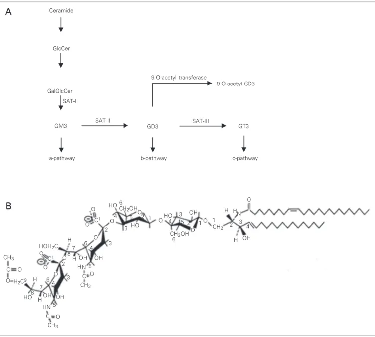
Gangliosides are a large group of sialyl-ated glycosphingolipids widely expressed in mammalian tissues and particularly abun-dant in the nervous system (14). Ganglio-sides possess a remarkable degree of struc-tural diversity, and numerous enzymes are involved in their synthesis, recycling, and turnover. The biosynthesis of gangliosides proceeds in a stepwise manner starting from lactosyl ceramide and involves several gly-cosyltransferases. Some of the genes coding for some of the enzymes have been recently cloned but several questions are still under active investigation regarding how the levels of glycosyltransferases are regulated and determined (Figure 1A) (for a review, see Ref. 14). An important modification of gan-gliosides is the O-acetylation of sialic acid residues on C-9, C-7 or C-9 and C-7 ob-served on restricted cells and tissues but the enzymes responsible for these modifications (O-acetyl transferases) have been particu-larly difficult to clone. It has been suggested that O-acetylation of gangliosides may serve as a cellular recognition signal and also as a tumor marker (12,15). Sialidases are the key enzymes for ganglioside degradation and the plasma membrane-bound sialidase has been implicated not only in the general catabo-lism of gangliosides but also in the modula-tion of cellular funcmodula-tion, such as prolifera-tion and differentiaprolifera-tion (16).
major species in the adult (for a review, see Ref. 18). Furthermore, the accumulation of gangliosides within the neurons in ganglio-side storage diseases results in extensive neurite outgrowth (19) and exogenously ad-ministered gangliosides accelerate the re-generation of neurons in the central nervous system in vivo and stimulate cellular differ-entiation with concomitant neurite sprouting
and extension in vitro (20). Recently, the
genes involved in the biosynthesis of gan-gliosides have been cloned and genetically engineered mice or cells lacking most of the gangliosides have been produced (21). Sur-prisingly, the mice and the cells appeared to be largely normal in their neuronal develop-ment. However, more recent studies have revealed that mice lacking complex
ganglio-CH3
O
O C
H H2C2
8 OH
A
A
A
A
A
OH OH
OH OH
OH
OH HO
HO
HO H
H HN
2 3 4 O
O O O
O
O
O O
O
O
O O O
C O C1
7
H
H
H
CH3
HN 6
4 3
8 7 6 4
2
2
C1
3
5 H
4 5
3 1 6
CH3
O C
2
6 2
CH2
CH2OH
CH2OH
HOH2C
Ceramide
GlcCer
GalGlcCer
GM3
a-pathway SAT-I
SAT-II
9-O-acetyl transferase
SAT-III
GT3
b-pathway c-pathway
B
B
B
B
B
CH3
C
O O
H2C9 H
HO8 5
9-O-acetyl GD3
GD3
3 5
4 1 1
N
sides exhibit axonal degeneration and myeli-nation defects (22).
Experiments in several laboratories have shown that glycosphingolipids, gangliosides in particular, are important components of membrane rafts, where they can mediate important physiological functions such as cell adhesion and signal transduction events and consequently affect cellular phenotypes and functions (for a review, see Ref. 23). Recently, such microdomains have been termed “glycosynapses” in analogy to “im-munological synapses” - the membrane as-sembly of immunocyte adhesion and signal-ing (24). Gangliosides are very abundant in glycosynapses and it has also been shown that anti-ganglioside antibodies can immu-noprecipitate glycosylphosphatidylinositol-anchored proteins (such as TAG-1), Src fam-ily kinases and caveolin (25,26). In addition, several studies have shown the association of gangliosides with integrins and their abil-ity to modulate integrin function (27).
Our studies have focused on a specific ganglioside recognized by the Jones mono-clonal antibody (mAb) (12). In our initial studies we have shown that the pattern of staining with this antibody in the developing rat nervous system correlates temporally and spatially with neuronal migration and neu-rite outgrowth (28,29). The biochemical
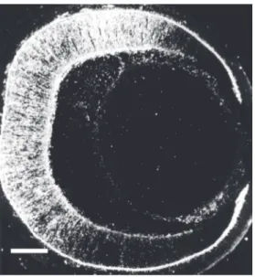
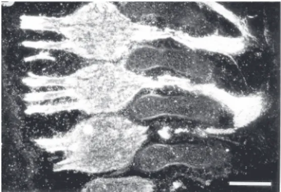
char-acterization of the antigen revealed that this mAb recognizes a ganglioside that migrates between GM1 and GM2 ganglioside stan-dards. Further characterization using over-lay assays on developed high-performance thin-layer chromatography plates indicates that the epitope recognized by the Jones mAb is expressed in several regions of the developing central and peripheral nervous system. In most regions examined the epi-tope resides in two bands (29). Further anal-ysis has revealed that the most abundant and frequent band is 9-O-acetyl GD3, a modifi-cation of GD3 ganglioside in which an acetyl ester is formed at position 9 of the terminal sialic acid residue (Figure 1B) (13). The particular epitope recognized by the Jones mAb depends upon the acetyl group at posi-tion 9 of sialic acid and the immunoreactiv-ity is abolished by mild base treatment since this treatment converts 9-O-acetyl GD3 to GD3 that is not recognized by the Jones mAb (13,30,31). This antigen is abundant in the nervous system and in tumor cells derived from the neural crest, and is absent in most of the other tissues examined (29). However, the same epitope was described in the im-mune system and was designated CD60b (32). The 9-O-acetyl GD3 is present in the retinas of all mammalian species studied so far but is not present in extracts of frog retinas (33). At least two other mAbs have been de-scribed (mAb D1.1 and RB13-2) that recog-nize the same epitope in nervous tissue (34,35). The immunoreactivity of the slower migrating band is also abolished by mild base treatment and it has been suggested that this band might correspond to 9-O-acetyl GQ1c (36). In the developing nervous system, we have shown that the expression pattern of this specific Jones mAb reactive ganglioside correlates with times of cell migration in the retina (Figure 2), superior colliculus, cer-ebellum, and telencephalon and in regions undergoing neurite extension, such as the developing optic tract, the white matter of the cerebellum, the dorsal roots (Figure 3),
the trigeminal system, and olfactory nerve (12,28,29,33,37-39). This has led us to sug-gest that the function of 9-O-acetyl GD3 is to modulate motility either directly or by modi-fying the efficacy of some other component of the system (39,40). The association of 9-O-acetylated gangliosides with cell migra-tion has been well characterized in cancer research, where it was found that tumors arising from neural crest-derived tissues ex-press high levels of 9-O-acetyl GD3. These gangliosides are concentrated in adhesion plaques and are involved in cell adhesion and migration in tumor cells (14,15). In ad-dition, disialogangliosides co-immunopre-cipitate with αVß3 integrin and GM1
co-immunoprecipitates with the epidermal growth factor receptor (for a review, see Ref. 24). Furthermore, it was also shown that cleavage of 9-O-acetyl groups by transgenic expression of influenza C virus hemaggluti-nin caused abnormalities in the development of the retinal layers in the mouse, suggesting a failure in cell migration concomitant with the absence of 9-O-acetyl GD3 (41). Re-cently, it was shown that down-regulation of 9-acetyl GD3 by stable transfection of O-acetylesterase cDNA and antisense vector against the GD3-synthase gene results in cell differentiation in melanoma cell lines (42). Based on our observations and the reports concerning other systems, we raised the hy-pothesis that 9-O-acetyl GD3 may play an important role in neuronal motility in the developing and adult nervous system. In the following sections we will describe the evi-dence showing a functional role for this ganglioside in neuronal migration and neu-rite outgrowth.
Gangliosides in glial-guided radial migration
In most cases, neuronal radial migration occurs on the processes of radial glial cells and has been studied extensively in the cor-tex and cerebellum although similar
arrange-ments for migrating neurons and specialized glia have been described in a number of other systems including the retina and the hippocampus (for a review, see Ref. 1). Re-cent studies have begun to provide a frame-work for the molecular mechanisms under-lying this mode of migration (for a review, see Ref. 43).
In the postnatal cerebellum, the postmi-totic neurons in the external granular cell layer also migrate radially but in this case away from the pia towards the internal granu-lar cell layer. This migration is also associ-ated with radial glia and this system has been extensively used as a model for studies of glial-guided migration (for a review, see Ref. 1). We have investigated the possible role of 9-O-acetyl GD3 in the glial-guided neuronal migration using the developing rat cerebel-lum as a model. In previous studies we had shown that the expression of this ganglioside is developmentally regulated in the develop-ing cerebellum (28). In E14-18 fetuses, im-munocytochemistry labeling with Jones mAb is present, extending from the ventricular to the pial surface of the cerebellar anlage. Most of the immunoreactivity is distributed in a radially oriented pattern that corresponds to the radial migration of Purkinje cell pre-cursors migrating from the ventricular zone towards the pial surface. Immunoreactivity, however, is present in the rhombic lip and in the developing external granular cell layer corresponding to the subpial tangential mi-gration of these cells. During the postnatal period in the rat, the cells in the external granular cell layer proliferate and migrate
radially away from the pia to form the inter-nal granular cell layer. At this stage, immu-nocytochemistry reveals a radially oriented pattern of Jones binding extending from the external granular cell layer towards the pre-sumptive granular cell layer (Figure 4). Elec-tron microscopic immunocytochemistry re-vealed that around the peak of cerebellar neuronal migration, 9-O-acetyl GD3 was lo-calized at the contact sites between migrat-ing granule cells and radial glia in the exter-nal granular cell layer and prospective mo-lecular layer (40). We have also observed that both cultured neurons from the rat cer-ebellum when in contact with glial cells that support cell migration (radial glial cells) and glial cells themselves express 9-O-acetyl GD3 (44). In contrast, glial cells with a stellate morphology, even when in contact with gran-ule neurons, do not express this antigen (44). To test the functional role of 9-O-acetyl GD3 in neuronal migration we have used cerebel-lar explants and cerebelcerebel-lar slices. We have shown that the Jones mAb blocks the migra-tion of neurons in a dose-dependent manner, suggesting that 9-O-acetyl GD3 is involved in the radial migration of presumptive gran-ule cells in the developing cerebellum (40). These results further support our view that this molecule is involved in cell migration.
The immunoreactivity for this ganglio-side is also present in the embryonic telen-cephalon, showing a radial organization cor-related with radial migration in this region (28). Blockage of the ganglioside with Jones antibody arrests neuronal migration on tis-sue slices of embryonic telencephalon (45).
Gangliosides in tangential migration
Glial-guided neuronal migration has been relatively well studied, whereas much less is known about tangential migration (for a re-view, see Ref. 11). Tangentially migrating neurons are found in the cortex, the cerebel-lum and the rhombencephalon and they mi-grate perpendicularly to the glial scaffolding (6,46,47). Some of the molecular cues that guide tangential migration have been recently identified and the picture that is emerging is that the same families of signals involved in the directional guidance of developing growth cones may also guide the translocation of cell bodies during tangential migration (9).
The migration of interneurons from the anterior subventricular zone to the olfactory bulb provides a useful system to study the cellular and molecular mechanisms that regu-late tangential neural migration. These neu-ronal precursors migrate through a distinct pathway within the subventricular zone called the rostral migratory stream, and this migra-tion occurs even in the adult. Molecular studies have shown that, in the particular case of the rostral migratory stream, the mech-anism of migration involves the sialylated form of the neural cell adhesion molecule (NCAM) and is guided, in part, by negative chemotropism (48).
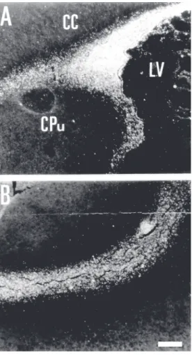
In view of the abundant evidence for a role of 9-O-acetyl GD3 in directional move-ments of neuronal soma or processes we have performed an immunohistochemical analysis of their expression along the route of tangential migrations of some olfactory bulb prospective interneurons in the devel-oping and adult rats. We have found that this ganglioside is highly expressed in the lateral ventricle subventricular zone and along the route of tangential migration (rostral migra-tory stream) into the olfacmigra-tory bulb during development (Figure 5). In the adult, we have found staining around the lateral ven-tricle and in chains of cells in the rostral migratory stream region (49). In a few
tions, individual stained cells are observed, with a morphology similar to that of migrat-ing cells. Despite the scarcity of stainmigrat-ing, this is one of the few regions where expres-sion of 9-O-acetyl GD3 is found in the adult nervous system. Interestingly, this region has been described as one of the regions in which neurogenesis persists in the adult brain and from which stem cells can be isolated in the adult brain. It would be interesting to determine whether stem cells isolated from adult brains also express 9-O-acetyl GD3.
To investigate the functional role of 9-O-acetyl gangliosides in tangential migration we have used explants of the subventricular zone region as a model. We have found that migrating chains similar to the ones formed
in vivo are also seen in this in vitro system.
The migrating chains from the subventricu-lar zone explant express 9-O-acetyl GD3 which is distributed in a punctiform manner in individual cells and treatment of the cul-tures with the antibody against 9-O-acetyl GD3 arrested neuronal migration in these cultures. These data suggest that 9-O-acetyl GD3 participates in neuronophilic as well as gliophilic migration (50,51).
Gangliosides and neurite outgrowth
During development, neuronal growth cones interact with physical and chemical cues in their environment and these interac-tions guide the growth cones along specific pathways to their appropriate targets (for a review, see Ref. 52). Many factors such as soluble or substrate-bound growth factors and components of the extracellular matrix provide favorable environments that allow or promote motility of growth cones and neurite elongation (53). Repulsive and in-hibitory factors have also been described and may participate in guidance of growth cones (for a review, see Ref. 52). Ganglio-sides, in particular disialoganglioGanglio-sides, have been implicated in adhesion systems and in cell to substrate adhesion of both melanoma
and neuroblastoma cells (15). Indeed, a num-ber of separate lines of evidence support the hypothesis that these molecules modulate the activity of the integrin family of cell surface adhesion molecules (for a review, see Ref. 23) and selectively induce the syn-thesis of MAP2 (high molecular weight mi-crotubule-associated protein 2) (54). Fur-thermore, numerous studies employing a variety of models have reported that ganglio-side treatment will increase the rates of neu-ritogenesis both in vitro and in vivo, and
axonal regeneration and sprouting following brain trauma to the central or peripheral nervous system, suggesting a role for gan-gliosides in neurite outgrowth.
In our studies of the expression of 9-O-acetyl GD3 in the developing nervous sys-tem we have found that, in addition to the pattern of staining coinciding with neuronal
migration, these gangliosides were also ob-served along pathways of axon extensions such as the optic tract, the olfactory nerve and the central and peripheral processes of dorsal root ganglia. This pathway staining is only observed while large numbers of growth cones are present at these positions and the staining disappears from most of the path-ways studied as soon as the axons reach their target regions (28,37). One interesting ex-ception to this generalization, however, is the olfactory system (37). In this system we have shown that the expression of 9-O-acetyl GD3 begins as early as embryonic day 13, when the olfactory epithelium and the mi-gratory mass were intensely stained. At em-bryonic day 19, the immunoreactivity had disappeared from the olfactory epithelium but remained in a few fascicles and some glomeruli of the olfactory bulb in the new-born and adult. We conclude that the expres-sion of 9-O-acetyl GD3 by olfactory axons and/or migrating cells may facilitate axonal outgrowth during development and might be involved in the formation of new glomeruli in the mature olfactory bulb (37).
We have recently investigated whether 9-O-acetyl gangliosides are also re-expressed during the regeneration of sciatic nerves that were submitted to crushing. We have found an up-regulation of 9-O-acetyl GD3 expres-sion starting five days after crushing, reach-ing a maximum at day 7, and decreasreach-ing afterwards to reach the level of the control animals at day 10 (Mendez-Otero R, Silva AO, Resende VTR and Hedin-Pereira C, unpublished data). These results suggested to us that 9-O-acetyl GD3 may play a func-tional role in neurite outgrowth during de-velopment and regeneration.
To test the functional role of 9-O-acetyl GD3 in neurite outgrowth, we used explants of dorsal root ganglia. In previous studies we have shown that embryonic dorsal root gan-glion neurons and their processes either in vivo (28), in explant cultures or as dissoci-ated cells present heavy immunoreactivity to
the antibody against the ganglioside. We then plated dorsal root ganglion explants from embryonic day 16 onto laminin, col-lagen or fibronectin. Neurites grew out of the explants and expressed high amounts of 9-O-acetyl GD3 in all substrates. We then investigated the functional role of this gan-glioside on neurite extension using embry-onic dorsal root ganglion explants grown on laminin substrates as a model. In these ex-periments, the behavior of individual growth cones was recorded using a time-lapse video-enhanced imaging system before and after the addition of antibodies that recognize spe-cific gangliosides known to be expressed on these growth cones. Using this system, it was possible to demonstrate that the advance of growth cones on laminin was halted in the presence of Jones mAb. The onset of the effect was rapid and signaled by an immedi-ate cessation of elongation, a loss of lamelli-podia and a retrieval of axoplasm (55). Block-ade of 9-O-acetyl GD3 also induces the ap-pearance of lateral spikes along the neurites in addition to the effect on the growth cone. We have shown that microfilaments and mi-crotubular rearrangements accompany these structural modifications (56). Our results suggest that 9-O-acetyl GD3 may play an important role in the extension of growth cones and consequently influence naviga-tion and pathway finding during develop-ment and regeneration.
Conclusion and future directions
subsets of moving cells and/or processes that express these molecules (Figure 6). Examples of such mechanism are the described inter-actions of gangliosides with integrin recep-tors (57,58) and with TAG-1 (an adhesion molecule) (25). Another possibility is that gangliosides may serve to modulate the avail-ability of divalent cations to a number of different calcium- or magnesium-dependent adhesion systems and consequently regulate ligand binding (57). Alternatively, it has been suggested that gangliosides may regulate the lateral diffusion and assembly of signaling complexes in membrane microdomains and therefore interfere with the functional role of these proteins, as proposed for integrin in focal adhesion points and TAG-1 (59). An-other possible model is based on the homo-philic interaction between gangliosides lo-cated on different cell membranes. A similar model has been proposed to explain the in-volvement of NCAM in regulating neural cell adhesion and axon growth. Finally, it has also been shown in several systems that selectins and/or galectins can function as receptors for specific gangliosides (60) and this interaction could trigger a cascade of reactions in both cells.
The ganglioside 9-O-acetyl GD3 could provide a new cell signaling mechanism in glial-guided neuronal migration in the devel-oping nervous system. Moreover, it is im-portant to note that most neurons migrate by extension of a leading, neurite-like process and that 9-O-acetyl GD3 has been
impli-Radial glia
Migrating neuron
Ca CaCa CaCa2+2+2+2+2+
9-O-GD3 9-O-GD3 9-O-GD3 9-O-GD3 9-O-GD3
Ca Ca Ca Ca Ca2+2+2+2+2+
9-O-GD3 9-O-GD3 9-O-GD3 9-O-GD39-O-GD3
α3 ß1 α3 ß1
cated in both neuronal migration and neurite outgrowth. The potential roles for this gan-glioside in identical mechanisms for neu-ronal migration and neurite outgrowth sug-gest an emerging framework in which gly-colipids are involved in cell movement in general.
Acknowledgments
The technical assistance of Felipe Marins is gratefully acknowledged.
Figure 6. Schematic diagram suggesting possible mechanisms for 9-O-acetyl GD3 action on neuronal motility. The ganglioside may modulate the integrin receptor through a Ca2+
-dependent mechanism and/or by a direct interaction between the ceramide moiety of the ganglioside and the integrin transmembrane helix. In a first stage, the integrin receptor is inactive and there is no recognition of extracellular matrix proteins. Later, gangliosides in the adjacent membrane become laterally packed around the integrin receptor (represented in the scheme by only one ganglioside), providing an optimal Ca2+-enriched
microenviron-ment for the activation and recognition of extracellular matrix proteins by this receptor. In addition, the gangliosides can interact directly with the integrin receptor through a con-served lysine located inside a 23-amino acid sequence at the carboxy-terminus of the integrin transmembrane helix. 9-O-GD3 = 9-O-acetyl GD3.
References
1. Hatten ME (1999). Central nervous system neuronal migration. An-nual Review of Neuroscience, 22: 511-539.
2. Parnavelas JG (2000). The origin and migration of cortical neurons: new vistas. Trends in Neurosciences, 23: 126-131.
3. Nadarajah B, Brunstrom JE, Grutzendler J, Wong ROL & Pearlman AL (2001). Two modes of radial migration in early development of the cerebral cortex. NatureNeuroscience, 4: 143-150.
4. de Diego I, Kyriakopoulou K, Karagogeos D & Wassef M (2002). Multiple influences on the migration of precerebellar neurons in the
caudal medulla. Development, 129: 297-306.
5. Norgren Jr RB & Lehman MN (1991). Neurons that migrate from the olfactory epithelium in the chick express luteinizing hormone-re-leasing hormone. Endocrinology, 128: 1676-1678.
6. Tamamaki N, Fujimori KE & Takauji R (1997). Origin and route of tangentially migrating neurons in the developing neocortical inter-mediate zone. Journal of Neuroscience, 17: 8313-8323.
8. Hynes RO, Patel R & Miller RH (1986). Migration of neuroblasts along preexisting axonal tracts during prenatal cerebellar develop-ment. Journal of Neuroscience, 6: 867-876.
9. Denaxa M, Chan C-H, Schachner M, Parnavelas JG & Karagogeos D (2001). The adhesion molecule TAG-1 mediates the migration of cortical interneurons from the ganglionic eminence along the corticofugal fiber system. Development, 128: 4635-4644. 10. Wichterle H, Garcia-Verdugo J & Alvarez-Buylla A (1997). Direct
evidence for homotypic, glia-independent neuronal migration. Neu-ron, 18: 779-791.
11. Corbin JG, Nery S & Fishell G (2001). Telencephalic cells take a tangent: non-radial migration in the mammalian forebrain. Nature Neuroscience, 4: 1177-1182.
12. Constantine-Paton M, Blum AS, Mendez-Otero R & Barnstable C (1986). A cell surface molecule distributed in a dorso-ventral gradi-ent in the perinatal rat retina. Nature, 324: 459-462.
13. Blum AS & Barnstable CJ (1987). O-acetylation of a cell-surface carbohydrate creates discrete molecular patterns during neuronal development. Proceedings of the National Academy of Sciences, USA, 84: 8716-8720.
14. Lloyd KO & Furukawa K (1998). Biosynthesis and functions of gangliosides: Recent advances. Glycoconjugate Journal, 7: 627-636.
15. Cheresh DA, Varki AP, Varki NM, Stallcup WB, Levine J & Reisfeld RA (1984). A monoclonal antibody recognizes an O-acetylated sialic acid in a human melanoma-associated ganglioside. Journal of Bio-logical Chemistry, 259: 7453-7459.
16. Reitzenstein C, Kopitz J, Schuhmann V & Cantz M (2001). Differen-tial functional relevance of a plasma membrane ganglioside sialidase in cholinergic and adrenergic neuroblastoma cell lines. European Journal of Biochemistry, 268: 326-333.
17. Rosner H (1994). Gangliosides and brain development. In: Nicolini M & Zatta PF (Editors), Glycobiology and the Brain. Pergamon Press, New York, 19-39.
18. Yu RK (1994). Developmental regulation of ganglioside metabolism.
Progress in Brain Research, 10: 3-44.
19. Purpura DP (1978). Ectopic dendritic growth in mature pyramidal neurons in human ganglioside storage disease. Nature, 276: 520-521.
20. Roisen F, Matta SG & Rapport MM (1986). The role of gangliosides in neurotrophic interaction in vitro. In: Tettamati G, Ledeen RW, Sadhoff K, Nagai Y & Toffano G (Editors), Gangliosides and Neuronal Plasticity. Liviana Press, Padova, Italy, 281-293.
21. Takamiya H, Yamamoto A, Furukawa K et al. (1996). Mice with disrupted GM2/GD2 synthase gene lack complex gangliosides but exhibit only subtle defects in their nervous system. Proceedings of the National Academy of Sciences, USA, 93: 10662-10667. 22. Chiavegatto S, Sun J, Nelson RJ & Schnaar RL (2000). A functional
role for complex gangliosides: motor deficits in GM2/GD2 synthase knockout mice. Experimental Neurology, 166: 227-234.
23. Pande G (2000). The role of membrane lipids in regulation of integrin function. Current Opinion in Cell Biology, 12: 569-574.
24. Hakomori SI (2002). The glycosynapse. Proceedings of the National Academy of Sciences, USA, 99: 225-232.
25. Kasahara K, Watanabe K, Takeuchi K, Kaneko H, Oohira A, Yama-moto T & Sanai Y (2000). Involvement of gangliosides in glycosyl-phosphatidylinositol-anchored neuronal cell adhesion molecule TAG-1 signaling in lipid rafts. Journal of Biological Chemistry, 275: 34701-34709.
26. Katagiri YU, Mori T, Nakajima H, Katagiri C, Taguchi T, Takeda T, Kiyokawa N & Fujimoto J (1999). Activation of Src family kinase Yes
induced by Shiga toxin binding to globotriaosyl ceramide (Gb3/ CD77) in low density, detergent-insoluble microdomains. Journal of Biological Chemistry, 274: 35278-35282.
27. Yates AJ & Rampersaud A (1998). Sphingolipids as receptor modu-lators: an overview. Annals of the New York Academy of Sciences, 845: 7-71.
28. Mendez-Otero R, Schlosshauer B, Barnstable CJ & Constantine-Paton M (1988). A developmentally regulated antigen associated with neural cell and process migration. Journal of Neuroscience, 8: 564-579.
29. Schlosshauer B, Blum AS, Mendez-Otero R, Barnstable CJ & Constantine-Paton M (1988). Developmental regulation of ganglio-side antigens recognized by the JONES antibody. Journal of Neuro-science, 8: 580-592.
30. Bonafede DM, Macala LJ, Constantine-Paton M & Yu RK (1989). Isolation and characterization of ganglioside 9-O-acetyl-GD3 from bovine buttermilk. Lipids, 24: 680-684.
31. Ritter G, Boosfeld E, Markstein E, Yu RK, Ren SL, Stallcup WB, Oettgen HF, Old LJ & Livingston PO (1990). Biochemical and sero-logical characteristics of natural 9-O-acetyl GD3 from human mela-noma and bovine buttermilk and chemically O-acetylated GD3. Can-cer Research, 50: 1403-1410.
32. Mason D, Andre P, Bensussan A et al. (2001). CD antigens 2001: Aims and results of HLDA workshops. Stem Cells, 19: 556-562. 33. Mendez-Otero R, Schlosshauer B & Constantine-Paton M (1992).
Role of acetylated gangliosides on neuronal migration and axonal outgrowth. In: Lent R (Editor), The Visual System from Genesis to Maturity. Birkhauser, Boston, MA, 49-62.
34. Levine JM, Beasly L & Stallcup WB (1984). The D1.1 antigen: a cell surface marker for germinal cells of the central nervous system.
Journal of Neuroscience, 4: 820-831.
35. Reinhardt-Maelicke S, Cleeves V, Kindler-Rohrborn A & Rajewsky MF (1990). Differential recognition of a set of O-acetylated ganglio-sides by monoclonal antibodies RB13-2, D1.1, and Jones during rat brain development. Developmental Brain Research, 51: 279-282. 36. Multani P, Bonafede DM, Yu RK & Constantine-Paton M (1988).
Biochemical characterization of Jones immunoreactive gangliosides in rat. Society for Neuroscience Abstracts, 14: 1016.
37. Mendez-Otero R & Ramon-Cueto A (1994). Expression of 9-O-acety-lated gangliosides during development of the rat olfactory system.
NeuroReport, 5: 1755-1759.
38. Mello LEAM & Mendez-Otero R (1996). Expression of 9-O-acety-lated gangliosides in the rat hippocampus. Neuroscience Letters, 213: 17-20.
39. Mendez-Otero R & Santiago MF (2001). Functional role of a gly-colipid in directional movements of neurons. Anais da Academia Brasileira de Ciências, 73: 221-229.
40. Santiago MF, Berredo-Pinho M, Costa MR, Gandra M, Cavalcante LA & Mendez-Otero R (2001). Expression and function of ganglio-side 9-O-acetyl GD3 in postmitotic granule cell development. Mo-lecular and Cellular Neurosciences, 17: 488-499.
41. Varki A, Hooshmand F, Diaz S, Varki NM & Hedrick SM (1991). Developmental abnormalities in transgenic mice expressing a sialic acid-specific 9-O-acetylase. Cell, 65: 65-74.
42. Birkle S, Gao L, Zeng G & Yu RK (2000). Down regulation of GD3 ganglioside and its O-acetylated derivative by stable transfection with antisense vector against GD3-synthase gene expression in hamster melanoma cells: effects on cellular growth, melanogen-esis, and dendricity. Journal of Neurochemistry, 74: 547-554. 43. Walsh CA & Goffinet AM (2000). Potential mechanisms of
Opinion in Genetics and Development, 10: 270-274.
44. Mendez-Otero R & Constantine-Paton M (1990). Granule cell induc-tion of 9-O-acetyl gangliosides on cerebellar glia in microcultures.
Developmental Biology, 138: 400-409.
45. Hedin-Pereira C, deMoraes ECP, Lent R & Mendez-Otero R (1997). Immunoblocking 9-O-acetylated gangliosides arrests cell migration in the developing cerebral cortex. Society for Neuroscience Ab-stracts, 23: 871.
46. Komuro H, Yacubova E, Yacubova E & Rakic P (2001). Mode and tempo of tangential cell migration in the cerebellar external granular layer. Journal of Neuroscience, 21: 527-540.
47. Ono K & Kawamura K (1989). Migration of immature neurons along tangentially oriented fibers in the subpial part of the fetal mouse medulla oblongata. Experimental Brain Research, 78: 190-201. 48. Hu H, Tomasiewics H, Magnuson T & Rutishauser U (1996). The
role of polysialic acid in migration of olfactory bulb interneuron precursors in the subventricular zone. Neuron, 16: 735-743. 49. Mendez-Otero R & Cavalcante LA (1996). Expression of
9-O-acety-lated gangliosides is corre9-O-acety-lated with tangential cell migration. Neu-roscience Letters, 204: 97-100.
50. Miyakoshi LM, Mendez-Otero R & Hedin-Pereira C (2001). The 9-O-acetyl GD3 ganglioside is expressed by migrating chains of subven-tricular zone neurons in vitro. Brazilian Journal of Medical and Bio-logical Research, 34: 669-673.
51. Miyakoshi LM, Mendez-Otero R & Hedin-Pereira C (2001). Role of 9-O-acetyl GD3 in the migration of subventricular zone neuronal pre-cursors. Journal of Neurochemistry, 78 (Suppl): 56 (Abstract). 52. Gallo G & Letourneau PC (1999). Axon guidance: a balance of
signals sets axons on the right track. Current Biology, 9: R490-R492.
53. Garcia-Abreu J, Mendes FA, Onofre GR, Freitas MS, Silva LCF, Moura Neto V & Cavalcante LA (2000). Contribution of heparan sulfate to the non-permissive role of the midline glia to the growth of midbrain neurites. Glia, 29: 260-272.
54. Ferreira A, Busciglio J, Landa C & Caceres A (1990). Ganglioside-enhanced neurite growth: evidence for a selective induction of high molecular weight MAP-2. Journal of Neuroscience, 10: 293-302. 55. Mendez-Otero R & Friedman JE (1996). Role of acetylated
ganglio-sides on neurite extension. European Journal of Cell Biology, 71: 192-198.
56. Araujo H, Menezes MA & Mendez-Otero R (1997). Blockage of 9-O-acetyl gangliosides induces depolymerization in growth cones and neurites. European Journal of Cell Biology, 72: 202-213.
57. Cheresh DA, Pierschbacher MD, Herzig MA & Mujoo K (1986). Disialogangliosides GD2 and GD3 are involved in the attachment of human melanoma and neuroblastoma cells to extracellular matrix proteins. Journal of Cell Biology, 102: 688-696.
58. Probstmeier R & Pesheva P (1999). Tenascin-C inhibits beta1 inte-grin-dependent cell adhesion and neurite outgrowth on fibronectin by a disialoganglioside-mediated signaling mechanism. Glycobiol-ogy, 9: 101-114.
59. Katz B-Z, Zamir E, Bershadsky A, Yamada KM & Geiger B (2000). Physical state of the extracellular matrix regulates the structure and composition of cell-matrix adhesions. Molecular Biology of the Cell, 11: 1047-1060.