FINE NEEDLE ASPIRATION CYTOLOGY OF THYROID LESIONS-10
YEAR RETROSPECTIVE STUDY AT CHHATTISGARH INSTITUTE OF
MEDICAL SCIENCES
Gupta R1, Bagde S2, Ganguly S3, Minj M4, Tembhurnikar P5
HOW TO CITE THIS ARTICLE:
Gupta R, Bagde S, Ganguly S, Minj M, Tembhurnikar P. “Fine Needle Aspiration Cytology of Thyroid Lesions-10 Year Retrospective Study at Chhattisgarh Institute of Medical Sciences”. Journal of Evidence based Medicine and Healthcare; Volume 2, Issue 41, October 12, 2015; Page: 6911-6915,
DOI: 10.18410/jebmh/2015/942
ABSTRACT: BACKGROUND: Fine needle aspiration cytology is a well-established simple, safe technique among diagnostic tests for evaluation of thyroid lesions. It helps for the preoperative diagnosis of benign and malignant lesions, thus minimizing and preventing the unwanted surgeries. OBJECTIVE: The aim of this retrospective study is to classify various cytomorphological lesions of the thyroid swellings in our setup & to know the age and sex incidence. METHOD: A retrospective study of 506 patients was conducted from January 2003 to December 2013 in pathology department of our hospital, Chhattisgarh Institute of Medical Sciences, Bilaspur. Fine needle aspiration of all cases was done after detailed clinical history, physical examination and thyroid function test. Then lesions were classified cytomorphologically. Age and sex incidence was calculated. RESULTS: FNAC results were classified as inadequate material, non-neoplastic, neoplastic and malignant. Out of total 506 cases studied. 416(82.2%) were non neoplastic 64(12.6%) were neoplastic 17(3.35%) were malignant lesions and 9(1.77%) cases were inadequate material. Age and sex incidence was calculated. Thyroid lesions showed female predominance, out of 506 cases, 428 were females and 78 were males. Commonest malignancy was papillary carcinoma of thyroid (7 cases). CONCLUSION: FNAC is a safe, simple less expensive widely accepted outpatient procedure for diagnosis of thyroid lesions. It also helps to avoid unwanted surgical interventions in patients with benign lesions.
INTRODUCTION: Fine needle aspiration cytology (FNAC) is a well-established, safe, non- invasive cost effective and efficient procedure used to differentiate between benign and malignant thyroid swelling.(1,2) Over the past 3 decades, confidence in FNAC as a reliable test has
grown considerably and it has emerged as a direct, accurate diagnostic procedure in the management of thyroid disease gaining worldwide acceptance.(3,4) Thyroid gland is in its
superficial and easily accessible position is an ideal tissue for fine needle aspiration cytology(5,2)
Important factor for the satisfactory test includes representative specimen from the swelling and an experienced cytopathologist to interpret the findings.(5,2) Thyroid swelling display range of
lesions from non-neoplastic to malignant. They are common lesions encountered in day to day surgical practice(2) To make an effective surgical intervention in these lesions, it is very vital to
make a preoperative assessment of the morphological nature of lesion.(2,6) This study was carried
MATERIALS AND METHODS: A cytological retrospective study of 506 cases from April 2003 to April 2013 of palpable thyroid swellings was carried out in Department of pathology, Chhattisgarh Institute of Medical Science (CIMS) Bilaspur. Detailed clinical history, physical Examination, thyroid function test and ultra sonographical examination were obtained in each case; all the patients were referred by clinicians to pathology department for cytopathological diagnosis.
A fine needle was then introduced using a 10ml plastic syringe fitted with a 23-24 gauge disposable needle. The aspirate contents of the needle were expelled on glass slides. Multiple smears were prepared 2-3 slides were immediately fixed in 95% ethyl alcohol and remaining was air dried.
The slides were stained with Haematoxylin and eosin, papanicolaus and MGG stain and examined with light microscope. And result were classified into four groups-
1. Inadequate material.
2. Non-neoplastic [Colloid goiter, colloid goiter with Hyper plastic change, colloid goiter with cystic change, Hashimotos thyroiditis, thyroiditis, thyroglossal cyst].
3. Neoplastic [Follicular neoplasm, hurthle cell neoplasm].
4. Malignant [Papillary carcinoma, medullary carcinoma and anaplastic carcinoma]
After the cytomorphological classification of thyroid swelling age and sex incidence was calculated.
RESULT: 506 patients with thyroid lesions were included in the study from April 2003 to April 2013 in all this, cases pre-operative FNAC was done. Out of 506 cases inadequate material was obtained in 9 cases and adequate material was obtained in 497 cases.
Out of 506 cases studied, 428(84.5%) were female while 78(15.5%) were male. The thyroid lesions showed female predominance with the female to male ratio of (5.5:1) Most of the cases were presented between 3rd to 5th decades of life (388 cases) [76.67%]. The youngest
patient in our study was 5 years old boy, while oldest is a 75 year old lady with a mean age of 38.5.
Benign lesions were found commonly in 3rd to 5th decade of life and malignant lesions
were found in 5th and 6th decade of life. In our study youngest patient diagnosed as follicular
neoplasm was of 16 years age and that diagnosed as papillary carcinoma was of 30 years age. Oldest patient was 65 years old which was diagnosed as medullary carcinoma. Out of 506 FNAC cases of thyroid swelling, studied at our set up, of 9 1.77% smears were reported as inadequate material. Remaining 497 cases were studied out of which 416(82.2%) patients showed non-neoplastic lesions, 64(12.6%) patients showed, non-neoplastic lesion and remaining 17(3.35%) cases were malignant.
17 cases were reported as malignant. Incidence of papillary carcinoma was highest, 7 (1.3%) cases while medullary and anaplastic carcinoma each were 5(0.9%) one case of papillary carcinoma was diagnosed with metastasis in the swelling over forehead.
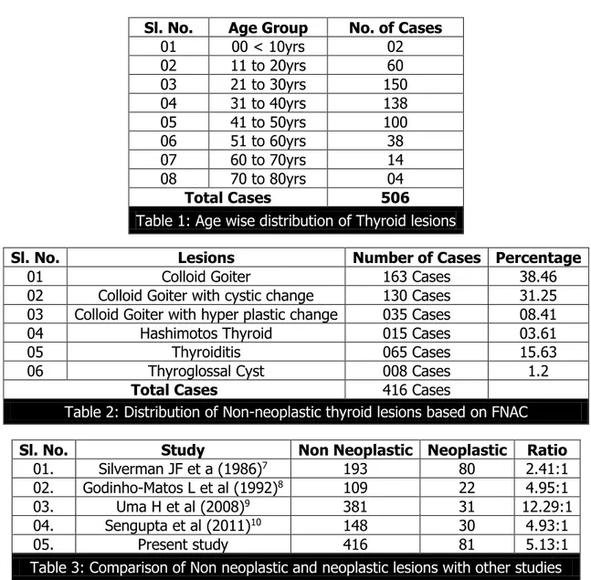
Sl. No. Age Group No. of Cases
01 00 < 10yrs 02
02 11 to 20yrs 60
03 21 to 30yrs 150
04 31 to 40yrs 138
05 41 to 50yrs 100
06 51 to 60yrs 38
07 60 to 70yrs 14
08 70 to 80yrs 04
Total Cases 506
Table 1: Age wise distribution of Thyroid lesions
Sl. No. Lesions Number of Cases Percentage
01 Colloid Goiter 163 Cases 38.46
02 Colloid Goiter with cystic change 130 Cases 31.25
03 Colloid Goiter with hyper plastic change 035 Cases 08.41
04 Hashimotos Thyroid 015 Cases 03.61
05 Thyroiditis 065 Cases 15.63
06 Thyroglossal Cyst 008 Cases 1.2
Total Cases 416 Cases
Table 2: Distribution of Non-neoplastic thyroid lesions based on FNAC
Sl. No. Study Non Neoplastic Neoplastic Ratio
01. Silverman JF et a (1986)7 193 80 2.41:1
02. Godinho-Matos L et al (1992)8 109 22 4.95:1
03. Uma H et al (2008)9 381 31 12.29:1
04. Sengupta et al (2011)10 148 30 4.93:1
05. Present study 416 81 5.13:1
Table 3: Comparison of Non neoplastic and neoplastic lesions with other studies
In our study the non-neoplastic to neoplastic ratio was comparable with the Godinho-matos L et al and Sengupta et al studies. The ratio in other studies varied from 2:41:1 to 12:29:1.
DISCUSSION: FNAC is regarded as the gold standard initial investigation in the diagnosis of thyroid swellings. The technique is safe, simple and quick with a low complication rate. Several other tests, such as uitrasonography, radioisotope scanning and FNA biopsy have been used for evaluation of thyroid swellings.(11,2) Studies have demonstrated that among all these diagnostic
In present study, the age of patient ranged from 5-72 years with a mean age of 38.5 years. This age range and mean incidence was slightly higher as compared with the studies.(3) We
have found majority of patients in 2nd decade of life 150 cases (20.8%) as compared to other
studies in which majority of patients (44%) were in their third decade of life.(5) In our study
thyroid swellings manifested at earlier age group as compared to other studies.
The study of 506 cases showed female to male ratio of [5.5:1] with 428 females and 78 males. Thyroid swellings were 11 times more common in females as compared to males.(5) Our
study showed thyroid swellings were 5-6 times common in females than males.
In the present study out of 497 cases 9 were inadequate material. Non neoplastic accounted for about 416 and neoplastic were 81 ratio about (5.13:1) which is comparable with sengupta at al (2011)10 and Godinho- Matos et al studied varied from 2.4:1 to 12.29:1.
In present study distribution of non-neoplastic thyroid lesions is colloid Goitre (162) cases (32.01%). Colloid Goitre with cystic change 130 cases (25.69%). Colloid Goitre with hyperplastic change 35 cases (6.91%) Hashimotos thyroiditis 15 cases (2.96%) Thyroidit 65cases (12.84%) and thyroglossal cyst 8 cases-(1.58%).
In addition to clinical information for diagnosis and treatment of thyroid lesions, ultrasonography, serology and radiographic evaluation of the swelling of neck (thyroid) and FNAC studies are essential. (12)
CONCLUSION: FNAC is regarded as the gold standard initial investigation in the diagnosis of thyroid swellings. Our primary aim of this study was to differentiate thyroid lesions into non-neoplastic and non-neoplastic categories. Depending upon the cytomorphological classifications of lesions, patients who were diagnosed as benign lesions were kept in close follow up and those diagnosed as malignant were subjected to surgery, thereby decreasing the rate of unnecessary surgery. In our study female to male ratio is 5.5:1, showing female predominance and the swellings were common in second decades. In non-neoplastic lesions colloid goitre is commonest while papillary carcinoma of thyroid is commonest in malignant category. The diagnostic accuracy of FNAC when it is done in conjection with advanced imaging techniques, immunologic analysis and electron microscopy and there by further reducing misdiagnosis.
REFERENCES:
1. H. A Nggada, A. Musa, B Gali and M.I. Khalil; Fine Needle Aspiration cytology of Thyroid nodule (s): A Nigerian Tertiary hospital Experience. The Internet journal of pathology. 2006 volume 5 number 1.
2. Parikh UR, goswami H.M, Saah A.M, Mehta N.P, Gonsai R.N: FNAC study of thyroid lesions (study of 240 cases)
3. Akhila sekhar, Inamdae S.S, V.S. Dombale, Prabhu M.H: FNAC study of Thyroid lesions_A 2
year prospective study in a Tertiary centre.
4. Clark DP, faquin WC Thyroid cyto pathology 2nd New York: springer 2010.
5. Manoj Gupta, Sarita Gupta and Ved bhushan Gupta Correlation of FNAC with Histopathology
in the diagnosis of solitary thyroid Nodule. Journal of “Thyroid Research, Vol. 2010; Article
6. Miller JM, Kini SR, Hamberg JI. The needle biopsy diagnosis of papillary thyroid carcinoma. Cancer 1985; 55: 2812-2817.
7. Silverman JF at al. the pole of fine needle aspiration biopsy in the rapid diagnoses and management of thyroid neoplasms cancer 1986; 57: 1164-70.
8. Godinho- Matos L, Kocjan G, Kurtz A, Contribution of fine needle aspiration cytology to diagnosis and management of thyroid disease. J clin pathol 1992; 45: 391-5.
9. Uma H, Sukant G, Harsh M, Nitin N, Role of fine needle aspiration cytology in the diagnosis and management of thyroid lesions: A study on 434 patient J cytol 2008; 25 (1) : 13-7. 10.Sengupta A, Pal R, Kar S, Zaman FA, Sengupta S, Pal S, fine needle aspiration cytology as
the primary diagnostic tool in thyroid enlargement J Nat SC Biol Med 2011; 2: 113-8
11.B. Mundaras, I. Macllister, J. Carson, P.C. Pyper. Accuracy of fine needle aspiration cytology in diagnosis of thyroid swellings. The internet journal of Endocrinology, 2006 Vol. 2 no. 2. 12.Murati A, Erdogan, Sevim S, Unal I, Akyuz S, Diagnositic efficacy and importance of
fine-Needle aspiration cytology of thyroid nodules, J cytol 2014: 31: 73-8.
5. Associate Professor, Department of Pathology, Chhattisgarh Institute of Medical Sciences, Bilaspur,
Chhattisgarh.
NAME ADDRESS EMAIL ID OF THE CORRESPONDING AUTHOR:
Dr. Gupta R, Assistant Professor, Department of Pathology,
Chhattisgarh Institute of Medical Sciences, Bilaspur-495001, Chhattisgarh.
E-mail: drrashmidsao@rediffmail.com
Date of Submission: 25/09/2015. Date of Peer Review: 26/09/2015. Date of Acceptance: 29/09/2015. Date of Publishing: 06/10/2015.
AUTHORS:
1. Gupta R. 2. Bagde S. 3. Ganguly S. 4. Minj M.
5. Tembhurnikar P.
PARTICULARS OF CONTRIBUTORS:
1. Assistant Professor, Department of Pathology, Chhattisgarh Institute of Medical Sciences, Bilaspur,
Chhattisgarh.
2. Associate Professor, Department of Pathology, Chhattisgarh Institute of Medical Sciences, Bilaspur,
Chhattisgarh.
3. Assistant Professor, Department of Pathology, Chhattisgarh Institute of Medical Sciences, Bilaspur,
Chhattisgarh.
4. Associate Professor, Department of Pathology, Chhattisgarh Institute of Medical Sciences, Bilaspur,