ELISA LOPES E LAGES
AVALIAÇÃO DE BIOMARCADORES INFLAMATÓRIOS
MOLECULARES EM PACIENTES COM CÂNCER
DE MAMA
Tese apresentada ao Programa de Pós- Graduação em Ginecologia, Obstetrícia e
Mastologia da Faculdade de Medicina de Botucatu-UNESP, para obtenção do título de Doutora.
Orientador: Prof. Dr. Agnaldo Lopes da Silva Filho Co-Orientadora: Dra. Andréa Teixeira de Carvalho
Dedico esta tese à minha família: H
Primeiramente ao Professor Agnaldo pelos ensinamentos e
exemplo, por quem tenho profunda admiração.
À Dra. Andrea Teixeira de Carvalho, pela constante orientação,
disponibilidade, e pontuações fundamentais.
Ao Dr. Olindo por disponibilizar seu laboratório para que este
estudo pudesse acontecer.
A toda equipe do Hospital Vera Cruz e Hospital da Baleia, em
especial à Dra. Renata Fernandino Garcia e ao Dr. Warne Pedro
de Andrade pela prontidão na triagem das pacientes.
A todos os alunos e apoio técnico do Laboratório de
Biomarcadores do Centro de pesquisas René Rachou-FIOCRUZ,
principalmente à Jordana, Marcela e Lorena, pela cortesia e
ajuda nos experimentos.
Aos colegas do Laboratório de Biomarcadores do Centro de
pesquisas René Rachou-FIOCRUZ: Matheus, Carol Campi, Daniele
e em especial à Fernanda Freire pela amizade e disponibilidade
À JJuliana Costa pela disponibilidade sempre que precisei.
À RRívia pelo carinho e conselhos.
À Fundação Oswaldo Cruz (FIOCRUZ), Conselho Nacional de
Desenvolvimento Científico e Tecnológico (CNPq), Coordenação de
Aperfeiçoamento de Pessoal de Nível Superior (CAPES) e
Fundação de Amparo à Pesquisa do Estado de Minas Gerais
(FAPEMIG) pelo apoio financeiro.
À Plataforma Tecnológica do Programa de Desenvolvimento
Tecnológico em Insumos para Saúde-PDTIS-FIOCRUZ pelo uso de
suas instalações.
Aos colaboradores do Setor de Pós-Graduação e do Departamento
de Ginecologia, Obsterícia e Mastologia da Faculdade de Medicina
de Botucatu-Unesp pela disponibilidade e preciosa ajuda.
Aos professores da Pós-graduação do Departamento de
Ginecologia e Obstetrícia da Faculdade de Medicina de Botucatu
da UNESP pelos ensinamentos. Em especial ao Professor PPaulo
Aos meus amigos pela constante torcida.
Aos meus familiares, presentes em todos os momentos e em
especial ao meu avô pelo exemplo e ensinamentos deixados.
Aos meus pais, MMarcos e CCristina e à minha irmã AAlice pelo amor
e apoio sempre.
Ao GGustavo, meu eterno companheiro.
À pequena HHelena que mesmo antes de nascer já me ensinou uma
Sumário: 110
Lista de abreviaturas
Resumo
1 Introdução
1.1 Câncer de Mama
1.2 Micropartículas e Câncer
1.3 Inflamação e Câncer 1.4 Justificativa
1.5 Referências
2 Objetivos
2.1 Objetivo geral
2.2 Objetivos específicos
3 Artigo I 4 Artigo II
5 Considerações Finais
°C grau Celsius
BC Breast Cancer
CAPES Coordenação de Aperfeiçoamento de Pessoal de Nível Superior
CBA Cytometric Bead Array
CCL (C-C motif) ligand
CD Cluster of differentiation CEP Comitê de Ética em Pesquisa
CNPq Conselho Nacional de Desenvolvimento Científico e Tecnológico
CT Chemotherapy
CXCL (C-X-C motif) ligand DNA ácido desoxirribonucléico
EDTA ácido etileno-diamino tetracético
ER Estrogen Receptor
FIOCRUZ Fundação Oswaldo Cruz FITC fluorescei isothiocyanate
FL Fluorescence
FSC Forward Scatter
g unidade de gravidade
HER-2 Human epidermal growth factor receptor 2 IFN Interferon
IL Interleucina
MAb Monoclonal antibody
min minute mL Microlitro MPs Micropartículas mRNA RNA mensageiro
ng nanograma
p53 proteína 53
PBS Tampão Fosfato Salino
PerCP Peridin Chlorophyll Protein pg picograma
Post-CT Post-chemotherapy
PR Progesterone Receptor
Pre-CT Pre-chemotherapy
PROs Produtos Reatores do Oxigênio RNA ácido ribonucléico
SSC Side scatter
TCLE Termo de concentimento livre e esclarecido TNF Fator de Necrose Tumoral
μm Micrometro
Resumo:
A alta prevalência do câncer de mama é um fator que nos instiga a investigar novos biomarcadores para a doença. Proteínas séricas ou plasmáticas já são utilizadas rotineiramente no rastreamento de algumas neoplasias. As micropartículas (MPs) são fragmentos da membrana plasmática liberadas por diversos tipos celulares e estão associadas com a resposta inflamatória. Estudos recentes mostram que a presença de MPs e citocinas/quimiocinas circulantes possuem uma relevante associação clínica com o câncer de mama. O objetivo deste estudo foi medir os níveis desses biomarcadores inflamatórios (MPs, citocinas e quimiocinas) no soro de mulheres com câncer de mama pré e pós-quimioterapia comparando com o grupo controle; assim como associar esses dados com diversos parâmetros clínicos e hemograma. Foi coletado sangue periférico de mulheres sem evidências de doenças (n=20) e com câncer de mama (n=38). Foi utilizada a citometria de fluxo para dosagens dos níveis séricos de citocinas (IL-1, IL-2, IL-4, IL-6 IL-10, IL-12, IL- 17A, TNF, IFN-gama), quimiocinas (CXCL-8, CXCL-9, CXCL-10, CCL-2, CCL-5) e micropartículas provenientes de diversas células (neutrófilos, leucócitos, monócitos, eritrócitos, endotélio, plaquetas, linfócitos). As diferenças entre os grupos foram avaliadas pelo teste de Mann-Whitney ou Kruskal- Walis. As diferenças com valor de p<0,05 foram consideradas significativas. Não houveram diferenças significativas nos níveis de micropartículas, citocinas e quimiocinas estudadas entre os grupos controle e câncer de mama. Entretanto houve uma diminuição dos níveis de micropartículas derivadas de plaquetas nas pacientes pós-quimioterapia e um aumento de níveis séricos de IL-6, CCL-5 e CXCL-10 pós-quimioterapia. Em associação com os dados clínicos foi demostrado que baixos níveis de micropartículas derivadas de monócitos e altos níveis de CCL-2 e CXCL10 estão associados a tumores mais avançados e metástase. Com esses dados podemos concluir que o câncer de mama possui uma resposta inflamatória mais localizada e induz a uma modulação da resposta imune sistêmica e que o tratamento quimioterápico é capaz de alterar essa configuração.
1. INTRODUÇÃO
1.1. Câncer de Mama
O câncer de mama é o tipo mais comum entre as mulheres e o segundo
tipo de câncer mais frequente no mundo. A cada ano, cerca de 20% a 29% dos
casos novos de câncer em mulheres são de mama [1]. São esperados para o
Brasil em 2014 57.120 novos casos desta patologia [2].
Os fatores de risco mais importantes para o câncer de mama estão
relacionados a eventos hormonais e reprodutivos, excetuando-se a presença
de câncer de mama em um parente de primeiro grau. São considerados fatores
de risco para o câncer de mama características ou comportamentos que
resultam em exposição prolongada a estrógenos, tais como menarca precoce,
nuliparidade, idade da primeira gestação a termo acima dos 30 anos,
anticoncepcionais orais, terapia de reposição hormonal e menopausa tardia [2,
3]. Mulheres que apresentam mutação nos genes BRCA1, BRCA2 e p53
possuem risco aumentado de desenvolvimento dessa doença, mostrando que
fatores genéticos também possuem uma grande associação a um maior risco
de desenvolvimento do câncer de mama [2].
Apesar do bom prognóstico se diagnosticado e tratado oportunamente,
as taxas de mortalidade por câncer de mama continuam elevadas no Brasil,
provavelmente devido ao diagnóstico tardio em estadios avançados da doença
diagnóstico e tratamento é de 85%, enquanto em países em desenvolvimento
está em torno de 60% [2]. A diferença de sobrevida nesses países está
provavelmente relacionada ao diagnóstico precoce e maior acesso ao
tratamento [4].
A quimioterapia neoadjuvante em câncer de mama é utilizada com a
finalidade de reduzir o volume tumoral e levar a uma cirurgia futura menos
agressiva [5]. A regressão da doença leva a uma cirurgia mais conservadora e
a maiores taxas de cura e sobrevida [6, 7].
A alta prevalência do câncer de mama é um fator que nos instiga a
investigar novos biomarcadores para um melhor rastreamento, diagnóstico e
posterior prognóstico da doença. Proteínas séricas ou plasmáticas já são
utilizadas rotineiramente no rastreamento de algumas neoplasias, como no
câncer de próstata e ovário e algumas biomoléculas séricas já vem sendo
estudadas quanto à sua eficácia na detecção do câncer de mama [8].
1.2. Micropartículas e Câncer
As micropartículas (MPs) são fragmentos da membrana plasmática de
algumas células, formadas por diversos tipos celulares em um processo de
vesiculação. Essas são definidas principalmente pelo seu tamanho (menor que
As MPs possuem na sua constituição as proteínas de membrana da sua
célula de origem [10]. Pela presença dessas proteínas é possível identificar a
origem das MPs. As mais comuns e numerosas são derivadas de plaquetas
[11], mas estas podem ser originadas de qualquer tipo celular como eritrócitos,
monócitos, neutrófilos, linfócitos células endoteliais e até mesmo células
cancerosas [12-15]
MPs carregam no seu interior uma vasta gama de biomoléculas, tais
como: quimiocinas, citocinas, enzimas, factores de crescimento, proteínas de
sinalização, lipídios e ácidos nucleicos (microRNAs, mRNAs, e até mesmo
DNAs) [15, 16]. MPs estão presentes no sangue periférico de indivíduos
saudáveis e podem ser formadas em condições fisiológicas ou quando a
homeostase do tecido é perturbada. [17]. Tem sido observado um aumento
significativo dessas moléculas em condições patológicas, tais como: doenças
autoimunes, diabetes, doenças cardiovasculares, doenças renais, doenças
inflamatórias, doenças infecciosas e diversos tipos de câncers [15, 17].
Investigações recentes apontaram uma possível relevância clínica na presença
de MPs séricas em diferentes tipos de doenças malignas, incluindo o câncer de
mama. No entanto, para utilização dessas moléculas como biomarcadores
tumorais ainda são necessários maiores estudos [18].
As MPs têm um importante papel na inflamação, coagulação e
homeostase vascular, possuindo várias funções fisiológicas, incluindo o
transporte de componentes da membrana da sua célula de origem para outras
19]. As micropartículas possuem um papel central na iniciação da coagulação e
formação de trombos [20]. A exposição celular a citocinas ou quimioterapia
resulta na secreção dessas MPs [21].
O tratamento quimioterápico leva a uma ativação de plaquetas e
consequente geração de micropartículas [22]. MPs de pacientes com câncer de
mama estão relacionadas a um maior risco de trombose, invasão tumoral e
angiogênese e o tratamento quimioterápico perturba o equilíbrio hemostático
dessas MPs [22, 23]. A resposta à quimioterapia é heterogênea e dinâmica,
envolve uma combinação de mecanismos moleculares independentes que são
regulados durante a progressão e tratamento de tumores [24]. Uma melhor
compreensão da ação de mediadores moleculares inflamatórios na resposta ao
tratamento quimioterápico ajuda a compreender a variabilidade terapêutica
observada em oncologia clínica [25].
Recentemente, tem sido sugerida uma relevância clínica para as MPs
circulantes em diferentes tipos de doenças malignas, incluindo o câncer de
mama [18]. Estudos mostram que a presença de micropartículas está
relacionada com câncer de mama avançado, invasão tumoral e metástase [15,
26, 27]. Dada à natureza sistêmica das MPs, estas poderiam ser usadas como
fatores de diagnóstico, prognóstico e predição de resposta terapeutica podendo
contribuir para estratégias de tratamento individualizado em pacientes com
1.3. Inflamação e Câncer
Observada no século XIX por Rudolf Virchow, a presença de leucócitos
ao redor de tumores foi a primeira sinalização de uma possível conexão entre
inflamação e câncer. Na última década, foram obtidas evidências elucidando o
papel crítico da inflamação no processo de carcinogênese [28, 29].
A inflamação exerce impacto em várias etapas da carcinogênese, desde
a iniciação tumoral até a instalação de doença metastática. Diversas evidências
conectam câncer e inflamação: doenças inflamatórias crônicas estão
associadas a risco aumentado de câncer; células e moléculas inflamatórias
estão presentes no microambiente tumoral; ausência de mediadores
inflamatórios inibe a progressão tumoral e metástase; e uso prolongado de
anti-inflamatórios reduz o risco de mortalidade por câncer [30].
O conceito de que apenas mutações são necessárias para o
desenvolvimento de um tumor é incompleto, células do sistema imune inato e
adaptativo também são necessárias para os tumores adquirirem características
teciduais malignas. Células do sistema imunológico afetam células malignas
por meio de produção de citocinas, quimiocinas, fatores de crescimento,
prostaglandinas, PROs e nitrogênio [31, 32].
O microambiente tumoral contém células do sistema imune associadas a
células neoplásicas. Estas células diversas comunicam-se por meio da
produção de citocinas ou quimiocinas, que são capazes de controlar e moldar o
crescimento tumoral. A interação dessas células e moléculas variadas no
imunidade anti-tumoral. Em tumores estabelecidos, a via dominante é a
inflamação pró-tumoral [28].
Portanto, citocinas e quimiocinas possuem um papel bem estabelecido
na resposta imune ao câncer e carcinogênese. Estas moléculas estão
envolvidas com a iniciação tumoral, progressão e metástase.[33]. O câncer de
mama é considerado como sendo fracamente imunogênico e pouco
reconhecido pelo sistema imune. Acredita-se que a resposta imunológica a
esse tipo de tumor está associada à ação de citocinas moduladoras presentes
no microambiente tumoral [34, 35].
Estudos recentes sugerem que o estabelecimento de um perfil de
resposta imunológica e inflamatória no câncer de mama pode fornecer
informações úteis para o prognóstico e tratamento da paciente [25]. Em
pacientes com câncer de mama, a expressão de níveis séricos de várias
citocinas parece estar associada a um subgrupo de alto risco de pacientes com
menores taxas de sobrevida em comparação com os pacientes que
apresentam baixos níveis de citocinas, podendo então ser utilizadas como
1.4. Justificativa
Medir a concentração sérica de micropartículas, citocinas e quimiocinas
é uma maneira pouco invasiva e indireta de se avaliar a atividade tumoral e sua
interação com a microcirculação e resposta inflamatória em pacientes com
câncer de mama. Evidências mostram que esses biomarcadores estão
associados com uma progressão e metástase no câncer de mama e poucos
estudos mostram esse perfil de resposta inflamatória em pacientes pré e
pós-quimioterapia. O estudo desses marcadores da inflamação pode propiciar
maior compreendimento sobre o comportamento biológico do câncer de mama,
assim como sinalizar para futuros marcadores de atividade de doença e
1.5. Referências
1. Siegel, R., D. Naishadham, and A. Jemal, Cancer statistics, 2012. CA Cancer J Clin, 2012. 62(1): p. 10-29.
2. INCA, Estimativa de câncer no Brasil, 2013, Ministério da Saúde: BRASIL.
3. Ambrosone, C.B., Oxidants and antioxidants in breast cancer. Antioxid Redox Signal, 2000. 2(4): p. 903-17.
4. Youlden, D.R., et al., The descriptive epidemiology of female breast
cancer: an international comparison of screening, incidence, survival and mortality. Cancer Epidemiol, 2012. 36(3): p. 237-48.
5. Beriwal, S., et al., Breast-conserving therapy after neoadjuvant
chemotherapy: long-term results. Breast J, 2006. 12(2): p. 159-64.
6. Abrial, S.C., et al., High prognostic significance of residual disease after
neoadjuvant chemotherapy: a retrospective study in 710 patients with operable breast cancer. Breast Cancer Res Treat, 2005. 94(3): p.255-63.
7. Tiezzi, D.G., et al., HER-2, p53, p21 and hormonal receptors proteins
expression as predictive factors of response and prognosis in locally advanced breast cancer treated with neoadjuvant docetaxel plus epirubicin combination. BMC Cancer, 2007. 7: p. 36.
9. Couper, K.N., et al., Parasite-derived plasma microparticles contribute
significantly to malaria infection-induced inflammation through potent macrophage stimulation. PLoS Pathog, 2010. 6(1): p. e1000744.
10. van der Heyde, H.C., et al., Flow cytometric analysis of microparticles. Methods Mol Biol, 2011. 699: p. 337-54.
11. George, J.N., et al., Isolation of human platelet membrane microparticles
from plasma and serum. Blood, 1982. 60(4): p. 834-40.
12. Scott, S., S.A. Pendlebury, and C. Green, Lipid organization in
erythrocyte membrane microvesicles. Biochem J, 1984. 224(1):p.285-90.
13. Satta, N., et al., Monocyte vesiculation is a possible mechanism for
dissemination of membrane-associated procoagulant activities and adhesion molecules after stimulation by lipopolysaccharide. J Immunol, 1994. 153(7): p. 3245-55.
14. Combes, V., et al., Circulating endothelial microparticles in malawian
children with severe falciparum malaria complicated with coma. JAMA, 2004. 291(21): p. 2542-4.
15. Barteneva, N.S., et al., Circulating microparticles: square the circle. BMC Cell Biol, 2013. 14: p. 23.
16. Bernimoulin, M., et al., Differential stimulation of monocytic cells results
17. Mause, S.F. and C. Weber, Microparticles: protagonists of a novel
communication network for intercellular information exchange. Circ Res, 2010. 107(9): p. 1047-57.
18. Tesselaar, M.E., et al., Microparticle-associated tissue factor activity: a
link between cancer and thrombosis? J Thromb Haemost, 2007. 5(3): p. 520-7.
19. Meziani, F., A. Tesse, and R. Andriantsitohaina, Microparticles are
vectors of paradoxical information in vascular cells including the endothelium: role in health and diseases. Pharmacol Rep, 2008. 60(1): p. 75-84.
20. Furie, B. and B.C. Furie, Role of platelet P-selectin and microparticle
PSGL-1 in thrombus formation. Trends Mol Med, 2004. 10(4): p. 171-8.
21. Lynch, S.F. and C.A. Ludlam, Plasma microparticles and vascular
disorders. Br J Haematol, 2007. 137(1): p. 36-48.
22. Pihusch, R., et al., Platelet flow cytometric findings in patients
undergoing conditioning therapy for allogeneic hematopoietic stem cell transplantation. Ann Hematol, 2002. 81(8): p. 454-61.
24. Ladoire, S., et al., In situ immune response after neoadjuvant
chemotherapy for breast cancer predicts survival. J Pathol, 2011. 224(3): p. 389-400.
25. Kristensen, V.N., et al., Integrated molecular profiles of invasive breast
tumors and ductal carcinoma in situ (DCIS) reveal differential vascular and interleukin signaling. Proc Natl Acad Sci U S A, 2012. 109(8): p. 2802-7.
26. Toth, B., et al., Platelet-derived microparticles and coagulation activation
in breast cancer patients. Thromb Haemost, 2008. 100(4): p. 663-9.
27. Toth, B., et al., Circulating microparticles in breast cancer patients: a
comparative analysis with established biomarkers. Anticancer Res, 2008. 28(2A): p. 1107-12.
28. Grivennikov, S.I., F.R. Greten, and M. Karin, Immunity, inflammation, and
cancer. Cell, 2010. 140(6): p. 883-99.
29. Balkwill, F. and A. Mantovani, Inflammation and cancer: back to
Virchow? Lancet, 2001. 357(9255): p. 539-45.
30. Balkwill, F., TNF-alpha in promotion and progression of cancer. Cancer Metastasis Rev, 2006. 25(3): p. 409-16.
31. van Kempen, L.C., K.E. de Visser, and L.M. Coussens, Inflammation,
32. DeNardo, D.G. and L.M. Coussens, Inflammation and breast cancer.
Balancing immune response: crosstalk between adaptive and innate immune cells during breast cancer progression. Breast Cancer Res, 2007. 9(4): p. 212.
33. Smyth, M.J., et al., Cytokines in cancer immunity and immunotherapy. Immunol Rev, 2004. 202: p. 275-93.
34. Allan, C.P., et al., The immune response to breast cancer, and the case for DC immunotherapy. Cytotherapy, 2004. 6(2): p. 154-63.
35. Rao, V.S., et al., Potential prognostic and therapeutic roles for cytokines in breast cancer (Review). Oncol Rep, 2006. 15(1): p. 179-85.
36. Tripsianis, G., et al., Coexpression of IL-6 and TNF-alpha: prognostic
significance on breast cancer outcome. Neoplasma, 2013.
2. OBJETIVOS
2.1. Objetivo Geral
• Avaliar os níveis de micropartículas e citocinas/quimiocinas séricas
em pacientes com câncer de mama.
2.2. Objetivos Específicos
x Comparar os níveis séricos de micropartículas e
citocinas/quimiocinas entre mulheres com câncer de mama e
mulheres saudáveis (grupo controle).
x Avaliar a associação dos níveis séricos de micropartículas e
citocinas/quimiocinas com fatores prognósticos.
x Avaliar a associação dos níveis séricos de micropartículas com o
hemograma.
x Avaliar o comportamento das micropartículas e citocinas/
quimiocinas séricas em mulheres com câncer de mama submetidas
Artigo I
Evaluation of plasma levels of microparticles and
cytokines/chemokines in women with breast cancer
compared to healthy women
Article Type: Original article
Abstract:
The high prevalence of breast cancer is a factor that stimulates the investigation of new biomarkers for the disease. The microparticles (MPs ) are released by many cell types and are associated with the inflammatory response. Recent studies show that the presence of circulating MPs and cytokines/chemokines have a relevant clinical association with breast cancer. The aim of this study was to compare the diferent levels of these inflammatory biomarkers (MPs, cytokines and chemokines) in the serum of women with breast cancer with the control group, and to correlate these data with various prognostic factors and hemogram. Peripheral blood of women with no evidence of disease (n = 20) and breast cancer (n = 38) was collected. Flow cytometry was used for determination of serum levels of cytokines (IL-1 , IL-2 , IL-4 , IL-6, IL-10 , IL-12 , IL -17A , TNF , IFN –gamma), chemokines (CXCL8 , CXCL -9 , CXCL 10 ,
CCL-2, CCL-5) and microparticles from different cells (neutrophils , leukocytes, monocytes, erythrocytes, endothelium , platelets, lymphocytes). A hierarchical network was developed to simulate the interaction between the serum immune microenvironment in healthy and disease. Differences between groups were evaluated by Mann- Whitney or Kruskal-Wallis test. Differences with p < 0.05 were considered significant. There were no significant differences in the levels of microparticles, cytokines and chemokines studied between control and breast cancer groups. Hemogram had no association with the levels of circulating microparticles. In association with the prognostic factors data has shown that lower levels of monocyte-derived microparticles and higher levels of CCL -2 and CXCL10 are associated with advanced disease and metastasis.The networks showed presence of more nodes and stronger edges on the breast cancer group compared with the control group as well as in the advanced disease compared with early disease. In conclusion, our data suggests no differences in the levels of MPs and inflammatory cytokines comparing breast cancer patients and healthy women, but breast cancer seems to have a more localized inflammatory response and induces a modulation of the immune system response. The network findings reinforce the importance of the study of biomarkers in association rather individually.
1. Introduction
Breast cancer (BC) remains the most common cancer and the leading
cause of cancer related death in women worldwide [1]. Although there was
some risk reduction with prevention, death rates from BC remain high,
especially in developing countries, probably because it is still diagnosed in
advanced stages [2]. Therefore, early detection to improve BC outcome and
survival remains a crucial issue for its control. Despite of nowadays reliable
tools used for diagnosis and prognosis of BC, more specific diagnostic,
prognostic and response to treatment biomarkers are important features in the
current management and for better understanding of the molecular mechanism
of BC development and metastasis.
Microparticles (MPs) are heterogeneous population of small fragments
(0.05–1 µm) released from cell membrane during cell activation and apoptosis.
Every type of eukaryotic cells are able to release MPs. It is clear its association
with thrombosis, inflammation, as well as to mediate cell–cell communication,
and currently MPs are considered potent tools in the cellular communication
network [3, 4]. Microparticles play a central role in coagulation initiation and
thrombus formation [5]. Cell exposure to cytokines or cytotoxic chemotherapy
results in the secretion of these microvesicles [6].
MPs contain a wide range of biomolecules such as chemokines,
(e.g. microRNA, mRNA, and even DNA) [7, 8]. They are present in the
peripheral blood of healthy individuals induced by both homeostatic activation
and apoptosis, but, a significant increase in pathological conditions, such as
autoimmune diseases, diabetes, cardiovascular diseases, cancer, and
infectious diseases has been observed [3, 8]. Recent investigations pointed out
a possible clinical relevance of circulating cell-derived MP´s in different kinds of
malignant diseases, including BC. However, if circulating MPs can be used as a
tumor marker is still unknown [9].
Recent studies suggest that the immune response profile and
inflammatory signature in breast cancer may provide useful information on
patient prognosis and treatment [10]. In patients with BC, the coexpression of
serum levels of several cytokines seems to be associated with a high-risk
subgroup of patients with significantly shorter survival and higher risk of death
compared with patients who present low levels of cytokines. Data shows that
cytokines could be used clinically as a useful tumor marker for the extension
and the outcome of the disease [11, 12].
In the present study, we compared the levels of MPs and
cytokines/chemokines on the serum of healthy women and women with breast
cancer, and the association of these biomarkers with prognostic factors and
hemogram. A hierarchical network was developed to simulate the interaction
2. Patients, Material and methods
2.1 Patients
This study was approved by the Ethics Committee (COEP)at
Universidade Federal de Minas Gerais (UFMG), CAAE #
01536112.3.0000.5149, and informed consent was obtained from all
participants.
The study enrolled 58 women: 20 healthy women, and 38 women with
breast cancer. These women were enrolled from Hospital Vera Cruz, Hospital
da Baleia and Hospital das Clínicas at UFMG, Belo Horizonte, Minas Gerais
State, Brazil. The patients answered a questionnaire encompassing clinical and epidemiological variables, and other clinical data were obtained from their medical records.
Women with no clinical evidence of any disease were included in the
control group. Patients with diagnosis of breast cancer were included in the
study groups. Exclusion criteria common to the two groups were chronic
hypertension, hemostatic abnormalities, diabetes, obesity, and cardiovascular,
2.2 Blood samples and hemogram
Blood samples were drawn in sodium citrate (0.129 mol/l) in a 9:1 volume
ratio for the microparticles analysis, and in EDTA-K (1.8 mg/mL) for flow
cytometric cytokine measurements. The samples were centrifuged at 2500×g
for 15 min to obtain plasma. Samples were aliquoted, and stored at -80 °C until
analysis. Fresh samples were also drawn in EDTA for hemogram analysis.
2.3 Determination of MPs plasma levels
MPs were prepared as described elsewhere [13]. Briefly, samples were
centrifuged at 13,000×g for 3 min to obtain platelet-free plasma, which was then
diluted 1:3 in citrated phosphate buffered saline (PBS) containing heparin and
centrifuged at 14,000×g for 90 min at 15 °C. The subsequent MP pellet was
resuspended in 1× annexin V binding buffer (Sigma-Aldrich, MO).
MPs isolated from plasma were gated on the basis of their forward (FSC)
and side (SSC) scatter distribution in density plots of synthetic 0.7–0.9 µm
SPHEROTM Amino Fluorescent Particles (Spherotech Inc., Libertyville, IL,
USA). Taking into account the presence of phosphatidylserine residues on the
MP surfaces, events present in the gate were assessed for their positive
staining for annexin V (Sigma-Aldrich) — a classical marker for microparticles
— using fluorescein isothiocyanate (FITC) conjugated monoclonal antibodies
against annexin V. Labeling with cell-specific monoclonal antibodies was
FITC-labeled immunoglobulin G1 (IgG1) and PE-labeled IgG1 isotype
controls, monoclonal antibodies directed against neutrophils (CD66-PE),
endothelial cells (CD51-PE), monocytes (CD14–PERCP), platelets (CD41–
PERCP), leukocytes (CD45–APC), and erythrocytes (CD235a–PECy5) were
purchased from BD Biosciences® (CA, USA). Monoclonal antibody directed
against T lymphocytes (CD3-PE) was purchased from Beckman Coulter
Immunotech (Marseille, France). The results are expressed as percentage of
Annexin V+-Cell marker+ MPs
2.4 Detection of plasmatic cytokine/chemokine levels by cytometric bead array immunoassay (CBA)
Cytokine/chemokine plasma levels were determined using commercially
available kits for Cytometric Beads Array– CBA (BD Biosciences Pharmingen,
USA), including the Human Inflammatory Cytokines Kit to quantify IL-1β, IL-6,
IL-10, TNF, and IL-12p70 along with the Human Th1/Th2/Th17 Kit to quantify
Interleukin IL-2, IL-4,, IFN-J, and IL-17A, and the Human Chemokine Kit to
quantify CXCL-8, CXCL-9, CXCL-10, CCL-5, and CCL-2.
The CBA immunoassay uses 7.5 µm polystyrene microbeads, assembled
in distinct fluorescent sets, unique on their type 4 fluorescence intensity (FL-4).
Each microbead is coupled to monoclonal antibody (MAb) against a given
cytokine/chemokine. Following incubation with the test sample, the
The method was carried out as recommended by the manufacturer,
modified as follows: briefly, 25 µl of undiluted plasma samples or standards
(previously diluted) were added to 15 µl of bead-mix and incubated for 90 min at
room temperature in the dark. The cytokine standard curves were run daily for
each assay. After incubation, the samples and standards were washed with 500
µl of wash buffer and centrifuged at 600g for 7 min at room temperature.
Subsequently, 20 µl of detection cocktail were added to each tube, and the
bead-mix re-incubated for 90 min at room temperature in the dark. Following
incubation, the samples and standards were washed again with 500 µl of wash
buffer and centrifuged at 600g for 7 min at room temperature to remove
unbound detector reagent. After washing, 250 µl of wash buffer was added to
each tube.
Data acquisition and analysis was performed in dual-laser
FACScalibur™flow cytometer (BDBiosciences Pharmingen, San Jose, CA,
USA), using the BDBioscience CBA software. Although the fluorescent labeled
particles in the BD CBA immunoassay are designed to be excited by the 488
nm and 532 nm lasers on other BD flow cytometers, they can also be excited by
the red diode laser 633 nm on dual-laser BD FACSCalibur instruments. The
detection of beads emission at FL-4 channel simplifies the instrument set-up
procedure and reduces the need for fluorescence compensation. Thus, a total
of 1,800 beads/tube were acquired after proper set-up of a flow cytometer.
2.5 Biomarker Network Analysis
Biomarker networks were assembled to assess the association between
levels of MPs, cytokines/chemokines, and prognostic factors in serum patients.
Spearman’s correlation test was performed to assess the association between
levels of these biomarkers. The positive and negative correlations were
significant when the p<0.05. To better represent the interactivity of the
molecules tested, the open source software, Cytoscape (version 2.8), was used
for composing networks of biomolecules interactions [14] Connecting edges
display underscore negative, ( ), moderate ( ) and strong ( ) as
proposed by Taylor, 1990. [15]
2.6 Statistical analysis
Data were analyzed using Graphpad Instat 4.0 and SPSS 15.0 statistical
software. Differences in the means of the frequencies between groups were
analyzed using two-tailed student’s t test or Mann-Whitney when data did not fit
a Gaussian distribution. The log-transformation of data was applied for
situations where variances normality assumptions failed followed by linear
regression to investigate the association between the clinical parameters and
MPs levels. Pairwise correlations were evaluated with Pearson’s correlation
coefficient r. Multiple linear regression models with stepwise backward deletion
were built to describe independent associations between covariates and the
presence of biomarkers. In all case, a p value < 0.05 was considered to be
3. Results
As shown in Table 1, patients included in this study had no significant
differences between the two groups regarding age, parity, breast-feeding,
menopause status, previous use of oral contraceptive or hormone therapy,
family history of breast cancer and smoking habits.
In the group of patients with breast cancer, 1 (2.6%) had stage I, 17
(44.7%) had stage II, 10 (26.3%) had stage III. and 10 (26.3%) had stage IV.
Regarding histological grade, 5 patients (13.2%) had grade I, 17 (44.7%) had
grade II, and 16 (42.1%) had grade III. Ten of the patients (26.3%) had
metastasis, 21 (55.3%) had positive estrogen receptor, 21(55.3%) had
positive progesterone receptor, and 15(39.5%) had positive Her-2.
(Table 1)
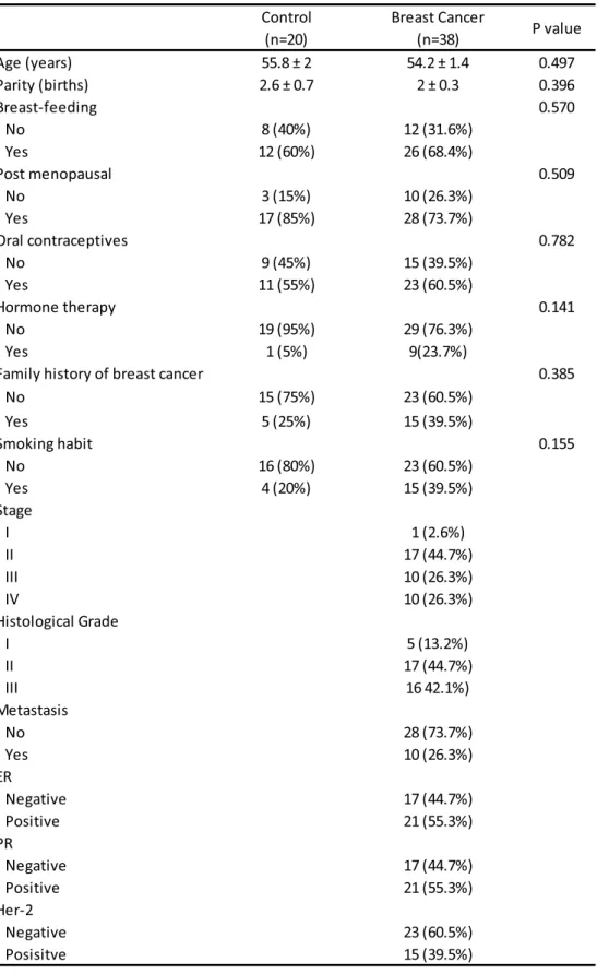
Figure 1 summarizes the cellular origin and number of circulating MPs
studied. No significant differences between control group and breast cancer
group were found for erythrocyte-, lymphocyte- neutrophil-, leucocyte-,
monocyte-, endothelial- and platelet-derived MPs. Although lower levels of
monocyte-derived MPs are associated with advanced breast cancer and
metastasis. Hemogram had no association with any type of MPs.
Also no differences between the two groups were observed for the
cytokines - IL-1- -2, IL-4, IL-6,, IL-10, IL-12, IL-17, TNF,
however, when associated with prognostic factors had interesting outcomes. In
patients with negative Her-2, levels of CXCL-10 were higher. However, higher
levels of IL-17 had positive correlation with RE. Elevated levels of CCL-2 and
CXCL-10 seem to be associated with advanced breast cancer.
MPs, cytokines/chemokines, and prognosis factors had important
4. Discussion:
The great advantage of cytokines/chemokines and microparticles
analysis by flow cytometry is the simultaneous measurement of multiple
biomarkers in a single sample [8, 16]. This methodology made possible in this
study to measure the levels of seven different MPs, nine different cytokines, and
five distinct chemokines.
Our data is the first to measure seven different cell-derived MPs. So far,
no data have been reported on circulating, lymphocyte- and erythrocyte-derived
MPs in breast cancer patients. In this study, the number of neutrophils-,
endothelial cells-, monocytes-, platelets-, leukocytes-, erythrocytes- and
lymphocytes-derived MPs in patients with breast cancer had no significant
differences to the number of MPs in the control group (Figure 1). Other studies
with breast cancer women showed the same findings regarding total number of
MPs, platelet-, endothelial cell- and neutrophil-derived MPs [9, 17], although,
leukocyte-derived MPs were higher in the breast cancer patients compared to
the controls [17]. These data discordance could be justified by the fact that the
MPs investigated are not specific for breast cancer, they are also released in
several diseases as well as in physiological conditions [8].
Our findings show that lower levels of monocyte-derived MPs are
correlated with advanced breast cancer and metastasis. Although, different
patients associated with tumor invasiveness [8, 18]. The patients with advanced
breast cancer had higher values of endothelial cell- and leukocyte-derived MPs
compared to the patients with earlier disease and healthy controls. Furthermore,
was detected higher levels of platelets-derived MPs on patients with bigger size
of the tumor [17].
In the majority of studies using patients with benign breast tumor as a
control group, platelet-derived–MPs were elevated in breast cancer patients as
compared to patients with benign breast tumor [18]. Also, patients with smaller
tumor size had significantly higher concentrations of neutrophil-derived MPs
than benign tumor. However, total MPs levels did not differ significantly between
patients with higher as compared with lower tumor stage [19].
Considering nodal status and MPs numbers, higher MPs total and
neutrophil-derived MPs levels were found in patients with lymph node
metastases compared to the benign tumor group [19]. Other study also shows
that the total numbers of circulating MPs and platelet-derived MPs were highest
in breast cancer patients with larger tumor size and distant metastases [18]. No
data have been reported on monocyte-derived MPs in association with cancer
spreading, but our findings showed a negative correlation between levels of this
type of MPs and metastasis. MPs released from cancer cells have been
implicated to contribute to tumor growth and metastasis. High levels of
circulating MPs may reflect cancer activity in patients [19].
Our data showed few correlations between levels of cytokines and
CXCL-10 had a negative correlation with Her-2. Most findings show higher
levels of cytokines/chemokines correlated with the presence of these prognosis
biomarkers [20, 21]. Positive correlations with disease progression were
detected with levels of CCL-2 and CXCL-10. Other studies show that high levels
of CCL-2 and CCL-5 correlate with advanced breast carcinoma, and these
findings may contribute to breast cancer progression. The role of CCL-2 as a
monocyte-attracting chemokine and its significant association with tumor
progression may explain the simultaneous positive correlation found in this
study between monocyte-derived MPs and CCL2 with advanced disease and
metastasis [22-24].
Cytokines and chemokines have a well-established role in cancer
immunity and carcinogenesis. These molecules are involved with tumor
initiation, growth and metastasis [25]. However, breast cancer is considered to
be weakly immunogenic, and poorly recognized by the immune system. This
immunological response is believed to be associated with the action of
modulatory cytokines in the tumor microenvironment [21, 26].
This prospective case–control study is the first report addressing the
association of the MPs with cytokines/chemokines and the development of a
network of these biomarkers in breast cancer. Despite the relatively small size
of the study population, our data showed considerable network connections
between MPs, cytokines/chemokines, and prognostic factors.
It was used the hierarchical network to simulate the microenvironment of
control group, and another of the breast cancer group. Comparing the two nets,
we can see a different profile between groups. The control group net presents
two isolated clusters. There were lesser and weaker connections between the
biomarkers, and also the majority of cytokines have a negative or no
association with the MPs. Analyzing the breast cancer group there were more
and stronger connections between the biomarkers. Most cytokines/chemokines
are present in the same cluster of MPs. We can see two nodes that play main
roles at the net because of their stronger and numerous associations with
lymphocyte-, and leucocyte-derived MPs.
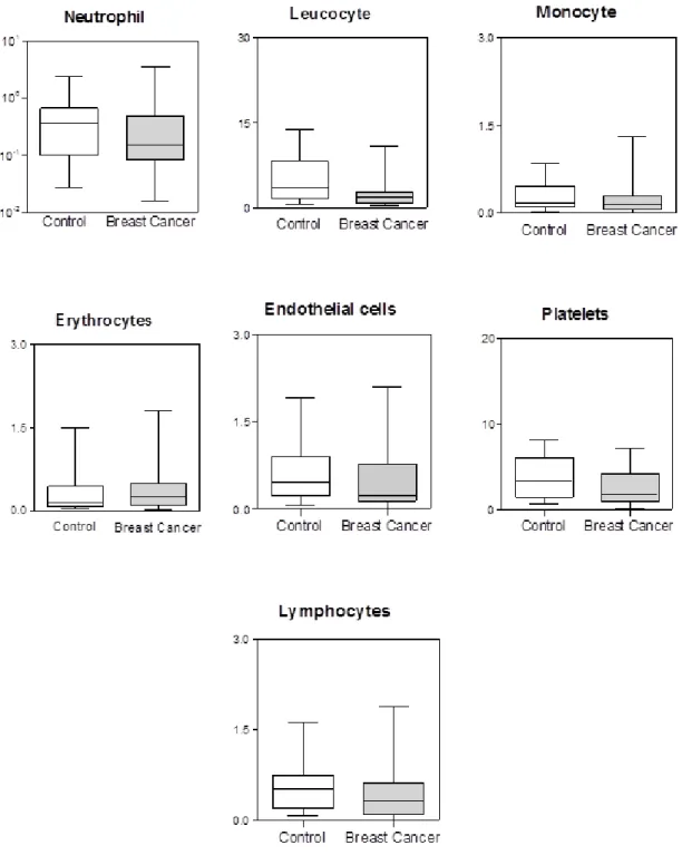
When divided the group of cancer in early disease (stages I and II), and
advanced disease (stages III and IV), there was a great difference between
networks (Figure 3). In early disease, we can see a balance between positive
and negative associations, and presence of just one cytokine. In contrast, what
we evaluated at the advanced disease network, there were stronger and
exclusively positives edges, whereas in the presence of more
cytokines/chemokines, the node balance is lost.
Despite the fact that our data showed no significant difference in the
levels of biomarkers between groups, the networks showed presence of more
nodes and stronger edges on the breast cancer group compared with the
control group as well as in the advanced disease compared with early disease.
These findings reinforce the importance of the study of biomarkers as a global
The present study has limitations due to the small number of women
evaluated. The detection of cancer serum biomarkers is a big challenge in
biomedical research. Our data suggest no differences in the levels of MPs and
inflammatory cytokines/chemokines comparing breast cancer patients and
healthy women. Our main contribution was to define a global biomarker network
in breast cancer. Future investigations could be focused on the possible role of
MPs as a prognostic factor in breast cancer.
Conflict of interest statement
There are no conflicts of interest.
Acknowledgments
This study was supported by Fundação Oswaldo Cruz (FIOCRUZ),
Conselho Nacional de Desenvolvimento Científico e Tecnológico (CNPq),
Coordenação de Aperfeiçoamento de Pessoal de Nível Superior (CAPES), and
Fundação de Amparo à Pesquisa do Estado de Minas Gerais (FAPEMIG). The
authors also thank the program for technological development in tools for
health-PDTIS-FIOCRUZ for the use of its facilities. OAMF and ATC thank CNPq
REFERENCES:
1. Basu, S., et al., Eicosanoids and adipokines in breast cancer: from
molecular mechanisms to clinical considerations. Antioxid Redox Signal, 2013. 18(3): p. 323-60.
2. Siegel, R., D. Naishadham, and A. Jemal, Cancer statistics, 2012. CA Cancer J Clin, 2012. 62(1): p. 10-29.
3. Mause, S.F. and C. Weber, Microparticles: protagonists of a novel
communication network for intercellular information exchange. Circ Res, 2010. 107(9): p. 1047-57.
4. Meziani, F., A. Tesse, and R. Andriantsitohaina, Microparticles are
vectors of paradoxical information in vascular cells including the endothelium: role in health and diseases. Pharmacol Rep, 2008. 60(1): p. 75-84.
5. Furie, B. and B.C. Furie, Role of platelet P-selectin and microparticle
PSGL-1 in thrombus formation. Trends Mol Med, 2004. 10(4): p. 171-8.
6. Lynch, S.F. and C.A. Ludlam, Plasma microparticles and vascular
disorders. Br J Haematol, 2007. 137(1): p. 36-48.
7. Bernimoulin, M., et al., Differential stimulation of monocytic cells results
8. Barteneva, N.S., et al., Circulating microparticles: square the circle. BMC Cell Biol, 2013. 14: p. 23.
9. Tesselaar, M.E., et al., Microparticle-associated tissue factor activity: a
link between cancer and thrombosis? J Thromb Haemost, 2007. 5(3): p. 520-7.
10. Kristensen, V.N., et al., Integrated molecular profiles of invasive breast
tumors and ductal carcinoma in situ (DCIS) reveal differential vascular and interleukin signaling. Proc Natl Acad Sci U S A, 2012. 109(8): p. 2802-7.
11. Tripsianis, G., et al., Coexpression of IL-6 and TNF-alpha: prognostic
significance on breast cancer outcome. Neoplasma, 2013.
12. Lyon, D.E., et al., Cytokine comparisons between women with breast
cancer and women with a negative breast biopsy. Nurs Res, 2008. 57(1): p. 51-8.
13. Combes, V., et al., Circulating endothelial microparticles in malawian
children with severe falciparum malaria complicated with coma. JAMA, 2004. 291(21): p. 2542-4.
14. Shannon, P., et al., Cytoscape: a software environment for integrated
models of biomolecular interaction networks. Genome Res, 2003. 13(11): p. 2498-504.
16. Tarnok, A., et al., Cytometric bead array to measure six cytokines in
twenty-five microliters of serum. Clin Chem, 2003. 49(6 Pt 1): p. 1000-2.
17. Toth, B., et al., Circulating microparticles in breast cancer patients: a
comparative analysis with established biomarkers. Anticancer Res, 2008. 28(2A): p. 1107-12.
18. Toth, B., et al., Platelet-derived microparticles and coagulation activation
in breast cancer patients. Thromb Haemost, 2008. 100(4): p. 663-9.
19. Liebhardt, S., et al., CEA-, Her2/neu-, BCRP- and Hsp27-positive
microparticles in breast cancer patients. Anticancer Res, 2010. 30(5): p. 1707-12.
20. Tripsianis, G., et al., Overall Survival and Clinicopathological
Characteristics of Patients with Breast Cancer in Relation to the Expression Pattern of HER-2, IL-6, TNF-alpha and TGF-beta1. Asian Pac J Cancer Prev, 2013. 14(11): p. 6813-20.
21. Rao, V.S., et al., Potential prognostic and therapeutic roles for cytokines
in breast cancer (Review). Oncol Rep, 2006. 15(1): p. 179-85.
22. Luboshits, G., et al., Elevated expression of the CC chemokine regulated
on activation, normal T cell expressed and secreted (RANTES) in advanced breast carcinoma. Cancer Res, 1999. 59(18): p. 4681-7.
24. Ben-Baruch, A., Host microenvironment in breast cancer development:
inflammatory cells, cytokines and chemokines in breast cancer progression: reciprocal tumor-microenvironment interactions. Breast Cancer Res, 2003. 5(1): p. 31-6.
25. Smyth, M.J., et al., Cytokines in cancer immunity and immunotherapy. Immunol Rev, 2004. 202: p. 275-93.
26. Allan, C.P., et al., The immune response to breast cancer, and the case
Tables and Figures:
Table 1: General characteristics of the patients
Control Breast Cancer
(n=20) (n=38)
Age (years) 55.8 ± 2 54.2 ± 1.4 0.497
Parity (births) 2.6 ± 0.7 2 ± 0.3 0.396
Breast-feeding 0.570
No 8 (40%) 12 (31.6%)
Yes 12 (60%) 26 (68.4%)
Post menopausal 0.509
No 3 (15%) 10 (26.3%)
Yes 17 (85%) 28 (73.7%)
Oral contraceptives 0.782
No 9 (45%) 15 (39.5%)
Yes 11 (55%) 23 (60.5%)
Hormone therapy 0.141
No 19 (95%) 29 (76.3%)
Yes 1 (5%) 9(23.7%)
Family history of breast cancer 0.385
No 15 (75%) 23 (60.5%)
Yes 5 (25%) 15 (39.5%)
Smoking habit 0.155
No 16 (80%) 23 (60.5%)
Yes 4 (20%) 15 (39.5%)
Stage
I 1 (2.6%)
II 17 (44.7%)
III 10 (26.3%)
IV 10 (26.3%)
Histological Grade
I 5 (13.2%)
II 17 (44.7%)
III 16 42.1%)
Metastasis
No 28 (73.7%)
Yes 10 (26.3%)
ER
Negative 17 (44.7%)
Positive 21 (55.3%)
PR
Negative 17 (44.7%)
Positive 21 (55.3%)
Her-2
Negative 23 (60.5%)
Posisitve 15 (39.5%)
Abbreviations: ER (estrogen receptor), PR (progesteron receptor) Her-2 (Human epidermal growth factor receptor 2) - Data are presented as mean ± SEM. Differences between groups were evaluated using t student or chi-square test.
Figure 2: Network analysis. Biomarkers networks in control or Breast cancer groups. Chemokine, cytokine and MPs nodes were assembled as well as the biomarkers correlation indexes amongst groups (negative ; moderate
Artigo II
Measurement of circulating microparticles and
cytokine/chemokine levels in women with breast cancer
submitted to chemotherapy
Article Type: Original article
Abstract:
Chemotherapy leads to platelet activation and generation of microparticles (MPs). The combination of microparticles and cytokine/chemokine levels could be used to predict chemotherapy response in breast cancer patients. The aim of this study was to measure the levels of inflammatory biomarkers ( MPs , cytokines and chemokines ) in the serum of women with breast cancer pre and post-chemotherapy. Peripheral blood of women with breast cancer ( n = 18 ) was collected pre-chemotherapy and post-chemotherapy. Flow cytometry was used for determination of serum levels of cytokines (IL-1, IL-2, IL-4, IL-6, IL-10, IL-12, IL-17A, TNF, IFN –gamma), chemokines (CXCL-8, CXCL-9, CXCL-10,
CCL-2 and CCL-5) and microparticles from different cells (neutrophils, leukocytes, monocytes, erythrocytes, endothelium, platelets and lymphocytes). A hierarchical network was developed to simulate the interaction between the serum immune microenvironment pre-chemotherapy and post-chemotherapy. Differences between groups were evaluated by Mann- Whitney or Kruskal-Wallis test. Differences with p < 0.05 were considered significant. Our data shows decreased levels of platelet -derived microparticles in postchemotherapy patients, and increased serum levels of IL6, CCL and CXCL 5 -10 in post-chemotherapy. In the correlation network we could identify presence of more biomarkers interactions in the pre-chemotherapy net. With these data we can conclude that chemotherapy is able to change the inflammatory response profile in patients with breast cancer.
Introduction:
Chemotherapy, radiotherapy and surgical resection are the current
approach in cancer treatment. Neoadjuvant chemotherapy in breast cancer is
used for the purpose of reducing tumor size leading to a less aggressive
surgery [1]. The regression of the disease leads to a more conservative surgery,
higher cure rates and survival [2, 3].
Cytotoxic chemotherapy kills tumor cells and the resulting apoptotic cell
death was previously considered an immunological null event. Chemotherapy
was thought to have a neutral or even immunosuppressive effect on the host
immune system by inducing neutropenia, lymphopenia, thrombocytopenia, and
anemia [4].
Chemotherapy leads to platelet activation and generation of
microparticles (MPs) associated with increased risk of thrombosis [5]. In
addition, chemotherapy leads to rebound angiogenesis facilitating tumor
re-growth. It was demonstrated that post-chemotherapy tumor-MPs are involved in
cancer thrombogenicity, tumor invasion and angiogenesis. MPs of patients with
breast cancer are thrombogenic, and chemotherapy administration disturb the
hemostatic balance on MPs [5, 6].
Given the systemic nature of MPs, there is therapeutic significance as
they could potentially be used diagnostically to predict therapeutic response and
microparticles and cytokine/chemokine levels could be used to create an
immune signature in breast cancer patients pre-chemotherapy and
post-chemotherapy [7].
The overall response to chemotherapy is heterogeneous and dynamic,
involving a combination of independent molecular mechanisms that are
functionally regulated during tumor progression and treatment [8]. An
understanding of the molecular mediators involved in drug responsiveness will
help to understand the basis of therapeutic variability observed in clinical
oncology [7].
In the present study, we observed the behavior of the serum levels of
MPs, cytokines, and chemokines in women with breast cancer
pre-chemotherapy and post-pre-chemotherapy, and the interactions between these
2. Patients, Material and methods
2.1 Patients
This study was approved by the Ethics Committee (COEP) at
Universidade Federal de Minas Gerais (UFMG), CAAE #:
01536112.3.0000.5149, and informed consent was obtained from all
participants.
The study included 18 women with diagnosis of breast cancer with
proposal of chemotherapy. These women were enrolled from Hospital Vera
Cruz, Hospital da Baleia, and Hospital das Clínicas, UFMG, Belo Horizonte,
Brazil. The patients answered a survey comprising clinical and epidemiological variables, and other clinical data were obtained from their medical records.
Exclusion criteria were chronic hypertension, hemostatic abnormalities,
diabetes, obesity, and cardiovascular, autoimmune, renal, hepatic diseases or
2.2 Blood samples
Peripheral blood was collected from patients before their first cycle of
chemotherapy, and 21 days after. Blood samples were drawn in sodium citrate
(0.129 mol/l) in a 9:1 volume ratio for the microparticles analysis, and in
EDTA-K (1.8 mg/mL) for flow cytometric cytokine/chemokine measurements. The
samples were centrifuged at 2500×g for 15 min to obtain plasma. Samples were
aliquoted, and stored at -80°C until analysis.
2.3 Determination of MP plasma levels
MPs were prepared as described elsewhere [9]. Briefly, samples were
centrifuged at 13,000×g for 3 min to obtain platelet-free plasma, which was then
diluted 1:3 in citrated phosphate buffered saline (PBS) containing heparin and
centrifuged at 14,000×g for 90 min at 15 °C. The subsequent MP pellet was
resuspended in 1× annexin V binding buffer (Sigma-Aldrich, MO).
MPs isolated from plasma were gated on the basis of their forward (FSC)
and side (SSC) scatter distribution in density plots of synthetic 0.7–0.9 µm
SPHEROTM Amino Fluorescent Particles (Spherotech Inc., Libertyville, IL,
USA). Taking into account the presence of phosphatidylserine residues on the
MP surfaces, events present in the gate were assessed for their positive
staining for annexin V (Sigma-Aldrich) — a classical marker for microparticles
against annexin V. Labeling with cell-specific monoclonal antibodies was
corrected for isotype-matched control antibodies.
FITC-labeled immunoglobulin G1 (IgG1) and PE-labeled IgG1 isotype
controls, monoclonal antibodies directed against neutrophils (CD66-PE),
endothelial cells (CD51-PE), monocytes (CD14–PERCP), platelets (CD41–
PERCP), leukocytes (CD45–APC), and erythrocytes (CD235a–PECy5), were
purchased from BD Biosciences® (CA, USA). Monoclonal antibody directed
against T lymphocytes (CD3-PE) was purchased from Beckman Coulter
Immunotech (Marseille, France).
2.4 Detection of plasmatic cytokine/chemokine levels by cytometric bead array immunoassay (CBA)
Cytokine/chemokine plasma levels were determined using commercially
available kits for Cytometric Beads Array – CBA (BD Biosciences Pharmingen,
USA), including the Human Inflammatory Cytokines Kit to quantify IL-1β, IL-6,
IL-10, TNF, and IL-12p70 along with the Human Th1/Th2/Th17 Kit to quantify
Interleukin IL-2, IL-4,, IFN-J, and IL-17A, and the Human Chemokine Kit to
quantify CXCL-8, CXCL-9, CXCL-10, CCL-5, and CCL-2.
The CBA immunoassay uses 7.5 µm polystyrene microbeads, assembled
in distinct fluorescent sets, unique on their type 4 fluorescence intensity (FL-4).
Each microbead is coupled to monoclonal antibody (MAb) against a given
bead-captured cytokines were detected by direct immuno assay using a ‘‘detection
cocktail’’ of distinct MAbs labeled with type 2 fluorescence, phycoerythrin- PE
(FL-2).
The method was carried out as recommended by the manufacturer,
modified as follows: briefly, 25 µl of undiluted plasma samples or standards
(previously diluted) were added to 15 µl of bead-mix and incubated for 90 min at
room temperature in the dark. The cytokine/chemokine standard curves were
run daily for each assay. After incubation, the samples and standards were
washed with 500 µl of wash buffer and centrifuged at 600g for 7 min at room
temperature. Subsequently, 20 µl of detection cocktail were added to each tube
and the bead-mix re-incubated for 90 min at room temperature in the dark.
Following incubation, the samples and standards were washed again with 500
µl of wash buffer and centrifuged at 600g for 7 min at room temperature to
remove unbound detector reagent. After washing, 250 µl of wash buffer was
added to each tube.
Data acquisition and analysis was performed in dual-laser
FACScalibur™flow cytometer (BDBiosciences Pharmingen, San Jose, CA,
USA), using the BDBioscience CBA software. A total of 1800 beads/tube were
acquired after proper set-up of a flow cytometer. Results were expressed as
2.5 Biomarker Network Analysis
Biomarker networks were assembled to assess the association between
levels of MPs and cytokines/chemokines, and prognostic factors. Spearman’s
correlation test was performed to assess the association between levels of
these biomarkers. The positive and negative correlations were significant when
the p<0.05. To better represent the interactivity of the molecules tested, the
open source software, Cytoscape (version 2.8), was used for composing
networks of biomolecules interactions [10] Connecting edges display
underscore negative, ( ), moderate ( ) and strong ( ) as proposed by
Taylor, 1990. [11]
2.6 Statistical analysis
Data were analyzed using Graphpad Instat 4.0 and SPSS 15.0 statistical
software. Differences in the means of the frequencies between groups were
analyzed using two-tailed student’s t test or Mann-Whitney when data did not fit
a Gaussian distribution. The log-transformation of data was applied for
situations where variances normality assumptions failed followed by linear
regression to investigate the association between the clinical parameters and
MPs levels. Pairwise correlations were evaluated with Pearson’s correlation
coefficient r. Multiple linear regression models with stepwise backward deletion
were built to describe independent associations between covariates and the
presence of biomarkers. In all cases, a p value < 0.05 was considered to be
3. Results
Patients included in this study had a mean age of 54 (±2.9) years,
ranging from 39 to 69 years. Parity had a mean of 2.2(±2.9) births. Eleven
(61.1%) patients had breast-feeding history, and seven (39.9%) family history of
breast cancer. Eleven (61.1%) patients were in post-menopause, six (33.3) had
previous use of hormone therapy, and nine (50%) previous use of oral
contraceptives. Eight (44.4%) patients have had or have smoking habits.
Regarding prognostic factors, in the group of patients with breast cancer,
1 (5.6%) had stage I, 8 (44.4%) stage II, 5 (27.8%) stage III, and 4 (22.2%) stage
IV. Regarding histological grade, 3 patients (16.7%) had grade I, 8 (44.4%)
grade II, and 7 (38.9%) grade III. Four patients (22.2%) had metastasis, 9
(50%) had positive estrogen receptor, 10 (55.6%) positive progesterone
receptor, and 8 (44.4%) positive Her-2.
Figure 1 summarizes the cellular origin and number of circulating MPs
studied. A lower number of platelet-derived MPs were observed in
post-chemotherapy measures. However, no significant differences between groups
were found for erythrocyte-, lymphocyte- neutrophil-, leucocyte-, monocyte- and
endothelial-derived MPs. Regarding the cytokines/chemokines assay, higher
levels of IL-6, CXCL-10 and CCL-5 were find in the post-chemotherapy
measurements as shown in Figure 2.
In the correlation network, a different profile of the biomarkers for the pre
Discussion:
Blood samples were collected from patients with breast cancer
pre-chemotherapy (pre-CT) and post-pre-chemotherapy (post-CT). Our data is the first
to measure 7 different cell-derived MPs (erythrocyte-, lymphocyte- neutrophil-,
leucocyte-, monocyte-, endothelial- and platelet), and 14 different
cytokine/chemokine (IL-1- , IL-2, IL-4, IL-6, IL-10, IL-12, IL-17, TNF,
IFN-CXCL-8, CXCL-9, CXCL-10, CCL-2, and CCL-5) in breast cancer patients
pre-CT and post-pre-CT.
Despite the relatively small size of the study population, our data showed
considerable differences in circulating cell-derived MPs and cytokines levels
post-CT.
MPs originated from platelets are the most numerous and seems to have
a key role in thrombosis, inflammation, angiogenesis, vascular dysfunction, and
can affect cancer patient prognosis [6]. Chemotherapy induces death of tumor
cells and disruption of tumor blood vessels what results in increased liberation
of MPs [6]. Differing from other studies, in our study, there was a significant
decreased in platelets-derived MPs post-CT (Figure 1). Another study in
patients with acute myeloid leukemia presented similar results, after
chemotherapy, with lower levels of MPs [12]. Rapidly growing cells tend to
secrete more MPs than cells with lower proliferation rate, what could be one of
Post-CT measures showed higher levels of IL-6, CXCL-10, and CCL-5
(Figure 2). In different types of cancer the response to chemotherapy is
increased in tumors[8]. The pro-inflammatory/modulatory profiles have
important role in chemotherapy response in breast cancer [7].
In Figure 3, it was used the hierarchical network to simulate the serum
patients microenvironment pre-CT and post-CT. Comparing the two nets, we
can see a different profile between groups. The pre-CT net presents more
connections, where we can identify presence of more correlations between
cytokines/chemokines. Analyzing the post-CT net, we can clearly see a great
difference in the microenvironment. There were fewer molecules involved, and
they were divided into two clusters. The presence of cytokines/chemokines was
greatly reduced, however, the MPS were still present. It seems that
cytokines/chemokines have a faster response to chemotherapy than MPs.
In summary, chemotherapy disturbs the balance on MPs and
cytokines/chemokines patterns in patients with breast cancer. This research
identifies systemic immune interactions in response to chemotherapy and has
potential for translation into clinical by identifying a response profile of breast
Conflict of interest statement
There are no conflicts of interest.
Acknowledgments
This study was supported by Fundação Oswaldo Cruz (FIOCRUZ),
Conselho Nacional de Desenvolvimento Científico e Tecnológico (CNPq),
Coordenação de Aperfeiçoamento de Pessoal de Nível Superior (CAPES), and
Fundação de Amparo à Pesquisa do Estado de Minas Gerais (FAPEMIG). The
authors also thank the program for technological development in tools for
health-PDTIS-FIOCRUZ for the use of its facilities. OAMF and ATC thank CNPq
References:
1. Beriwal, S., et al., Breast-conserving therapy after neoadjuvant
chemotherapy: long-term results. Breast J, 2006. 12(2): p. 159-64.
2. Abrial, S.C., et al., High prognostic significance of residual disease after
neoadjuvant chemotherapy: a retrospective study in 710 patients with operable breast cancer. Breast Cancer Res Treat, 2005. 94(3): p.255-63.
3. Tiezzi, D.G., et al., HER-2, p53, p21 and hormonal receptors proteins
expression as predictive factors of response and prognosis in locally advanced breast cancer treated with neoadjuvant docetaxel plus epirubicin combination. BMC Cancer, 2007. 7: p. 36.
4. Pages, F. and G. Kroemer, Prognostic impact of anticancer immune
responses: an introduction. Semin Immunopathol, 2011. 33(4): p. 317-9.
5. Pihusch, R., et al., Platelet flow cytometric findings in patients
undergoing conditioning therapy for allogeneic hematopoietic stem cell transplantation. Ann Hematol, 2002. 81(8): p. 454-61.
6. Aharon, A. and B. Brenner, Microparticles, thrombosis and cancer. Best Pract Res Clin Haematol, 2009. 22(1): p. 61-9.
7. Kristensen, V.N., et al., Integrated molecular profiles of invasive breast
8. Ladoire, S., et al., In situ immune response after neoadjuvant
chemotherapy for breast cancer predicts survival. J Pathol, 2011. 224(3): p. 389-400.
9. Combes, V., et al., Circulating endothelial microparticles in malawian
children with severe falciparum malaria complicated with coma. JAMA, 2004. 291(21): p. 2542-4.
10. Shannon, P., et al., Cytoscape: a software environment for integrated
models of biomolecular interaction networks. Genome Res, 2003. 13(11): p. 2498-504.
11. Taylor, R., Interpretation of the Correlation Coefficient: A Basic Review. Journal of Diagnostic Medical Sonography, 1990. 6(1): p. 35-39.
12. Van Aalderen, M.C., et al., Procoagulant myeloblast-derived
microparticles in AML patients: changes in numbers and thrombin generation potential during chemotherapy. J Thromb Haemost, 2011. 9(1): p. 223-6.
13. Ratajczak, J., et al., Membrane-derived microvesicles: important and