Published Ahead of Print 26 October 2012.
2013, 195(1):85. DOI: 10.1128/JB.01442-12.
J. Bacteriol.
Ferreira
Bolzani, J. Belasque Jr., L. V. S. Sacramento and H.
I. C. Silva, L. O. Regasini, M. S. Petrônio, D. H. S. Silva, V. S.
citri
against Xanthomonas citri subsp.
http://jb.asm.org/content/195/1/85
Updated information and services can be found at:
These include:
SUPPLEMENTAL MATERIAL
Supplemental material
REFERENCES
http://jb.asm.org/content/195/1/85#ref-list-1
at:
This article cites 50 articles, 12 of which can be accessed free
CONTENT ALERTS
more»
articles cite this article),
Receive: RSS Feeds, eTOCs, free email alerts (when new
http://journals.asm.org/site/misc/reprints.xhtml Information about commercial reprint orders:
http://journals.asm.org/site/subscriptions/ To subscribe to to another ASM Journal go to:
on February 7, 2014 by UNESP - Universidade Estadual Paulista
http://jb.asm.org/
Downloaded from
on February 7, 2014 by UNESP - Universidade Estadual Paulista
http://jb.asm.org/
Antibacterial Activity of Alkyl Gallates against
Xanthomonas citri
subsp.
citri
I. C. Silva,aL. O. Regasini,bM. S. Petrônio,bD. H. S. Silva,bV. S. Bolzani,bJ. Belasque, Jr.,cL. V. S. Sacramento,dH. Ferreiraa Faculdade de Ciências Farmacêuticas (FCF), Departamento de Ciências Biológicas, Universidade Estadual Paulista (UNESP), Araraquara, Sao Paulo, Brazila
; Departamento de Química Orgânica, Instituto de Química, UNESP, Araraquara, Sao Paulo, Brazilb
; Departamento Científico, Fundecitrus, Araraquara, Sao Paulo, Brazilc
; Departamento de Princípios Ativos Naturais e Toxicologia, FCF, UNESP, Araraquara, Sao Paulo, Brazild
The plant-pathogenic bacteriumXanthomonas citrisubsp.citriis the causal agent of Asiatic citrus canker, a serious disease that
affects all the cultivars of citrus in subtropical citrus-producing areas worldwide. There is no curative treatment for citrus can-ker; thus, the eradication of infected plants constitutes the only effective control of the spread ofX. citrisubsp.citri. Since the
eradication program in the state of São Paulo, Brazil, is under threat, there is a clear risk ofX. citrisubsp.citribecoming
en-demic in the main orange-producing area in the world. Here we evaluated the potential use of alkyl gallates to preventX. citri
subsp.citrigrowth. These esters displayed a potent anti-X. citrisubsp.citriactivity similar to that of kanamycin (positive
con-trol), as evaluated by the resazurin microtiter assay (REMA). The treatment ofX. citrisubsp.citricells with these compounds
induced altered cell morphology, and investigations of the possible intracellular targets usingX. citrisubsp.citristrains labeled
for the septum and centromere pointed to a common target involved in chromosome segregation and cell division. Finally, the artificial inoculation of citrus withX. citrisubsp.citricells pretreated with alkyl gallates showed that the bacterium loses the
ability to colonize its host, which indicates the potential of these esters to protect citrus plants againstX. citrisubsp.citri
infection.
B
razil is the main producer of concentrate orange juice in the world. Exporting 98% of its production, Brazil holds the lead-ing position in the orange juice business, representlead-ing 85% of the international market in this sector (1). Brazilian citriculture, an economic activity that generates over $2 billion/year in export revenues, contributes to more than half of the orange juice pro-duced globally. Despite great success, the culturing of citrus faces constant threats such as diseases, which in the past decade forced the eradication of 40 million trees, resulting in the suppression of the spread of the disease.Among the threats, citrus canker represents one of the most serious diseases of citrus, which is present in the main sweet-or-ange-producing areas of the world, the state of São Paulo, Brazil, and Florida. The etiological agent is the Gram-negative bacterium
Xanthomonas citrisubsp.citri(2).X. citrisubsp.citriis responsible for Asiatic citrus canker, a severe form of the disease that affects all the commercially important citrus species and cultivars in use (reviewed in reference3). Infected trees exhibit crater-like lesions on aerial tissues, and with time, infection decreases orange pro-duction. In susceptible citrus hosts and in the absence of disease control measures, it causes premature fruit drop, defoliation, and shoot dieback.X. citri subsp.citripenetrates the host plant by natural openings (stomata) and wounds (3). Bacteria spread in the orchards through the action of wind-blown rain, and the coloni-zation of the leaf mesophyll is exacerbated by the concomitant presence of the citrus leaf miner Phyllocnistis citrella Stainton (Lepidoptera: Gracillariidae) (4).
The state of São Paulo, which produces 75% of the oranges in Brazil, had the first official detection ofX. citrisubsp.citriin 1957 (5), and since then, it adopted a strict eradication program still considered the only effective strategy to avoid the advance of citrus canker in areas where it is not endemic (6). From 1999 until 2009, growers in the state of São Paulo were obligated to eliminate all the plants in an orchard if the incidence of symptomatic trees
ex-ceeded 0.5%; below this figure, the growers had to eradicate only the affected trees and the neighboring ones contained in a radius of 30 m. From mid-2009, the legislation in this state was relaxed, and only the 30-m radius is being applied. As a consequence, 6 months after the legislation change, the number of new cases of the disease increased 80%, and there is now a real risk of citrus canker becoming endemic in the state of São Paulo. Citrus canker is already endemic in other orange-planting areas in the south of Brazil. There, as well as in Argentina, producers use integrated management approaches such as the cultivation of less-suscepti-ble citrus genotypes produced inX. citrisubsp.citri-free nurseries, the planting of windbreaks as barriers against the spread of the disease, and the spraying of copper bactericides (7). These prac-tices increase the cost of production and are not as effective as eradication to prevent new infections and the spread ofX. citri
subsp.citri; besides, copper sprays are known to leave residuals on fruits and soil, and the emergence of resistant strains is an issue (8). Current research is focused on developing new strategies to minimize losses and to make citriculture profitable. The use of a combination of disease control measures is recommended, which includes the induction of innate plant defenses (9,10), the gener-ation of more resistant citrus varieties (11,12), and, perhaps, the
Received9 August 2012 Accepted19 October 2012
Published ahead of print26 October 2012
Address correspondence to H. Ferreira, [email protected].
I.C.S. and L.O.R. contributed equally to this work.
Supplemental material for this article may be found athttp://dx.doi.org /10.1128/JB.01442-12.
Copyright © 2013, American Society for Microbiology. All Rights Reserved.
doi:10.1128/JB.01442-12
January 2013 Volume 195 Number 1 Journal of Bacteriology p. 85–94 jb.asm.org 85
on February 7, 2014 by UNESP - Universidade Estadual Paulista
http://jb.asm.org/
development/amelioration of chemical inhibitors to fight the bac-terium and yet display reduced environmental impacts.
Supporting this idea, we initiated a search for natural and syn-thetic compounds that are able to perturb the growth ofX. citri
subsp.citri. We evaluated the anti-X. citrisubsp.citriactivity of gallic acid (3,4,5-trihydroxybenzoic acid), an intermediate of the hydrolysable tannin biosynthesis pathway in plants (13–15) which, together with its natural and semisynthetic derivatives, has been associated with a broad spectrum of biological actions (16–
21). Moreover, gallic acid and derivatives demonstrated antimi-crobial properties. Octyl gallate exhibited fungicidal activity againstSaccharomyces cerevisiaeandZygosaccharomyces bailiiat any stage of their growth (22,23). Leal et al. previously reported the potent fungitoxicity of nonyl gallate against yeasts, dermato-phytes, and hialohyphomycetes (24). Lauryl gallate was found to show antibacterial activity against Gram-positive and Gram-neg-ative bacteria, including methicillin-resistantStaphylococcus au-reus(MRSA) (25,26).
Here we demonstrate that alkyl gallates are potent inhibitors of
X. citrisubsp.citri. The anti-X. citrisubsp.citriactivity of such compounds can be correlated with the size of their carbon side chain. We show evidence for the possible intracellular targets of these compounds, as bacteria treated with alkyl gallates exhibited a disruption of the divisional septum and/or the bacterial centro-mere. We also show that the treatment of the bacterium with selected alkyl gallates reduces viability and host infection and that these compounds may possess postinfection activity, indicating their potential for use in disease management.
MATERIALS AND METHODS
Synthesis of the alkyl gallates.Synthetic esters of gallic acid were pre-pared as described previously, with minor modifications (16, 17). A 3.0-ml solution ofN,N=-dicyclohexylcarbodiimide (DCC) (1.0 mmol) in driedp-dioxane was added to a cooled (5°C) solution of 0.2 mmol gallic acid (Sigma) (compound 1) (Table 1) and 20 mmol alkyl alcohol in 6.0 ml of driedp-dioxane. After 48 h, the solvent was removed under reduced pressure. The residue was partitioned 3 times with ethyl acetate (EtOAc) and filtered. The filtrate was washed successively with a saturated aqueous citric acid solution (3 times) and saturated aqueous NaHCO3(3 times),
dried over anhydrous MgSO4, and evaporated under reduced pressure.
The crude products were purified over a silica gel column eluted with mixtures of hexanes and EtOAc, furnishing alkyl gallates (compounds 2 to 16) (Table 1). The molecular structures of these compounds were estab-lished by1H and13C nuclear magnetic resonance (NMR) spectral analysis
(16,17).
Bacterial strains and media.Wild-typeXanthomonas citrisubsp.citri
(2) strain 306 (IBSBF 1594), formerly designatedX. axonopodispv.citri, was used in the present work. TheX. citrisubsp.citri306 mutant strains expressing green fluorescent protein (GFP)-ZapA (27) and ParB-GFP (A. P. Ucci and H. Ferreira, unpublished data) wereX. citrisubsp.citri amy::pPM2a-zapAand X. citrisubsp. citri parB::pPM7g-parB, respec-tively. Cells were cultivated at 30°C under rotation (200 rpm) in LB/LB agar medium (28), and kanamycin was added, when required, at 15.6
g/ml.
REMA.The resazurin microtiter assay (REMA) plate method was per-formed as previously described (29,30), with modifications. Stock solu-tions of chemicals at 2 to 5 mg/ml were prepared by dissolving the alkyl gallates (dried-powder samples) in 10% dimethyl sulfoxide (DMSO) (di-luted in LB). Test suspensions of gallates were prepared by diluting the stock solutions in LB using a 2-fold scheme; after serial dilution, the most concentrated sample had⬃1% DMSO (which did not alterX. citrisubsp.
citrigrowth in preliminary analyses) and an initial concentration of a given compound at 1,000g/ml. The concentration range prepared for
each of the compounds was 15 to 1,000g/ml. In 96-well microtiter plates, 100l of each of the compound dilutions was added to a mixture of 90l of LB and 10l of bacterial inoculum (standardized to 105CFU/
well). The negative control consisted of 190l of LB and 10l of cell suspension; the positive control had the addition of kanamycin (15.6g/ ml). Upon the incubation of the test plates at 30°C for 24 h, cell viability was determined by the addition of 15l of a 0.01% (wt/vol) resazurin solution to each of the wells, following an extra incubation period of 2 h at 30°C. Viable microorganisms reduced the blue dye to a pink color, which was detected by fluorescence scanning using a SPECTRAfluor Plus (Tecan) microfluorimeter set to an excitation/emission profile of 530 nm/ 590 nm. Three independent experiments were conducted, and the data were used to construct plots of chemical concentration versus cell growth inhibition in order to determine the MIC50and MIC80.
Microscopy.Cells of wild-type and mutant strains ofX. citrisubsp.
citriwere treated with the alkyl gallates for 6 h at 30°C and immobilized on agarose-covered slides for observations by microscopy, as previously de-scribed (27). Cells were visualized by using an Olympus BX-61 micro-scope and documented with a monochromatic XM-10 camera.Image processing and analyses were conducted by using Cell-F (Olympus).
Pathogenicity tests.The host citrus used was cultivar Pera Rio of sweet orange (Citrus sinensisL. Osbeck), kept under greenhouse condi-tions at 25°C to 35°C. For the infiltration tests,X. citrisubsp.citricells were cultivated in LB until the optical density (OD) at 600 nm reached⬃1; cells were diluted to 105CFU/ml in LB and treated with the compounds at the
MIC80for 6 h at 30°C. After this period, cell suspensions were infiltrated
on the abaxial surface of leaves by using hypodermic syringes. Symptoms were observed during the course of 3 weeks. All the tests were performed in triplicates.
For the postinfection test,X. citrisubsp.citricell suspensions were diluted to 108CFU/ml in LB; leaves were inoculated by the puncture
TABLE 1Anti-Xanthomonas citriactivities of gallic acid and alkyl gallates
Compound Name Rc
MIC (g/ml) (MIC inM)
C log
Pa
1 Gallic acid H 500 (2,940) 0.89 2 Methyl gallate CH3 62.5 (340) 0.92
3 Ethyl gallate CH2CH3 62.5 (315) 1.27
4 n-Propyl gallate (CH2)2CH3 62.5 (295) 1.73
5 i-Propyl gallate CH(CH3)2 62.5 (295) 1.55
6 n-Butyl gallate (CH2)3CH3 62.5 (276) 2.13
7 i-Butyl gallate CH2CH(CH3)2 62.5 (276) 1.95
8 n-Pentyl gallate (CH2)4CH3 31.2 (130) 2.53
9 n-Hexyl gallate (CH2)5CH3 31.2 (123) 2.92
10 n-Heptyl gallate (CH2)6CH3 31.2 (116) 3.32
11 n-Octyl gallate (CH2)7CH3 31.2 (110) 3.72
12 n-Nonyl gallate (CH2)8CH3 15.6 (53) 4.11
13 n-Decyl gallate (CH2)9CH3 15.6 (50) 4.51
14 n-Undecyl gallate (CH2)10CH3 15.6 (48) 4.90
15 n-Dodecyl gallate (CH2)11CH3 62.5 (185) 5.30
16 n-Tetradecyl gallate (CH2)13CH3 1,000 (2,732) 6.09
Kanamycinb
15.6 (32)
aTheoretical lipophilicity (20). bPositive control.
cR, radical.
on February 7, 2014 by UNESP - Universidade Estadual Paulista
http://jb.asm.org/
method, in which needles are rapidly immersed into the cell suspension following the perforation of the leaves from the abaxial surface. After the development of symptoms, in up to 4 weeks,⬃5l of the compounds at 2 mg/ml was dropped onto the lesions, and exposure took place for 30 min at room temperature. Forty-eight hours after the treatment, lesions were removed individually from the leaves and macerated in 1 ml of 1⫻ phos-phate-buffered saline (PBS), and the number of viable cells in the suspen-sions was determined by plating onto NYG agar (5 g/liter peptone, 3 g/liter yeast extract, and 20 g/liter glycerol).
Statistics.Statistical analysis was performed by using one-way analysis of variance (ANOVA) followed by a Tukey posttest (P⬍0.05). MIC values were determined by using logarithmic regression analyses.
RESULTS
Alkyl gallates inhibitX. citrisubsp.citri growth.In order to
evaluate the potential of alkyl gallates to inhibit the growth ofX. citrisubsp. citri, cells were cultivated and exposed to different concentrations of gallic acid (compound 1) (Table 1) and a col-lection of gallic acid alkyl esters (3,4,5-trihydroxybenzoates) (compounds 2 to 16), followed by an evaluation of their inhibitory activities using the REMA (see Fig. S1 in the supplemental mate-rial). InTable 1, we also summarize the MICs of these compounds, which were determined within the concentration range of 3.9 to 1,000g/ml. Of the 16 compounds tested, 14 alkyl gallates (com-pounds 2 to 15) were able to inhibit bacterial growth. Gallic acid itself was not active againstX. citrisubsp.citri, exhibiting an MIC of 500 g/ml (2,940 M). On the other hand, methyl gallate (compound 2) showed an MIC of 62.5g/ml (340M), which was approximately 8 times more potent than gallic acid (com-pound 1). This observation supported the idea that the esterifica-tion of the carboxyl group on gallic acid structure was an impor-tant criterion for the anti-X. citri subsp. citri activity, which encouraged us to further investigate other derivatives esterified with linear and ramified alcohols from C2to C14. Among these, the compounds that carried side chains ranging from 9 to 11 carbon atoms (compounds 12 to 14) displayed the most potent activities againstX. citrisubsp.citri. Undecyl gallate (compound 14), for instance, showed a more prominent effect, with an MIC of 15.6 g/ml (48M), which resembles the inhibitory activity of our positive control, kanamycin (MIC⫽15.6g/ml, or 32M). Al-together, our results indicate a clear and positive correlation among MIC values, alkyl chain length, and lipophilicity.
Considering that lipophilicity is an important property for the development of bioactive compounds (31), the theoretical lipo-philicity values of the alkyl gallates were expressed as calculated partition coefficients (C logP), as previously reported (20) (Table 1). By comparisons of the determined MIC values with the C logP
values, we noticed that the anti-X. citrisubsp.citriactivity of the alkyl gallates was associated with their lipophilicity. Highly li-pophobic gallates (compounds 2 to 7), which exhibit C logP val-ues ranging from 0.92 to 1.95, as well as the highly lipophilic do-decyl gallate (compound 15) (C logP⫽5.30) had an overall weak antibacterial effect. The anti-X. citrisubsp.citriactivity then in-creased in conjunction with an increase in the number of side-chain carbons until reaching a peak, here represented by undecyl gallate (compound 14) (C logP⫽4.90). Subsequently, we docu-mented a diminished effect for dodecyl gallate (compound 15) (C logP⫽5.30), which preceded the approach of a cutoff repre-sented by tetradecyl gallate (compound 16) (C logP⫽6.09).
Finally, in order to test if the compounds had bacteriostatic or bactericidal activities, we attempted to propagate the cells in LB
agar after the treatments. As a result, we could not detect cellular growth following treatment with chemicals in concentrations where the REMA indicated 100% inhibition; hence, we conclude that the alkyl gallates eliminated bacterial viability.
X. citrisubsp.citricells treated with alkyl gallates are unable
to induce citrus canker lesions.The colonization of the host is a vital process to the survival of any pathogen; therefore, we wanted to evaluate if the treatment ofX. citrisubsp.citricells with the alkyl gallates would impair their ability to multiply in artificially inoc-ulated plant tissues and to produce the typical symptoms of citrus canker. Leaves of sweet orange, cultivar Pera Rio, a susceptible host, were infiltrated withX. citrisubsp.citricell suspensions (105 CFU/ml) after treatment with the substances at the MIC80and monitored for a period of 3 weeks in order to score for the appear-ance of symptoms. The reason for the use of the MIC80was to ensure that after treatment, there would be enough live cells, al-though exposed to a fairly high concentration of the compounds, to be evaluated in their ability to colonize the host citrus.
The typical canker lesions induced by the untreated cells (con-trol) can be characterized by hyperplasia and hypertrophy on the region of the inoculum, which is frequently surrounded by a chlo-rotic halo (Fig. 1A). Six of the substances tested (compounds 6 to 10 and 12) (Table 1) completely precluded the ability ofX. citri
subsp.citrito produce disease symptoms (Fig. 1B). Alkyl gallate compound 11, although active, induced only a partial reduction of the symptoms. The remaining chemicals did not induce any de-tectable alteration of the colonization/virulence phenotype ofX. citrisubsp.citri. Note that the infiltration of the compounds alone or with 1% DMSO did not produce any lesions on leaves. In ad-dition, the treatment ofX. citrisubsp.citriwith DMSO (without any alkyl gallate) did not reduce the virulence of the bacterium (Fig. 1A).
Irrespective of their mechanisms of action, alkyl gallate com-pounds 6 to 10 and 12 directly or indirectly impaired the ability of
X. citrisubsp.citrito colonize a susceptible host, demonstrating that these compounds confer substantial protection againstX. citrisubsp.citriinfection. In order to evaluate their potential to heal diseased leaves, we treated citrus canker lesions individually with compounds 6 to 10 and 12 (Fig. 2). Host leaves were perfo-rated with needles previously immersed inX. citrisubsp.citricell suspensions, and upon the appearance of symptoms,⬃5l of alkyl gallate compounds 6 to 10 and 12 (at 2 mg/ml) was dropped onto the cankers. At 48 h posttreatment, alkyl gallates 6, 7, 9, 10, and 12 caused a significant reduction of the bacterial population inside the lesions. Compounds 6, 7, 10, and 12 showed a reduction of the bacterial count of practically 1 order of magnitude and were as effective on the lesions as the positive control, kanamycin (also at 2 mg/ml) (compare the log CFU/ml determined for the controls and DMSO-treated and untreated lesions with the ones for kana-mycin and the compounds). Compound 9 was the most promi-nent of the alkyl gallates tested, showing a reduction of the bacte-rial population of almost 2 orders of magnitude. The only exception was compound 8, which was active at precluding host colonization and unable to act postinfection. Altogether, the data show the potential of alkyl gallates to treat affected plant tissues.
Alkyl gallates induce an increase in cell length.To investigate the possible mechanisms of action of the alkyl gallates, we studied the effects of these compounds on cell morphology. In these anal-yses, we looked for signs of division arrest and/or chromosome Gallates Induce Division Arrest inXanthomonas citri
January 2013 Volume 195 Number 1 jb.asm.org 87
on February 7, 2014 by UNESP - Universidade Estadual Paulista
http://jb.asm.org/
segregation defects, which can normally be translated as in-creased-length and cell filamentation phenotypes.
Wild-type cells ofX. citrisubsp.citriwere treated for 6 h with the 14 compounds for which we observed a concentration-re-sponse pattern in the REMA described above (compounds 2 to 15)
(Table 1). The treatment ofX. citrisubsp.citriwith the alkyl gal-lates generated aberrant phenotypes, such as cells showing septum misplacement, judged by the fact that constrictions were formed in places other than in the middle of the rod, and some rods with extremely increased cellular volumes (data not shown). Since FIG 1Effects of alkyl gallates on the ability ofX. citrisubsp.citrito induce citrus canker symptoms. Cells were subjected to treatment with the compounds at concentrations corresponding to the MIC80for a period of 6 h and subsequently infiltrated into leaves ofCitrus sinensisL. Osbeck (Pera Rio). (A) Leaf infiltrated
with untreatedX. citrisubsp.citricells andX. citrisubsp.citricells treated with the vehicle 1% DMSO. Results correspond to a period of 3 weeks of incubation. (B) Schematics of infiltration for the remaining leaves. Xac, untreatedX. citrisubsp.citricells; Substance, alkyl gallate dissolved in 1% DMSO; Treated Xac, treated
X. citrisubsp.citricells. The compounds used are indicated below each picture. Here we show a representative experiment from three independent tests.
on February 7, 2014 by UNESP - Universidade Estadual Paulista
http://jb.asm.org/
these phenotypes were documented rarely following an exhaustive inspection of the samples, we decided to use a more tractable measurement to detect morphological variations. First, we per-formed pairwise visual comparisons of treated and untreated cells, which enabled us to detect alterations in cell length (see Fig. S2 in the supplemental material). Second, to facilitate the evaluations, we measured individually 100 cells for each concentration of the chemical compounds tested (the concentrations used for mor-phological analyses varied from 3.9 to 60g/ml, the last one being the maximum concentration for which we could detect cells under phase-contrast microscopy without the accumulation of cell de-bris). Measurements were performed in two independent experi-ments, giving a total of 200 cells, and used to calculate the average size of the cells (Table 2). Treatment with substances 2 to 5 and 9 to 14 led to a significant increase in the average cell size. For each value marked with an asterisk inTable 2, compare the value ex-pressed as the average cell size for the internal experimental con-trol (IC) (untreatedX. citrisubsp.citricells) with that for treat-ment (note that each treattreat-ment had its own IC to avoid wrong estimates due to small fluctuations in cell size that may occur naturally; ICs were performed alongside and under the same con-ditions of treatment without the compounds). Alkyl gallates 6 to 8 and 15 were the only ones that did not differ statistically compared with their internal controls. Taken together, these results show that 10 out of 14 substances that inhibited the growth ofX. citri
subsp.citriin REMA (⬃72% of the alkyl gallate series) were able to induce morphological changes in this bacterium, detected as sig-nificant increases in cell length. The observation thatX. citrisubsp.
citricontinued to grow after treatment with the alkyl gallates ar-gues against an inhibition of the transcription/translation pro-cesses in these cells, and it might be indicative of perturbations of other cellular processes, such as cell division and/or chromosomal segregation. On the other hand, compounds 6 to 8 and 15 might cause growth inhibition by acting on other targets.
Alkyl gallates may target the chromosome segregation and cell division machineries.As described above, we raised the pos-sibility that some alkyl gallates could perturb the processes of cell division and/or chromosome segregation based on the observa-tion thatX. citrisubsp.citricells treated with these compounds exhibited an increased cell length. To further investigate this, we looked at the ability of such compounds to disrupt the centromere and/or septum structures with the help of fluorescent markers. TwoX.citrisubsp.citrimutant strains were used for these analy-ses: one was theX. citri subsp.citri amy::pPM2a-zapA mutant expressing extra copies of ZapA as GFP fusions (GFP-ZapA) (27), FIG 2Bacterial population is reduced in lesions treated with the alkyl gallates. Lesions of citrus canker were treated with the alkyl gallates at 2 mg/ml for 30 min; 48 h after exposure to the compounds, lesions were removed individually from the leaves and macerated in 1⫻PBS, and the number of viable cells in the suspensions was determined by plating onto NYG agar. Each bar corresponds to the average of the bacterial population per milliliter (log CFU/ml) from 6 lesions per treatment (⫾standard deviations). Kan, kanamycin (2 mg/ml); DMSO (1%), vehicle in which the compounds were diluted; Control, untreated lesions.
TABLE 2Morphological analysis of cells treated with the alkyl gallatesa
Compoundb
Avg cell length (m)⫾SD
ICc Treatedd
2 1.44⫾0.31 1.74⫾0.32* 3 1.44⫾0.31 1.65⫾0.65* 4 1.44⫾0.31 1.57⫾0.21* 5 1.44⫾0.31 1.61⫾0.28* 6 1.49⫾0.23 1.52⫾0.21 7 1.49⫾0.23 1.47⫾0.18 8 1.72⫾0.3 1.85⫾0.48 9 1.72⫾0.3 1.97⫾0.62* 10 1.39⫾0.27 1.62⫾0.3* 11 1.39⫾0.27 1.71⫾0.32* 12 1.39⫾0.27 1.71⫾0.38* 13 1.39⫾0.27 1.6⫾0.29* 14 1.52⫾0.24 1.62⫾0.32* 15 1.57⫾0.29 1.60⫾0.30
an⫽200. Data correspond to averages⫾standard deviations. b
Numbers correspond to the numbers shown in Table 1.
cAverage length of the internal control (IC) (untreated cells). d
Cells were treated for 6 h at 30°C. *,P⬍0.05 by one-way ANOVA with a Tukey posttest.
Gallates Induce Division Arrest inXanthomonas citri
January 2013 Volume 195 Number 1 jb.asm.org 89
on February 7, 2014 by UNESP - Universidade Estadual Paulista
http://jb.asm.org/
and the other was theX.citrisubsp.citri parB::pPM7g-parB mu-tant, which expresses ParB-GFP alongside the native ParB pro-teins (A. P. Ucci and H. Ferreira, unpublished).
ZapA is a conserved bacterial factor that acts as a positive reg-ulator of the cell division protein FtsZ (32). Considering that the site of ZapA action is right at the site of Z-ring formation, GFP-ZapA constitutes an excellent septum marker. In order to evaluate if the alkyl gallates could perturb septum assembly, we subjected
X.citrisubsp.citri amy::pPM2a-zapAcells to treatment with the compounds that induced altered cell morphology (compounds 2 to 5 and 9 to 14) (Table 1). Normally growingX.citrisubsp.citri amy::pPM2a-zapAcells, or even cells treated with the vehicle 1% DMSO, exhibited the standard GFP-ZapA localization pattern, which corresponds to a fluorescent septum between dividing rods (Fig. 3A, arrows). When the cells were exposed to the alkyl gallates, compounds 9, 10, and 11 induced a complete disruption of the septa (Fig. 3B), where the GFP-ZapA fluorescence signal was dis-persed throughout the cytoplasm. Note that treatment with some compounds (e.g., alkyl gallate 11) induced an apparent contrac-tion of the cytoplasmic content in a few cells (Fig. 3B, asterisks). Only cells with a normal aspect (Fig. 3B, black and white crosses) were considered in our analyses. The time course of exposure/ documentation was 6 h of total exposure with observations every 30 min; all the compounds mentioned above disrupted the sep-tum within the first 30 min. As usual for a division arrest pheno-type, the cells treated with alkyl gallates 9, 10, and 11 continued to grow (see above), which indicates that they are metabolically ac-tive but not dividing normally.
Given the fact that, by REMA, we noticed a clear peak of anti-X.
citrisubsp.citriactivity around compound 13, which could be related to its carbon side chain, we wondered if it was possible to extend our window of compounds targeting the septum by eval-uating alkyl gallates with shorter carbon side chains as well. As a result, we found that compound 8 was also able to disrupt the septal structure (Fig. 3B), even though this substance did not pro-mote any significant alteration in cell length (see above).
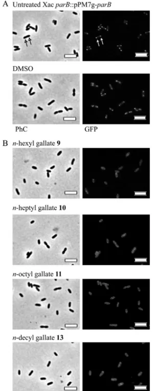
Rod elongation may also occur in response to chromosome segregation defects, and in this sense, the ParB-GFP/DNA com-plex, which is known to form a centromere-like structure in bac-teria, may serve as a marker for the disruption of the segrosome (the chromosome segregation machinery). The bacterial centro-mere is constituted by ParB bound to DNA at specificcis-acting elements (parS) located around the origin of replication of the chromosomes (e.g.,Bacillus subtilis[33]). As proposed previously, ParB functions by interacting with other cellular factors to facili-tate chromosome partitioning toward the polar regions, enabling cell division to proceed (34–36). Thus, ParB-GFP colocalizes with the origins, and the centromere structure formed can be seen by fluorescence microscopy as two foci, each close to a cell pole (37,
38).X.citrisubsp.citrialso exhibits a ParB-GFP localization pro-file (Fig. 4A, arrows) (Ucci and Ferreira, unpublished) that resem-bles the ones documented previously forB. subtilisand, perhaps,
Caulobacter crescentus(37–39). Upon treatment with the alkyl gal-lates, we found that four substances (compounds 9 to 11 and 13) (Table 1) showed activity against the ParB-GFP/DNA complex (Fig. 4B). The time course of treatment showed that this complex is fairly stable, taking an average of 4 h to be disrupted by the compounds (the equivalent of two doubling times for X. citri
subsp.citri). This is consistent with the observation that in B. subtilis, Spo0J-GFP (a ParB-like protein) can be seen throughout
FIG 3Alkyl gallates disrupt the divisional septum ofX. citrisubsp.citri. Mu-tant cells ofX. citrisubsp.citriexpressing GFP-ZapA were cultivated until the OD at 600 nm reached⬃0.3 and then subjected to treatments with the alkyl gallates at concentrations corresponding to the MIC50. The presence or
ab-sence of GFP-ZapA septal labeling was monitored by fluorescence microscopy after 30 min of exposure to the alkyl gallates. (A) Cells cultivated in the absence (untreated) or in the presence of 1% DMSO. Septa are marked with arrows. (B) Cells treated with the alkyl gallates (we show only data for the compounds that perturbed septa). Note that in our analyses, we excluded cell types which resembled the ones labeled with asterisks and considered those similar to the ones marked with crosses. DMSO, vehicle in which substances were dissolved; PhC, phase-contrast microscopy. The scale bars correspond to 4m. Magni-fication,⫻100.
on February 7, 2014 by UNESP - Universidade Estadual Paulista
http://jb.asm.org/
the cell cycle, and occasionally, a Spo0J-GFP focus splits into two, which happens when an origin of replication fires (40). When we compared the pictures taken using phase-contrast microscopy with the GFP ones after treatment with the alkyl gallates, we saw that cells looked fairly normal, although enlarged, but without a clear sign of death and/or an accumulation of inclusion bodies. Our results indicate an action of the alkyl gallates on the centro-mere complex instead of a simple aggregation of ParB-GFP inX. citrisubsp.citricells.
Among the four compounds that disrupted the septum (com-pounds 8 to 11), three also acted on the centromere (com(com-pounds 9 to 11), which raises the possibility that they may target a factor(s) or system(s) that is common to both processes of chromosome segregation and cell division.
DISCUSSION
The plant pathogenXanthomonas citriis the causal agent of citrus canker, a severe disease that affects cultivars of citrus worldwide and represents a major threat to one of the most important eco-nomic activities in Brazil. The eradication of entire orchards of trees infected byX. citrisubsp.citriis the only effective control to prevent the spread of the disease to new areas; however, the obli-gation to do so based on legal governmental actions has led to many disputes, generating too much discontent, especially among orange growers (3). Citriculture in the largest orange-producing area in the world, the state of São Paulo, Brazil, seems to be shifting from eradication to an integrated management system (7), where any help to control citrus canker will make a difference. Here we show the potential use of alkyl gallates to preventX. citrisubsp.
citrigrowth and dissemination, and in addition, some of the com-pounds tested displayed an ability to reduce the bacterial popula-tion within lesions of diseased leaves. The compiled activities of gallic acid and the alkyl gallates againstX. citrisubsp.citriare summarized inTable 3. The colorimetric REMA indicated that the alkyl gallates are potent anti-X. citrisubsp.citriagents exhibiting MICs comparable to those of our positive control, kanamycin FIG 4Alkyl gallates target the centromere ofX. citrisubsp.citri.X. citrisubsp.
citrimutants expressing ParB-GFP were cultivated until the OD at 600 nm reached⬃0.3 and then treated with the alkyl gallates at the MIC50for a period
of 6 h at 30°C. The presence or absence of ParB-GFP foci was monitored by fluorescence microscopy every 30 min from the start of exposure to the com-pounds and documented after 2 h. (A) Cells cultivated in the absence (un-treated) or in the presence of 1% DMSO. The ParB-GFP signal is marked with white arrows on the GFP panel. (B) Cells exposed to the alkyl gallates. Data are relative only to compounds that disrupted the centromere. DMSO, vehicle in which substances were dissolved; PhC, phase-contrast microscopy. The scale bars correspond to 4m. Magnification,⫻100.
TABLE 3Summary of the activities displayed by alkyl gallates againstX. citrisubsp.citri
Compound
Presence of activity
Growth inhibition
Impairs host colonization
Increases cell length
Septum/centromere disruption
Septum Centromere
1 ⫺ ⫺ ⫺ ⫺ ⫺
2 ⫹ ⫺ ⫹ ⫺ ⫺
3 ⫹ ⫺ ⫹ ⫺ ⫺
4 ⫹ ⫺ ⫹ ⫺ ⫺
5 ⫹ ⫺ ⫹ ⫺ ⫺
6 ⫹ ⫹ ⫺ ⫺ ⫺
7 ⫹ ⫹ ⫺ ⫺ ⫺
8 ⫹ ⫹ ⫺ ⫹ ⫺
9 ⫹ ⫹ ⫹ ⫹ ⫹
10 ⫹ ⫹ ⫹ ⫹ ⫹
11 ⫹ ⫺ ⫹ ⫹ ⫹
12 ⫹ ⫹ ⫹ ⫺ ⫺
13 ⫹ ⫺ ⫹ ⫺ ⫹
14 ⫹ ⫺ ⫹ ⫺ ⫺
15 ⫹ ⫺ ⫺ ⫺ ⫺
16 ⫺ ⫺ ⫺ ⫺ ⫺
Gallates Induce Division Arrest inXanthomonas citri
January 2013 Volume 195 Number 1 jb.asm.org 91
on February 7, 2014 by UNESP - Universidade Estadual Paulista
http://jb.asm.org/
(⬍50g/ml). Furthermore, our data are in agreement with data from many other studies of homologous series (41–43) showing that the biological behavior of the alkyl gallates could be correlated with the cutoff phenomenon, which has frequently been attrib-uted to their amphiphilic properties. Structurally, the amphiphile of the alkyl gallates is associated with the presence of two groups, phenolic hydroxyls (hydrophilic moiety) and an alkyl side chain (lipophilic tail). Therefore, we believe that the length of the carbon side chain determines the release of the bioactive portion of the alkyl gallates (the gallic acid unit) inside the cells.
At present, the only substances used in the field to confer some degree of protection againstX. citrisubsp.citriare copper-based formulations. Copper sprays confer a relative control of citrus canker, reducing new infections on young, immature plant tissues (leaves, stems, and fruits) (44, 45). The toxic effect of copper againstX. citrisubsp.citritakes place only at the plant surface, without any internal and/or systemic action (46). As a conse-quence, infected tissues may act as a source of bacterial inoculum, even though plants receive frequent sprays of copper formulations (44,46). Although useful and frequently applied in agriculture, copper accumulates in the environment and may lead to the de-velopment of copper-resistant bacterial populations (46, 47). Here we showed that the exposure ofX. citrisubsp.citricells to alkyl gallates (compounds 6 to 12, except compound 11) (Table 3) prevents the pathogen from colonizing and/or inducing citrus canker symptomsin planta, which has a potential disinfectant action. To our knowledge, this is the first report showing the effect of organic compounds againstX. citrisubsp.citri. Moreover, the treatment of citrus canker lesions on the leaves of a susceptible host with alkyl gallates 6, 7, 9, 10, and 12 impaired the prolifera-tion of the pathogen within the plant tissue. Further analyses are now required in order to evaluate the leaf surface retention and subsequent penetrability of the compounds into the mesophyll, which are important measures to judge their efficacy in the field. There have been several reports in the literature of cell division inhibitors that were identified and/or developed based on assisted screenings for natural, semisynthetic, and synthetic compounds that are able to disrupt FtsZ function in bacteria (for a compre-hensive review, see reference48). Several of these compounds have been included in patent-claiming processes, since cell divi-sion has become one of the ideal targets for the development of antibacterial agents. What supports this view is the fact that pro-tein factors that operate on bacterial cell division, an essential process for life, are in general unique toBacteria. Moreover, these factors are widespread and conserved among this domain of life, and for those proteins which show some degree of homology to eukaryotic counterparts (e.g., bacterial FtsZ and eukaryotic tubu-lins [49,50]), there are still considerable differences among them to allow for the development of bacterium-specific inhibitors. Here we found that compounds 8 to 11 were able to disrupt sep-tum formation in X. citrisubsp. citri(Table 3). Moreover, we demonstrated thatX. citrisubsp.citricells treated with such sub-stances were unable to colonize the host citrus, which showed their potential to protect against this plant pathogen. At the mo-ment, it remains unknown whether the disappearance of the sep-tum induced by these compounds was a direct effect on FtsZ or indirect by targeting any of the other cell division proteins that are recruited to the Z ring during cytokinesis (51). Future experi-ments to assess the direct action of alkyl gallates on FtsZ are needed and will be carried out shortly.
Three out of the four gallates that disrupted the septum also targeted the centromere ofX. citrisubsp.citri(compounds 9 to 11) (Table 3). To show this, we used anX. citrisubsp.citrimutant strain expressing ParB-GFP (Ucci and Ferreira, unpublished) in which the ParB-GFP/DNA complex (the bacterial centromere) has an easily recognizable localization pattern of 2 foci per rod, each occupying a region close to a cell pole (Fig. 4). Upon treat-ment with alkyl gallates 9 to 11, the two discrete centromere foci disappeared, and only dispersed fluorescence could be detected within the cells. This observation raised the possibility that a fac-tor/system common to both chromosome segregation and cell division could be the real target for these compounds inX. citri
subsp.citri. Chromosome segregation and cell division are two essential and interlinked processes in bacteria (34,52,53). Direct links between components of the two processes (e.g., FtsZ and ParB via an FtsZ inhibitor such as MipZ inC. crescentus) have been demonstrated (34,36). Curiously, we found recently thatX. citri
subsp.citrihas an overall chromosome segregation/cell division scheme that resembles what was proposed previously for the modelC. crescentus(54; Ucci and Ferreira, unpublished). In the latter, the correct site for septum assembly within the cell is de-fined by the action of MipZ, which in turn has its localization dependent on the centromere (ParB/DNA) complex. The disrup-tion of MipZ and/or the centromere funcdisrup-tion leads to a cell divi-sion defect inC. crescentus(34). Finally, cytoskeletal proteins, such as the actin-like protein MreB, contribute to chromosome parti-tioning in bacteria (55). Based on these observations, we speculate that compounds 9 to 11 may disrupt cell division by targeting the chromosome segregation machinery ofX. citrisubsp.citri, which also requires further investigation.
The present study shows that the alkyl gallates could be grouped according to two main different biological profiles, which probably reflect their actions on distinct targets. The first group is represented by those compounds that induced altered cell morphology (compounds 2 to 5) (Table 3), but they were unable to interfere with host colonization; furthermore, they showed no detectable influence on the processes of chromosome segregation and/or cell division. The second group (compounds 9 to 11) pre-cluded the ability ofX. citrisubsp.citrito colonize the host citrus, where chromosome segregation and cell division were implicated as the intracellular targets for such compounds.
ACKNOWLEDGMENTS
I.C.S. received a Ph.D. scholarship from FAPESP (2010/02667-4). This work was supported by FAPESP research grants 2004/09173-6, 2010/ 05099-7, and 2011/07458-7.
We thank Dirk-Jan Scheffers and Franklin Behlau for helpful com-ments on the manuscript.
REFERENCES
1.Neves MF, Trombin VG, Milan P, Lopes FF, Cressoni F, Kalaki RB.
2010. O retrato da citricultura brasileira, vol 1. Marcos Fava Neves, Ribeirão Preto, Sao Paulo, Brazil.
2.Schaad NW, Postnikova E, Lacy GH, Sechler A, Agarkova I, Stromberg PE, Stromberg VK, Vidaver AK.2005. Reclassification ofXanthomonas campestrispv.citri(ex Hasse 1915) dye 1978 forms A, B/C/D, and E asX. smithiisubsp.citri(ex Hasse) sp. nov. nom. rev. comb. nov.,X. fuscans
subsp.aurantifolii(ex Gabriel 1989) sp. nov. nom. rev. comb. nov., andX. alfalfaesubsp.citrumelo(ex Riker and Jones) Gabriel et al., 1989 sp. nov. nom. rev. comb. nov.;X. campestrispv. malvacearum (ex smith 1901) dye 1978 asX. smithiisubsp.smithiinov. comb. nov. nom. nov.;X. campestris
pv. alfalfae (ex Riker and Jones, 1935) dye 1978 asX. alfalfaesubsp.alfalfae
on February 7, 2014 by UNESP - Universidade Estadual Paulista
http://jb.asm.org/
(ex Riker et al., 1935) sp. nov. nom. rev.; and “var. fuscans” ofX. campestris
pv. phaseoli (ex Smith, 1987) dye 1978 asX. fuscanssubsp.fuscanssp. nov. Syst. Appl. Microbiol.28:494 –518.
3.Gottwald TR, Graham JH, Schubert TS.2002. Citrus canker: the pathogen and its impact. Plant Health Prog. doi:10.1094/PHP-2002-0812-01-RV.http: //www.plantmanagementnetwork.org/pub/php/review/citruscanker/. 4.Chagas MCM, Parra JRP, Namekata T, Hartung JS, Yamamoto P.2001.
Phyllocnistis citrellaStainton (Lepidoptera: Gracillariidae) and its rela-tionship with the citrus canker bacteriumXanthomonas axonopodispv.
citriin Brazil. Neotrop. Entomol.30:55–59.
5.Bitancourt AA.1957. O cancro cítrico. Biologico23:101–111.
6.Belasque J, Jr, Fernandes NG, Massari CA.2009. The success of eradi-cation campaign of citrus canker in São Paulo States, Brazil. Summa Phy-topathol.35:91–92.
7.Belasque J, Jr, Behlau F.2011. Current status of citrus canker control in São Paulo state, Brazil: a new chapter in a 50-year book?, p 13–15.In
Ferreira H, Belasque J, Jr (ed), Proceedings of the International Workshop on Xanthomonas citri/Citrus Canker. UNESP/Fundecitrus, Ribeirão Preto, Sao Paulo, Brazil. http://www.fcfar.unesp.br/wxc/download /workshop_Xanthomonas.pdf.
8.Canteros BI.1999. Copper resistance inXanthomonas campestrispv.citri, p 455– 459.InMahadevan A (ed), Plant pathogenic bacteria. Proceedings of the International Society of Bacteriology, Centre for Advanced Study in Botany. University of Madras, Chennai, India.
9.Miller AM, Barreto TP, Silva MRL, Leite RP, Jr.2011. Control of citrus canker mediated by neonicotinoids in combination with acibenzolar-S-methyl, p 49 –51.InFerreira H, Belasque J, Jr (ed), Proceedings of the International Workshop onXanthomonas citri/Citrus Canker. UNESP/ Fundecitrus, Ribeirão Preto, Sao Paulo, Brazil.http://www.fcfar.unesp.br /wxc/download/workshop_Xanthomonas.pdf.
10. Graham JH, Myers ME.2011. Soil drenches of imidacloprid, thia-methoxam and acibenzolar-S-methyl for induction of SAR to control citrus canker in young citrus trees. Plant Dis.95:725–728.
11. Yang L, Hu C, Li N, Zhang J, Yan J, Deng Z.2011. Transformation of sweet orange [Citrus sinensis(L.) Osbeck] with pthA-nls for acquiring resistance to citrus canker disease. Plant Mol. Biol.75:11–23.
12. Fu XZ, Chen CW, Wang Y, Liu JH, Moriguchi T.2011. Ectopic expres-sion of MdSPDS1 in sweet orange (Citrus sinensisOsbeck) reduces canker susceptibility: involvement of H(2)O(2) production and transcriptional alteration. BMC Plant Biol.11:55. doi:10.1186/1471-2229-11-55. 13. Grundhofer P, Niemetz R, Schilling G, Gross GG.2001. Biosynthesis
and subcellular distribution of hydrolyzable tannins. Phytochemistry57: 915–927.
14. Muir RM, Ibanez AM, Uratsu SL, Ingham ES, Leslie CA, McGranahan GH, Batra N, Goyal S, Joseph J, Jemmis ED, Dandekar AM. 2011. Mechanism of gallic acid biosynthesis in bacteria (Escherichia coli) and walnut (Juglans regia). Plant Mol. Biol.75:555–565.
15. Werner I, Bacher A, Eisenreich W.1997. Retrobiosynthetic NMR studies with 13C-labeled glucose. Formation of gallic acid in plants and fungi. J. Biol. Chem.272:25474 –25482.
16. Ximenes VF, Lopes MG, Petronio MS, Regasini LO, Silva DH, da Fonseca LM.2010. Inhibitory effect of gallic acid and its esters on 2,2= -azobis(2-amidinopropane)hydrochloride (AAPH)-induced hemolysis and depletion of intracellular glutathione in erythrocytes. J. Agric. Food Chem.58:5355–5362.
17. Morais MC, Luqman S, Kondratyuk TP, Petronio MS, Regasini LO, Silva DH, Bolzani VS, Soares CP, Pezzuto JM.2010. Suppression of TNF-alpha induced NFkappaB activity by gallic acid and its semi-synthetic esters: possible role in cancer chemoprevention. Nat. Prod. Res.
24:1758 –1765.
18. Dodo K, Minato T, Noguchi-Yachide T, Suganuma M, Hashimoto Y.
2008. Antiproliferative and apoptosis-inducing activities of alkyl gallate and gallamide derivatives related to (⫺)-epigallocatechin gallate. Bioorg. Med. Chem.16:7975–7982.
19. Locatelli C, Rosso R, Santos-Silva MC, de Souza CA, Licinio MA, Leal P, Bazzo ML, Yunes RA, Creczynski-Pasa TB.2008. Ester derivatives of gallic acid with potential toxicity toward L1210 leukemia cells. Bioorg. Med. Chem.16:3791–3799.
20. Rosso R, Vieira TO, Leal PC, Nunes RJ, Yunes RA, Creczynski-Pasa TB.
2006. Relationship between the lipophilicity of gallic acid n-alquil esters’ derivatives and both myeloperoxidase activity and HOCl scavenging. Bioorg. Med. Chem.14:6409 – 6413.
21. Regasini LO, Fernandes DC, Castro-Gamboa I, Silva DHS, Furlan M,
Barreiro EJ, Cardoso-Lopes EM, Young MCM, Torres LB, Vellosa JCR, Oliveira OMM.2008. Constituintes químicos das flores dePterogyne ni-tens(Caesalpinioideae). Quim. Nova31:802– 806.
22. Kubo I, Xiao P, Fujita K.2001. Antibacterial activity of octyl gallate: struc-tural criteria and mode of action. Bioorg. Med. Chem. Lett.11:347–350. 23. Fujita K, Kubo I.2002. Antifungal activity of octyl gallate. Int. J. Food
Microbiol.79:193–201.
24. Leal PC, Mascarello A, Derita M, Zuljan F, Nunes RJ, Zacchino S, Yunes RA.2009. Relation between lipophilicity of alkyl gallates and anti-fungal activity against yeasts and filamentous fungi. Bioorg. Med. Chem. Lett.19:1793–1796.
25. Kubo I, Xiao P, Fujita K.2002. Anti-MRSA activity of alkyl gallates. Bioorg. Med. Chem. Lett.12:113–116.
26. Kubo I, Fujita K, Nihei K.2003. Molecular design of multifunctional antibacterial agents against methicillin resistantStaphylococcus aureus
(MRSA). Bioorg. Med. Chem.11:4255– 4262.
27. Martins PM, Lau IF, Bacci M, Belasque J, Jr, do Amaral AM, Taboga SR, Ferreira H.2010. Subcellular localization of proteins labeled with GFP inXanthomonas citrissp.citri: targeting the division septum. FEMS Microbiol. Lett.310:76 – 83.
28. Sambrook J, Fritsch EF, Maniatis T.1989. Molecular cloning: a laboratory manual, 2nd ed. Cold Spring Harbor Laboratory Press, Cold Spring Harbor, NY.
29. Martin A, Camacho M, Portaels F, Palomino JC. 2003. Resazurin microtiter assay plate testing ofMycobacterium tuberculosissusceptibilities to second-line drugs: rapid, simple, and inexpensive method. Antimicrob. Agents Chemother.47:3616 –3619.
30. Palomino JC, Martin A, Camacho M, Guerra H, Swings J, Portaels F.
2002. Resazurin microtiter assay plate: simple and inexpensive method for detection of drug resistance inMycobacterium tuberculosis.Antimicrob. Agents Chemother.46:2720 –2722.
31. Waring MJ.2010. Lipophilicity in drug discovery. Exp. Opin. Drug Dis-cov.5:235–248.
32. Gueiros-Filho FJ, Losick R.2002. A widely conserved bacterial cell divi-sion protein that promotes assembly of the tubulin-like protein FtsZ. Genes Dev.16:2544 –2556.
33. Lin DC, Grossman AD.1998. Identification and characterization of a bacterial chromosome partitioning site. Cell92:675– 685.
34. Thanbichler M, Shapiro L.2006. MipZ, a spatial regulator coordinating chromosome segregation with cell division inCaulobacter.Cell126:147– 162.
35. Marston AL, Errington J.1999. Dynamic movement of the ParA-like Soj protein ofB. subtilisand its dual role in nucleoid organization and devel-opmental regulation. Mol. Cell4:673– 682.
36. Donovan C, Schwaiger A, Kramer R, Bramkamp M.2010. Subcellular localization and characterization of the ParAB system from Corynebacte-rium glutamicum.J. Bacteriol.192:3441–3451.
37. Glaser P, Sharpe ME, Raether B, Perego M, Ohlsen K, Errington J.1997. Dynamic, mitotic-like behavior of a bacterial protein required for accurate chromosome partitioning. Genes Dev.11:1160 –1168.
38. Lin DC, Levin PA, Grossman AD.1997. Bipolar localization of a chro-mosome partition protein inBacillus subtilis.Proc. Natl. Acad. Sci. U. S. A.
94:4721– 4726.
39. Mohl DA, Gober JW.1997. Cell cycle-dependent polar localization of chro-mosome partitioning proteins inCaulobacter crescentus.Cell88:675– 684. 40. Sharpe ME, Errington J.1998. A fixed distance for separation of newly
replicated copies of oriC inBacillus subtilis: implications for co-ordination of chromosome segregation and cell division. Mol. Microbiol.28:981–990. 41. Kubo I, Fujita K, Kubo A, Nihei K, Ogura T.2004. Antibacterial activity
of coriander volatile compounds againstSalmonella choleraesuis.J. Agric. Food Chem.52:3329 –3332.
42. Birnie CR, Malamud D, Schnaare RL.2000. Antimicrobial evaluation of N-alkyl betaines and N-alkyl-N,N-dimethylamine oxides with variations in chain length. Antimicrob. Agents Chemother.44:2514 –2517. 43. Hammond DG, Kubo I.2000. Alkanols inhibit respiration of intact
mi-tochondria and display cutoff similar to that measured in vivo. J. Pharma-col. Exp. Ther.293:822– 828.
44. Behlau F, Belasque J, Jr, Graham JH, Leite RP, Jr. 2010. Effect of frequency of copper applications on control of citrus canker and the yield of young bearing sweet orange trees. Crop Prot.29:300 –305.
45. Gottwald TR, Timmer LW.1995. The efficacy of windbreaks in reducing the spread of citrus canker caused byXanthomonas campestrispv.citri. Trop. Agric.72:194 –201.
Gallates Induce Division Arrest inXanthomonas citri
January 2013 Volume 195 Number 1 jb.asm.org 93
on February 7, 2014 by UNESP - Universidade Estadual Paulista
http://jb.asm.org/
46. Behlau F, Canteros BI, Jones JB, Graham JH.2012. Copper resistance genes from different xanthomonads and citrus epiphytic bacteria confer resistance toXanthomonas citrisubsp.citri.Eur. J. Plant Pathol.133:949 –963. 47. Graham JH, Dewdney MM, Myers ME.2010. Streptomycin and copper
formulations for control of citrus canker on grapefruit. Proc. Fla. State Hortic. Soc.123:92–98.
48. Awasthi D, Kumar K, Ojima I. 2011. Therapeutic potential of FtsZ inhibition: a patent perspective. Expert Opin. Ther. Pat.21:657– 679. 49. Erickson HP, Stoffler D.1996. Protofilaments and rings, two
conforma-tions of the tubulin family conserved from bacterial FtsZ to alpha/beta and gamma tubulin. J. Cell Biol.135:5– 8.
50. Lowe J, Amos LA.1998. Crystal structure of the bacterial cell-division protein FtsZ. Nature391:203–206.
51. Errington J, Daniel RA, Scheffers DJ. 2003. Cytokinesis in bacteria. Microbiol. Mol. Biol. Rev.67:52– 65.
52. Lau IF, Filipe SR, Soballe B, Okstad OA, Barre FX, Sherratt DJ.2003. Spatial and temporal organization of replicatingEscherichia coli chromo-somes. Mol. Microbiol.49:731–743.
53. Lemon KP, Grossman AD.2001. The extrusion-capture model for chro-mosome partitioning in bacteria. Genes Dev.15:2031–2041.
54. Ptacin JL, Lee SF, Garner EC, Toro E, Eckart M, Comolli LR, Moerner WE, Shapiro L.2010. A spindle-like apparatus guides bacterial chromo-some segregation. Nat. Cell Biol.12:791–798.
55. Gitai Z, Dye NA, Reisenauer A, Wachi M, Shapiro L. 2005. MreB actin-mediated segregation of a specific region of a bacterial chromosome. Cell120:329 –341.