Article
Printed in Brazil - ©2012 Sociedade Brasileira de Química0103 - 5053 $6.00+0.00
A
*e-mail: [email protected]
Cytotoxic Cordiaquinones from the Roots of
Cordia polycephala
Hozana Patrícia S. Freitas,a Ana Isabel V. Maia,a Edilberto R. Silveira,a
José Delano B. Marinho Filho,b Manoel O. Moraes,b Cláudia Pessoa,b
Letícia V. Costa Lotufob and Otília Deusdênia L. Pessoa*,a
aDepartamento de Química Orgânica e Inorgânica, Universidade Federal do Ceará,
CP 12.200, 60021-940 Fortaleza-CE, Brazil
bDepartamento de Farmacologia e Fisiologia, Universidade Federal do Ceará,
CP 3157, 60430-270 Fortaleza-CE, Brazil
A investigação química das raízes de Cordia polycephala resultou no isolamento de duas novas naftoquinonas terpenoídicas, 6-[10-(12,12-dimetil-13α -(22-metil-21-butenoilóxi)-16-metenilciclohexil)etil]-naftaleno-1,4-diona e (6-[10-(12,12-dimetil-13α -(tigloilóxi)-16-metenilciclohexil)etil]-naftaleno-1,4-diona, designadas de cordiaquinonas N (1) e O (2), respectivamente. As cordiaquinonas já conhecidas, B (3), L (4) e E (5), foram também isoladas. Suas estruturas foram elucidadas após detalhada análise dos dados de RMN 1H e 13C (1D e 2D) e EMAR (espectrometria de massas de alta resolução). Todas as cordiaquinonas (1-5) foram avaliadas contra quatro linhagens de células cancerígenas: HCT-8 (colo), HL-60 (leucemia), MDA-MB-435 (melanoma) e SF295 (glioblastoma), mostrando IC50 na faixa de 1,2 a 11,1 µmol L
-1. As novas
cordiaquinonas 1 e 2 foram as mais ativas contra todas as linhagens de câncer, enquanto 3 foi mais seletiva para células leucêmicas (IC50 2,2 µmol L
-1).
The chemical investigation of roots of Cordia polycephala resulted in the isolation of two new terpenoid naphtoquinones, 6-[10-(12,12-dimethyl-13α -(22-methyl-21-butenoyloxy)-16-methenylcyclohexyl)ethyl]-naphtalene-1,4-dione and (6-[10-(12,12-dimethyl-13α -(tigloyloxy)-16-methenylcyclohexyl)ethyl]-naphtalene-1,4-dione, named as cordiaquinone N (1) and O (2), respectively. The known cordiaquinones B (3), L (4) and E (5) were also isolated. Their structures were elucidated after detailed 1D and 2D NMR and high resolution electrospray ionization mass spectrometry (HRESIMS) data analysis. All cordiaquinones (1-5) were evaluated against four human cancer cell lines: HCT-8 (colon), HL-60 (leukemia), MDA-MB-435 (melanoma) and SF295 (glioblastoma), showing IC50 values in the range of 1.2 to 11.1 µmol L
-1. The new cordiaquinones
1 and 2 were the most active against all cancer cell lines, while compound 3 was selective to leukemia HL-60 cells (IC50 2.2 µmol L
-1).
Keywords: Cordia polycephala, cordiaquinones, cytotoxic activity
Introduction
The genus Cordia, one of the major and most important of the Boraginaceae family, is known as a prolific source of structurally diverse secondary metabolites such as sesquiterpenes,1 diterpenes,2 triterpenes,3 triterpene
saponins,4 and their ability to biosynthesize meroterpenoid
benzoquinones and naphthoquinones, including their respective reduced forms (hydroquinones).5-10 These
quinoid compounds, known as cordiaquinones, could be
considered as chemotaxinomical markers for the Cordia
genus. Nevertheless, the occurrence in their producing plants is limited to the roots and rarely to the trunk wood. As part of a search for bioactive compounds from Cordia, the chemical composition of several Cordia species native to the Brazilian flora, including its pharmacological and biological effects have been performed.11,12 Previous
investigations have demonstrated that cordiaquinones display anticancer activity, inducing reactive oxygen species generation and apoptosis in cancer cells.13,14 The
Johnston (syn.: Cordia ulmifolia Juss, Cordia wickstroemii
Steudel, Varronia polycephala Lam. Varronia paniculata
Wakström), a flowering and aromatic shrub native to Brazil, Colombia, French Guiana and Suriname. In addition, the cytotoxic effect of all isolated compounds was also evaluated against four cancer cell lines.
Results and Discussion
Compound 1 was isolated as a yellow resin. Its molecular formula (C26H30O4)was established by HRESIMS (observed
m/z 407.2252 [M + H]+; calc. for C
26H31O4 407.2222)
and NMR spectral analysis. The IR spectrum exhibited absorption bands for carbon-oxygen (1712 and 1666 cm-1)
and carbon-carbon (1600 cm-1) double bonds. The 13C NMR
spectrum displayed 26 carbon signals, identified through DEPT and HSQC experiments as eight methines (six sp2
and two sp3), one methylidene, four methylenes, four
methyls and nine non-hydrogenated carbons (eight sp2
and one sp3). The 1H and 13C NMR features of 1 (Table 1)
suggested that this compound was a 1,4-naphthoquinone, especially due to the presence of the signals at d 185.6 and 185.1.8,12 Comparison of the 1H and 13C NMR data of 1
with those reported for cordiaquinone L,8 showed high
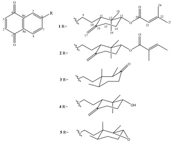
similarity, except for the presence of the additional signals at d 166.4, 156.9, d 116.5/5.65 (br s), d 27.6/1.89 (s) and d 20.4/2.17 (s) in 1, which were assigned to a 3-methyl-2-butenoyl moiety. The position of this group at C-13 was established by the HMBC correlation exhibited between H-13 (d 4.70) and C-20 (d 166.4) (Figure 1), while its equatorial position was deduced from the multiplicity and coupling constant values displayed by H-13 (dd, J 8.5 and 4.5 Hz). The relative stereochemistry of 1 was established by analysis of the NOESY spectrum, which showed NOE correlations among the axial protons H-13, H-11 and Me-18 (Figure 1). Thus, the structure of 1 was established as the 6-[10-(12,12-dimethyl-13α -(22-methyl-21-butenoyloxy)-16-methenylcyclohexyl)ethyl]-naphtalene-1,4-dione). Terpenoid naphthoquinones have been previously isolated from Cordia species and designated as cordiaquinones, an allusion to the genus. Since cordiaquinones A-M were already reported,8 compound 1 was designated as
cordiaquinone N.
Compound 2 was also obtained as a yellow resin. The molecular formula (C26H30O4)determined by HRESIMS
(observed m/z 407.2206 [M+H]+; calc. for C 26H31O4,
407.2222), was identical to that of 1. Its IR spectrum exhibited absorption bands for carbon-oxygen (1703 and 1667cm-1) and carbon-carbon (1600 cm-1) double bonds.
Comparative analyses of the 1H and 13C NMR data of 2 with
those of 1 (Table 1) showed very close structural similarity,
except for the signals related to the ester side chain (C-21 to C-24). Besides the doublet at d 1.76 (d, J 7.0 Hz) for a vinyl methyl (C-23) and the correspondent vinyl hydrogen at d 6.81 (q, J 7.0 Hz), the HMBC spectra displayed the long range correlations of the methyl protons at d 1.81 (s, H3-24) with the carbons at d 167.8 (C-20), 129.1 (C-21) and 137.2 (C-22) (Figure 2), revealing that the ester side chain was indeed a tigloyloxy moiety. The aforementioned data allowed to establish the structure of 2 as the 6-[10-(12, 12-dimethyl-13α-(tigloyloxy)-16-methenylcyclohexyl) ethyl]-naphtalene-1,4-dione, named cordiaquinone O. In addition to new cordiaquinones N and O (1-2), three known cordiaquinones, B (3), L (4), E (5)8,10 were isolated (Figure 3).
Figure 1. Relevant HMBC (HàC) and NOESY correlations for 1.
Figure 2. Relevant HMBC (H→C) correlations for 2.
The MTT analysis showed that all compounds (1-5) exhibited cytotoxic activity against tested cancer cell lines (Table 2). Compounds 1 and 2 were the most active among all, with IC50 values ranging from 1.2 to 3.4 µmol L-1,
whereas for the known cordiaquinones B, E and L, IC50
ranged from 2.2 to over 15 µmol L-1. It’s worthwhile to
mention that cordiaquinone E (3) was selective towards leukemia cells (IC50 2.2 µmol L-1), since it displayed a
weak activity to cells from different histological origins (IC50 > 15 µmol L-1 in MDA-MB-435 and SF-295 cells,
and IC50 of 11.1 µmol L
-1 in HCT-8 cells). None of the
tested compounds (1-5) showed hemolytic activity in mice
erythrocytes (EC50 > 500 µmol L
-1), suggesting that the
cytotoxicity of cordiaquinones is not related to non-specific membrane damage (Table 2). Among cordiaquinones, one could speculate about the higher activity of compounds 1
and 2 referring to the presence of the extra α-β-conjugated carbonyl of the end tigloyloxy side chains.
Several molecules possessing a quinone moiety show antiproliferative effects on tumor cell growth.15 The cytotoxic
activity of quinones is related to inhibition of electron transporters,16 uncouple of oxidative phosphorylation,17 ROS
generation,18 protein adducts formation,19 especially with
enzyme SH groups and DNA damage.15 In a previous work,
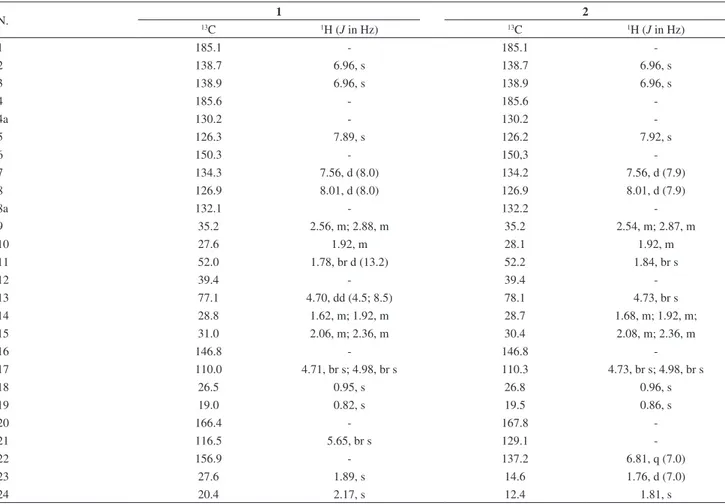
Table 1.1H (500 MHz) and 13C (125 MHz) NMR data of cordiaquinones 1 and 2 in CDCl 3
N. 13 1 2
C 1H (J in Hz) 13C 1H (J in Hz)
1 185.1 - 185.1
-2 138.7 6.96, s 138.7 6.96, s
3 138.9 6.96, s 138.9 6.96, s
4 185.6 - 185.6
-4a 130.2 - 130.2
-5 126.3 7.89, s 126.2 7.92, s
6 150.3 - 150,3
-7 134.3 7.56, d (8.0) 134.2 7.56, d (7.9)
8 126.9 8.01, d (8.0) 126.9 8.01, d (7.9)
8a 132.1 - 132.2
-9 35.2 2.56, m; 2.88, m 35.2 2.54, m; 2.87, m
10 27.6 1.92, m 28.1 1.92, m
11 52.0 1.78, br d (13.2) 52.2 1.84, br s
12 39.4 - 39.4
-13 77.1 4.70, dd (4.5; 8.5) 78.1 4.73, br s
14 28.8 1.62, m; 1.92, m 28.7 1.68, m; 1.92, m;
15 31.0 2.06, m; 2.36, m 30.4 2.08, m; 2.36, m
16 146.8 - 146.8
-17 110.0 4.71, br s; 4.98, br s 110.3 4.73, br s; 4.98, br s
18 26.5 0.95, s 26.8 0.96, s
19 19.0 0.82, s 19.5 0.86, s
20 166.4 - 167.8
-21 116.5 5.65, br s 129.1
-22 156.9 - 137.2 6.81, q (7.0)
23 27.6 1.89, s 14.6 1.76, d (7.0)
24 20.4 2.17, s 12.4 1.81, s
Table 2. Cytotoxic activity of compounds 1-5
Compounds IC50 / (µmol L-1) (95% confidence interval) Hemolysis
EC50 / (µmol L-1)
HL-60 HCT-8 SF-295 MDA-MB-435
1 1.5 (1.0-2.0) 2.3 (1.5-3.7) 1.8 (1.5-2.2) 2.9 (2.4-3.7) > 500
2 1.2 (0.7-2.0) 1.2 (1.0-1.5) 2.0 (1.7-2.4) 3.4 (2.9-3.9) > 500
3 2.2 (0.9-4.3) 11.1 (10.5-12.0) > 15 > 15 > 500
4 5.0 (4.2-5.8) 5.0 (4.3-5.8) 4.6 (4.0-5.2) 7.5 (6.5-8.6) > 500
it was described for the first time the mechanisms underlying the cytotoxic effects of cordiaquinones. Cordiaquinone J, isolated from C. leucocephala, demonstrated a rapid induction of apoptosis and necrosis in a mechanism dependent of the generation of reactive oxygen species in leukemia cells.14
Experimental
General experimental procedures
Optical rotations were measured on a Perkin-Elmer 341 digital polarimeter. IR (4000 to 650 cm-1) spectra
were obtained on a Perkin-Elmer 100 FT-IR spectrum. The high resolution electrospray ionization mass spectra (HRESIMS) were acquired using a LCMS-IT-TOF (SHIMADZU) spectrometer. 1H (500 MHz) and 13C NMR
(125 MHz) spectra were performed on a Bruker DRX-500 spectrometer. HPLCanalysis was carried out using a UFLC (SHIMADZU) system equipped with a SPD-M20A diode array UV-Vis detector and a Phenomenex LC-Silica column, 5 µm (4.6 × 250 mm). The mobile phase was consisted of hexane/CHCl3 (65:35 v/v) with a 4.72 mL min-1 flow
rate, and the chromatograms were acquired at 247 nm. Column chromatography was carried out on silica gel 60 (70-230 mesh, Vetec or 230-400 mesh, Merck), TLC was performed on precoated silica gel aluminium sheets (kieselgel 60 F254, 0.20 mm, Merck). Fractions and pure
compounds were monitored by TLC, and the spots were visualized by the color reaction by spraying with a solution of vanillin/perchloric acid/EtOH followed by heating (ca. 100 °C).
Plant material
Roots of Cordia polycephala were collected in August 2009 along the road margins at Pico-Alto, Guaramiranga-CE, Brazil, and identified by Maria Iracema B. Loiola, botanist of the Federal University of Ceará, Brazil. The voucher specimen (IAC # 44.582) has been deposited at the Herbário Prisco Bezerra, at the Departamento de Biologia of the Universidade Federal do Ceará, Brazil.
Extraction and isolation
Roots of C. polycephala (2.0 Kg), dried and pulverized, were extracted with hexane at room temperature. The solvent was removed under reduced pressure to yield a dark extract (20 g), which was fractioned over silica gel by elution with hexane, hexane/EtOAc (9:1, 8:2, 6:4, 4:6, 2:8), EtOAc and MeOH to yield 63 fractions (30 mL
each), which after TLC analysis were combined into eight fractions (F1-F8). F4 (6.9 g, obtained by elution with hexane/EtOAc 6:4) was subjected to repeated column chromatography using hexane/EtOAc mixtures as eluents. The sub-fraction hexane/EtOAc 9:1 (250 mg) was applied to HPLC separation using a Gemini Phenomenex LC-Silica column (250 mm × 4.6 mm i.d.) in the isocratic mode (hexane/CHCl3 65:35, v/v) at a flow rate of 4.72 mL min
-1,
with an injection volume (‘loop’) of 200 µL, to yield compounds 1 (7.5 mg) and 2 (11.0 mg). F5 (4.8 g, obtained with hexane/EtOAc 4:6) was fractioned over silica gel eluting with hexane/EtOAc (9:1, 8:2, 6:4, 4:6, 2:8) and EtOAc. From the hexane/EtOAc 9:1 fraction (523.4 mg), the cordiaquinone E (3, 39.2 mg) was isolated. The sub-fraction hexane/EtOAc 6:4 (2.2 g) was subjected to silica gel column chromatography using the same solvent system. From the hexane/EtOAc 8:2 the cordiaquinone B (4, 133.6 mg) was obtained. The fraction obtained with hexane/ EtOAc 6:4 (135 mg) was submitted to semipreparative HPLC using the same column and the isocratic mode, as above, but using hexane/isopropanol (95:05, v/v) at a flow rate of 4.0 mL min-1, with an injection volume (‘loop’) of
200 µL, to yield cordiaquinone L (5, 28.4 mg).
Cell lines and cells culture
The human cell lines used in this work were HL-60 (promyelocytic leukemia), HCT-8 (colon), MDA-MB-435 (melanoma) and SF-295 (brain), which were all obtained from the National Cancer Institute (Bethesda, MD, USA). The cells were maintained in RPMI 1640 medium supplemented with 10% fetal bovine serum, 2 mmol L-1
glutamine, 100 U mL-1 penicillin, and 100 µg mL-1
streptomycin at 37 ºC with 5% CO2.
Inhibition of cancer cells proliferation
The cytotoxicity of all cordiaquinones were tested against the HL-60 (promyelocytic leukemia), HCT-8 (colon), MDA-MB-435 (melanoma) and SF-295 (brain) cell lines. For all experiments, cells were plated in 96-well plates (105 cells per well for adherent cells or 0.3 × 10⁵ cells per
well for suspended cells in 100 µL of medium). After 24 h, all cordiaquinones (0.06-77.0 µmol L-1) dissolved in 1% DMSO
3-(4,5-dimethyl-2-thiazolyl)-2,5-diphenyl-2H-tetrazoliumbromide (MTT) to a purple formazan product.20
At the end of the incubation, the plates were centrifuged and the medium was replaced with fresh medium (150 µL) containing MTT (0.5 mg mL-1). Three hours later, the plates
were centrifuged, the MTT formazan product was dissolved in 150 µL DMSO, and the absorbance was measured using a multiplate reader (Spectra Count, Packard, Ontario, Canada). The drug effect was quantified as the percentage of the control absorbance of the reduced dye at 595 nm.
Hemolytic activity
Membrane disruption was performed in 96-well plates. Briefly, each well received 100 µL of a 0.85% NaCl solution containing 10 mmol L-1 CaCl
2 and 100 µL of a 2%
suspension of mouse erythrocytes in the same medium. The cordiaquinones were tested at concentrations ranging from 12 to 500 µmol L-1. Triton X-100 (Isofar, Brazil) at 0.1% (in
0.85% NaCl) was used as a positive control. After incubation for 60 min at room temperature, the plate was centrifuged, the supernatant was removed and the liberated hemoglobin was measured at 540 nm (DTX-880, Beckman Coulter®).21
Conclusions
From roots of C. polycephala were isolated five terpenoid naphtoquinones, two of which are new. The isolation of additional new terpenoid quinones from
Cordia species reinforces the importance of this type of compounds as possible chemomarkers for the genus. All naphthoquinones showed antiproliferative effects, particularly the new compounds (1 and 2). The investigation of plants from Cordia species as a source of anticancer agents could improve the development of new drugs.
Supplementary Information
Supplementary information for compounds 1 and 2 is available free of charge at http://jbcs.sbq.org.br as PDF file.
Acknowledgments
This work was supported by grants from the Brazilian Governmental Agencies: CAPES, CNPq, PRONEX, FUNCAP and INCT.
References
1. Menezes, J. E. S.; Machado, A. F. E. A.; Lemos, T. L. G.; Silveira, E. R.; Braz-Filho, R.; Pessoa, O. D. L.; Z. Naturforsch., C: Biosci.2004, 59,19.
2. Siddqui, B. S.; Perwaiz, S.; Begum, S.; Tetrahedron2006, 62, 10087.
3. Kuroyanagi, M.; Kawahara, N.; Sekita, S.; Satake, M.; Hayashi, T.;
J. Nat. Prod.2003,66, 1307.
4. Santos, R. P.; Viana, F. A.; Lemos, T. L. G.; Silveira, E. R.; Braz-Filho, R.; Pessoa, O. D. L.; Magn. Reson. Chem.2003,
41, 735.
5. Bieber, L. W.; Messana, I.; Lins, S. C. N.; da Silva Filho, A. A.; Chiappeta, A. A.; Méllo, J. F.; Phytochemistry 1990, 29, 1955. 6. Ioset, J. R.; Marston, A.; Gupta, M. P.; Hostettmann, K.; J. Nat.
Prod. 2000, 63, 424.
7. Mori, K.; Kawano, M.; Fuchino, H.; Ooi, T.; Satake, M.; Agatsuma, Y.; Kusumi, T.; Sekita, S.; J. Nat. Prod. 2008, 71, 18.
8. Diniz, J. C.; Viana, F. A.; Oliveira, O. F.; Maciel, M. A. M.; Torres, M. C. M.; Braz-Filho, R.; Silveira, E. R.; Pessoa, O. D. L.;
Magn. Reson. Chem.2009, 47, 190.
9. Dettrakul, S.; Surerum, S.; Rajviroongit, S.; Kittakoop, P.;
J. Nat. Prod.2009, 72, 861.
10. Ioset, J. R.; Marston, A.; Gupta, M. P.; Hostettmann, K.;
Phytochemistry1998, 47, 729.
11. Lemos, T. L. G.; Monte, F. J. Q.; Santos, A. K. L.; Santos, H. S.; Oliveira, M. F.; Costa, S. M.; Pessoa, O. D. L.; Braz-Filho, R.;
Nat. Prod. Res.2007, 21, 529.
12. Ioset, J. R.; Marston, A.; Gupta, M. P.; Hostettmann, K.; J. Nat. Prod.2000, 53, 613.
13. Menezes, J. E. S. A.; Lemos, T. L. G.; Pessoa, O. D. L.; Braz-Filho, R.; Montenegro, R. C.; Wilke, D. V.; Costa-Lotufo, L. V.; Pessoa, C.; De Moraes, M. O.; Silveira, E. R.; Planta Med.
2005, 71, 54.
14. Marinho-Filho, J. D. B.; Bezerra, D. P.; Araújo, A. J.; Montenegro, R. C.; Pessoa, C.; Diniz, J. C.; Viana, F. A.; Pessoa, O. D. L; Silveira, E. R.; Moraes, M. O.; Costa-Lotufo, L. V.;
Chem. Biol. Interact.2010, 183, 369. 15. Asche, C.; Mini Rev. Med. Chem.2005, 5, 449.
16. Vennerstrom, J. L.; Vennerstrom, J. W.; J. Med. Chem.1988,
31, 1269.
17. Howland, J. L.; Biochim. Biophys. Acta1963, 77, 419. 18. Monks, T. J.; Hanzlik, R. P.; Cohen, G. M.; Ross, D.; Graham,
D. G.; Toxicol. Appl. Pharmacol.1992, 112, 2.
19. Kleiner, H. E.; Rivera, M. I.; Pumford, N. R.; Monks, T. J.; Lau, S. S.; Chem. Res. Toxicol.1998, 111, 282.
20. Mosmann, T.; J. Immunol. Methods 1983,65, 55.
21. Jimenez, P. C.; Fortier, S. C.; Lotufo, T. M. C.; Pessoa, C.; Moraes, M. E. A.; Moraes, M. O. Costa-Lotufo, L. V.; J. Exp. Mar.Biol. Ecol.2003, 287, 93.