1. Department of Orthopedics and Traumatology, Universidade Federal de São Paulo, São Paulo, SP, Brazil.
Study conducted by the Spine Group, Department of Orthopedics and Traumatology, Universidade Federal de São Paulo (Unifesp), SP, Brazil. Correspondence: Rua Borges Lagoa, 783 – 5º andar, Vila Clementino – 04038-032 – São Paulo, SP. Brasil. [email protected]
ABSTRACT
Objective: To conduct cross-sectional study identifying the profile of the Brazilian spinal surgeon. Methods: Data were collected through a questionnaire with multiple alternatives during two major events for spine surgery at national level in 2011, the Congresso da Sociedade Brasileira de Coluna (Congress of the Brazilian Spine Society) and Simpósio Internacional de Coluna (International Spine Symposium, SINCOL). The data were submitted to statistical analysis comparing and stratifying the information obtained according to the profile. Results: We obtained 182 questionnaires answered by orthopedists and neurosurgeons with peculiarities and similarities on their medical manage-ment. Conclusions: The data obtained in this study may be important for the development of health policies in the spine surgery in Brazil. Keywords: Spine; Orthopedics; Neurosurgery; Cross-sectional studies; Brazil.
RESUMO
Objetivo: Realizar estudo transversal identificando o perfil do cirurgião de coluna no Brasil. Métodos: Foram coletados dados por meio de questionários com múltiplas alternativas, em dois eventos de relevância para a cirurgia de coluna no âmbito nacional em 2011, o Congresso da Sociedade Brasileira de Coluna (SBC) e o Simpósio Internacional de Coluna (SINCOL). Os dados foram submetidos a análise estatística comparando e estratificando as informações obtidas conforme o perfil encontrado. Resultados: Obtivemos 182 questionários respondidos por ortopedistas e neurocirurgiões com particularidades e semelhanças em suas condutas médicas. Conclusões: Os dados obtidos nessa pesquisa podem ser importantes para o desenvolvimento de políticas de saúde na área de cirurgia de coluna no Brasil.
Descritores: Coluna vertebral; Ortopedia; Neurocirurgia; Estudos transversais; Brasil.
RESUMEN
Objetivo: Llevar a cabo estudio transversal para identificar el perfil del cirujano de columna en Brasil. Métodos: Los datos fueron recolectados por medio de cuestionarios con opciones múltiples, en dos eventos de relevancia para la cirugía de columna vertebral, a nivel nacional en 2011, Congreso Brasileño de la Sociedad Brasileña de Columna (SBC) y el Simposio Internacional de Columna (SINCOL). Los datos fueron sometidos a análisis estadístico, comparándose y estratificándose las informaciones obtenidas, de acuerdo con el perfil encontrado. Resultados: Se obtuvieron cuestionarios respondidos por 182 ortopedistas y neurocirujanos, con particularidades y similitudes en sus actividades médicas. Conclusiones: Los datos obtenidos en este estudio pueden ser importantes para el desarrollo de políticas de salud en el área de la cirugía de columna en Brasil.
Descriptores: Columna vertebral; Ortopedia; Neurocirugía; Estudios transversales; Brasil.
BRAZILIAN SPINE SURGEON PROFILE
PERFIL DO CIRURGIÃO DE COLUNA BRASILEIRO
PERFIL DEL CIRUJANO BRASILEÑO DE COLUMNA
O
RIGINALA
RTICLE/A
RTIGOO
RIGINAL/A
RTÍCULOO
RIGINALPEDRO LUZ ALVES1, FERNANDO TADASHI SALVIONI UETA1, RENATO HIROSHI SALVIONI UETA1, DAVID DEL CURTO1, DÉLIO EULÁLIO MARTINS1,
MARCELO WAJCHENBERG1, EDUARDO BARROS PUERTAS1
Received on 03/12/2012, accepted on 07/25/2013.
INTRODUCTION
Spine surgery is a complex specialty that treats a wide variety of diseases, and which can be approached by several areas of medicine, such as orthopedics, rheumatology, and neurosurgery, among others.
Various epidemiological studies1-3 are continuously performed
around the world to define the best therapeutic approaches and new lines of research. However, most studies are regional and relate to disease epidemiology alone without defining the geographical distribution of the surgeon and the surgical techniques performed.
After performing searches in the major scientific portals MEDLINE (via PubMed), LILACS, EMBASE (via Ovid), we encountered a national epidemiological study4 that sought to identify surgical
techniques that have no longer been used in spinal surgery among Brazilian spine surgeons.
Nationally, no other study was found seeking to define the char-acteristics of the specialty and the spine surgeon. In Brazil there are about two thousand neurosurgeons registered in the Brazilian Soci-ety of Neurosurgery (SBN), and approximately 12,500 orthopedists
in the Brazilian Society of Orthopedics and Traumatology (SBOT), divided into various sectors within their specialties. The Brazilian Spine Society (SBC) had 730 members at this time.
Due to the complexity of this specialty, there is an imminent need to collect and interpret information regarding the activity of surgeons to trace the characteristic profile of the Brazilian spine surgeon, in order to effectively plan the application of resources for the health sector, academia, and the professional sector.
METHODS
Data were collected through multiple choice questionnaires in two events relevant to spine surgery in 2011, the SBC Congress and the SINCOL.
The above events were selected in order to achieve similar representation from the number of neurosurgeons and orthopedists who work in the field of spine surgery, given that the SINCOL is organized primarily by neurosurgeons and the Brazilian Spine Congress by orthopedists.
The questionnaires were administered individually and voluntarily
answered during lectures, and collected at the end. There were 23 simple and straightforward questions based on the everyday work of the spine surgeon.
After collection, the data were statistically analyzed, and the infor-mation was compared and stratified according to the profile found.
STATISTICAL ANALYSIS
The characteristics evaluated in the spine surgeons were de-scribed using absolute and relative frequencies.
Each characteristic evaluated was described according to physician’s specialty and the existence of an association between the specialty and characteristics was determined using the chi--square test or Fisher’s exact test or the likelihood ratio test when it was not possible to apply the chi-square test.
RESULTS
One hundred eighty-two questionnaires were completed by spine surgeons during the two events and significant associations were observed in some respects. The various characteristics evaluated are shown below after the analysis and correlation of data.
Of the surgeons who answered the question about their spe-cialty, 74.7% are orthopedists, 24.7% are neurosurgeons, and 0.5% reported no specialty.
Concerning geographical distribution, the Southeast had the highest concentration of surgeons; the state of São Paulo had the highest rate of participation, at 36.3%. (Figure 1)
The number of young spine surgeons is significantly higher among orthopedists, contrary to what was observed in the group of neurosurgeons. (Figures 2 and 3)
Surgeons have a diverse profile regarding the distribution of the workplace locations. (Figure 4) A percentage of doctors work in
philanthropic associations that were classified as other workplaces. Professionals working in polytrauma centers totaled 68.1%, 30.2% do not see such patients and 1.6% did not answer.
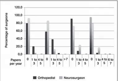
Surgeons were asked whether they subscribe to any scientific jour-nal, 73.6% subscribe, 18.1% do not subscribe, and 8.2% did not answer. Orthopedists publish most papers in the Brazilian Journal of Orthopaedics (RBO) and the Coluna/Columna Journal, while neu-rosurgeons publish in PUBMED and LILACS. (Figure 5)
The percentage of surgeries performed by surgeons in the Uni-fied Health System (SUS) is 34.41%, and 26.62% are performed in the private sector, which is less than the number of procedures per-formed through health insurance plans, which is 38.97%. (Figure 6) The number of surgeries performed per week, such as degen-erative, minimally invasive spine surgery (MISS), tumors, deformity, trauma, and osteotomies, was similar among surgeons. Orthope-dists had the highest number of surgeries performed on deformities, 87.7%, with p < 0.001#.
The most commonly used classification of deformities was that of Lenke et al.,5 but almost the same proportion use Moe,6 King
and Moe,7 and Lenke et al.,5 with no difference among surgeons,
as shown in Figure 7.
Concerning the type of instrument used, 67.6% of surgeons use pedicle screws with the derotation technique, 1.6% use subla-minar wire loop fixation with rods, 30.8% use pedicle screws with the translation technique, 6% use a hybrid system, 1.1% use other instrumentation, and 32.1% did not answer. Percentages exceed 100% because some surgeons use more than one technique. The technique with posterior instrumental suture loop fixation was only used by orthopedists.
Figure 1. Regional distribution of surgeons.
Figure 2. Time orthopedists have been in practice.
Figure 3. Time neurosurgeons have been in practice.
Figure 4. Workplace of surgeons.
North
Northeast Midwest Southeast Regions of Brazil
P
ercentage
Orthopedist Neurosurgeon
South
Percentage Time
0 10 20 30 40 50 60 70 >15 years
11 to 15 years
6 to 10 years
0 to 5 years
Result of the chi-square test p < 0.001.
Percentage Time
0,0 10,0 20,0 30,0 40,0 50,0 60,0 >15 years
11 to 15 years
6 to 10 years
0 to 5 years
* Result of the chi-square test p < 0.001.
Other
Percentage
Workplace
Federal University Municipal University
Public Clinic State University
61.9% of orthopedists use it in idiopathic scoliosis, but only 34.3% use it for neuromuscular scoliosis. Thirty-six point one percent of neurosurgeons use it for tumors, while only 19.4% of orthopedists do, with p < 0.034* in the chi-square test. Sixty-two point four per-cent of professionals do not use monitoring in surgery.
Regarding the application of magnetic resonance imaging, in ado-lescent idiopathic scoliosis with typical curves, orthopedists request it in 37.4% of cases while neurosurgeons do in 58.3%. (Figure 8)
Concerning the classification of cervical and thoracolumbar fractures, most surgeons use only the AO method8-10,78.6%
in the cervical region and 89.1% in the thoracolumbar region. Orthopedists tend to complement with other classifications, such as Louis,11 Denis,12 and Harms,13 SLICS (The Sub-axial Cervical
Spine Injury Classification System) and TLICS (Thoracolumbar Injury Classification and Severity Score)14 and especially Allen et al.15 with
p < 0.002*. (Figure 9)
Regarding the use of MRI in trauma without neurological deficit, 63.7% request resonance, 31.3% do not, and 4.9% did not answer.
Concerning the treatment of cervical dislocation, the anterior approach alone without the use of traction had the highest percen-tage of use, with 34.6%, while 12.6% use an anterior and posterior approach without traction, 15.4% use the posterior approach with traction, 9.9% use the posterior approach alone without traction, 12.6% use the anterior approach with traction, 4.9% use the anterior and posterior approach with traction, and 9.9% did not answer. Figure 10 illustrates the methods of treatment in relation to type of access and use of traction.
Figure 7. Classification of deformities.
Figure 8. MRI in idiopathic scoliosis.
Figure 9. Differences in the use of the Allen-Ferguson rating. Regarding the use of traction in severe scoliotic deformities,
18.7% use traction, 69.2% do not use it, and 12.1% did not answer. Concerning the approach used in severe scoliosis, 26.9% use an anterior and posterior approach in two surgical times, 36.8% use a posterior approach plus osteotomies, 8.2% use an anterior and posterior approach at the same time, 12.1% do not use an anterior approach or osteotomies and 15.9% did not answer, with p < 0.02#.
Respecting the use of intraoperative monitoring for deformities,
Figure 6. Percentage of surgeries performed in the SUS, through health insur-ance plans, and in private institutions.
P
ercentage
45
40
35
30
25
20
15
10
5
0
SUS Health insurance plan Private Surgeries
Figure 5. Number of publications in scientific journals. Papers
per year
P
ercentage of surgeons
0 1 to 0 >7 0 0
3 120,0
100,0
80,0
60,0
40,0
20,0
0,0
1 to 3
1 to 3
1 to 3 4 to
5
4 to 5
4 to 5
4 to 5
6 to 7
Orthopedist Neurosurgeon
Orthopedist Neurosurgeon
P
ercentage
60,0
50,0
40,0
30,0
20,0
10,0
0,0
Lenke King Lenke+King Other Classification
P
ercentage
100,0
90,0
80,0
70,0
60,0
50,0
40,0
30,0
20,0
10,0
0,0
AF Cerv. Class. Yes AF Cerv. Class. No Orthopedist Neurosurgeon Surgeon
# p < 0,005.
P
ercentage
70,0
60,0
50,0
40,0
30,0
20,0
10,0
0,0
Requests MRI Does not request Orthopedist Neurosurgeon Surgeon
In regards to classifications of spinal cord metastases, 31.9% use the Enneking classification,16 54.9% use the Tomita
classifica-tion,17 9.3% use other classifications,18 18.1% do not and 5.5% did
not answer, with p < 0.001* for the physicians that use Enneking classification and those that do not use spinal tumor classifica-tions. (Figure 11)
The autologous graft is the most used in spinal arthrodesis, with 81.9% using an autograft, 11% using a BMP graft, 1.1% using a graft from bone bank, 1.1% not using grafts, 1.1% using BMP alone and 3.8% did not answer. (Figure 12)
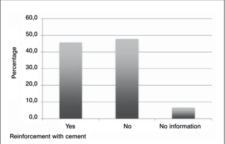
Respecting the technique used in osteoporotic fractures, 47.8% perform vertebroplasty, 47.8% use kyphoplasty, 25.8% use
arthrod-esis with a posterior approach, 3.3% use the cage, and 6% did not answer. Regarding reinforcement with cement of posterior arthrod-esis with pedicle screws in osteoporotic fractures, 83 (45.6%) use it, 87 (47.8%) do not, and 12 (6.6%) did not answer. (Figure 13) Navigation in spinal surgery is used by 22%, 75.3% do not use it, and 2.7% did not answer.
Figure 10. Method of treatment of cervical dislocations.
Figure 11. Use of classifications for tumors.
Figure 12. Types of graft in spine arthrodesis.
Figure 13. Reinforcement with cement in osteoporotic fractures.
DISCUSSION
The research results should be analyzed considering the sample size and methodology. Thus we can define the profile of the profes-sional who operates in spine surgery in Brazil.
Regarding the profile of the surgeon and their regionalization of performance, there are no differences between specialties, but we note that the largest number of surgeons operate respectively in the Southeast and South regions of Brazil, probably because they are the regions with the highest physician to inhabitant ratio.19
Concerning the age of the surgeons, the number of young spine surgeons is proportionally significantly higher among orthopedists, contrary to what is observed in the group of neurosurgeons. In ad-dition, interest in the medical and scientific improvement is higher among younger spine surgery professionals, seeing that they sub-scribe to and produce more periodicals and papers.
Due to the Brazilian health policy and its recent history,20 with
diminishing incentives to the public service provider and the public service sector with low wages, poor working conditions, and ex-cessive increase in the number of health insurance plans, we note that despite spine surgeons in Brazil working in the SUS, in private clinics and through health insurance plans, the largest percentage of surgeries are performed through health insurance plans.
There are no previous data on what types of surgeries are per-formed by orthopedists and neurosurgeons. Due to the history of development of spinal surgery for deformities, orthopedists still per-form this procedure more often than neurosurgeons, considering that the vast majority of publications on this subject are conducted by orthopedists.5-7,21-29
Several studies21,22 on the reproducibility of the Lenke and King
classifications of deformities have been performed, highlighting the complexity and lack of awareness of the Lenke classification. The King classification has good reproducibility, but is limited because it only evaluates the coronal plane. In our study, the Lenke classifica-tion is more often used for deformities, almost in the same proporclassifica-tion of those who use the two classifications, King and Lenke, with no difference among surgeons. Moreover, although there is no consen-sus on the best instrumentation and correction technique,23,24 the
technique with posterior instrumentation and suture loop fixation25 is
used only among orthopedists, perhaps because deformities have been treated for many decades by these surgeons.
The use of traction in deformities is not routine in the surgeon’s
P
ercentage
Reinforcement with cement
Yes No No information 60,0
50,0
40,0
30,0
20,0
10,0
0,0
P
ercentage
Treatment 45,0 40,0 35,0 30,0 25,0 20,0 15,0 10,0 5,0 0,0
A alone without... A + P without traction
P + traction A + traction
A + P + traction P alone without...
0,0 10,0 20,0 30,0 40,0 50,0 60,0 70,0 Does not use
Other
Tomita
Enneking
Orthopedist
Neurosurgeon
P
ercentage
Type of graft
Graft...
BMP + graft Bone bank Does not use
BMP
100,0 90,0 80,0 70,0 60,0 50,0 40,0 30,0 20,0 10,0 0,0
daily practice, but in more severe deformities, it becomes necessary.26
Although there is no consensus in the literature regarding the best surgical approach due to the variety of deformities,27,28 most
surgeons use the anterior and posterior approach in two surgical times, or the posterior approach with use of osteotomies.
Puertas et al.29 reported results that suggest that intraoperative
monitoring with somatosensory evoked potential is effective in pre-venting neurological injury in corrective surgery for rigid curves in idiopathic scoliosis. In those times, motor potential was not used. In our study, most orthopedists use sensory and motor evoked potential for intraoperative monitoring of deformities, except in neu-romuscular scoliosis.
The role of intraoperative monitoring in spinal tumors has not been well defined,30,31 but most Brazilians neurosurgeons use it in
their surgeries. The routine use of monitoring is still a challenge as regards the availability and heterogeneous structure of our country. It is a resource that is not available throughout the country due to the small number of trained professionals and its high cost, and so it is not so used by a large portion of physicians.
The request for magnetic resonance imaging has not yet reached a consensus among surgeons. Unlike neurosurgeons, orthopedists tend not to request such examination for typical curves, although there is evidence of up to 10% intra-channel changes in idiopathic scoliosis with typical curves.32 Freitas et al.33 published a study
showing the presence of 14.5% of syringomyelia on MRI in patients with scoliosis that was considered to be idiopathic.
Several studies34,35 were conducted to define the
reproduci-bility of cervical and thoracolumbar fracture classifications. Most neurosurgeons use only the AO method, while orthopedists tend to complement it with other classifications such as the Denis, Harms, SLICS, TLICS, and especially Allen-Ferguson.
Some authors36 have suggested that with the exception of
pa-tients with a neurologic deficit, MRI is not useful for finding unstable injuries in the spines of patients that are conscious or already have a normal tomography. However, there are data in the literature37,38
reporting additional injuries that radiographs and CT scans are not able to demonstrate, so most surgeons choose to request MRI in the presence of a cervical dislocation without neurological deficit.
There is a wide variety of opinions regarding the approach and use of traction for the treatment of cervical trauma,39 but surgeons
prefer management with the use of the anterior approach alone without traction, probably because it is the simplest approach.
A trend towards the use of the Tomita and Enneking classifi-cations for vertebral tumors by orthopedists and only the Tomita classification by neurosurgeons was observed. Though there is not much evidence40 for comparing the reproducibility of spinal tumor
classifications, perhaps it will be possible to obtain a more reliable and reproducible classification with more comparative studies.
One of the goals of the spine surgeon is to be able to perform good spinal fusion. There are several studies41,42 comparing the
efficacy of grafts, but because of their osteogenic, osteoinductive, and osteoconductive properties, the graft is still the best option and the preferred choice of the Brazilian surgeon.
Vertebroplasty and kyphoplasty are safe43 techniques that are
widely used by national surgeons in osteoporotic fractures. Our study showed that the use of cementation in the posterior approach as reinforcement for instrumentation44 is still not preferred by spine
surgeons, but may be an option for avoiding an anterior approach. Neuronavigation is not yet widespread in our environment due to its high cost and low availability, though it can assist in the surgical planning of difficult to access tumors, as has been demonstrated by some authors.45
CONCLUSION
The spine surgery is a complex specialty that treats a variety of diseases, so it is important to know the specifics among Brazilian spine surgeons independently from their specialty.
This study shows the profile of the professionals who work in spine surgery in Brazil with the aim of serving as a source of data for the development of health programs and performance improve-ments in this field of work in Brazil.
All authors declare no potential conflict of interest concerning this article.
REFERENCES
1. Schoenfeld AJ, Sielski B, Rivera KP. Epidemiology of cervical spine fractures in the US military. 2012;12(9):777-83.
2. Li J, Liu G, Zheng Y. The epidemiological survey of acute traumatic spinal cord injury (ATSCI) of 2002 in Beijing municipality. Spinal Cord. 2011;49(7):777-82.
3. Yousefzadeh CS, Safaee M. Epidemiology of traumatic spinal injury: a descriptive study. Acta Med Iran. 2010;48(5):308-11.
4. Defino HL, Herrero CF, Zardo EA. O que eu não faço mais na cirurgia da coluna vertebral: pesquisa entre cirurgiões de coluna brasileiros. Coluna/Columna. 2011;10(4):336-42. 5. Lenke LG, Betz RR, Harms J. Adolescent idiopathic scoliosis: a new classification to
determine extent of spinal arthrodesis. J Bone Joint Surg Am. 2001;83(8):1169-81. 6. Moe JH. Methods and techniques in scoliosis. In: AAOS. Symposium on the spine. St.
Louis: CV Mosby; 1969. p. 169-240.
7. King HA, Moe JH, Bradford DS, Winter RB. The selection of fusion levels in thoracic idiopathic scoliosis. J Bone Joint Surg Am. 1983;65(9):1302-13.
8. Magerl F, Aebi M, Gertzbein SD, Harms J, Nazarian S. A comprehensive classification of thoracic and lumbar injuries. Eur Spine J. 1994;3(4):184-201.
9. Aebi M, Nazarian S. Klassifikation der Halswirbelsäulenverletzungen. Orthopäde. 1987;16(1):27-36.
10. Blauth M, Kathrein A, Mair G. Classification of injuries of the lower cervical spine. In: Aebi M, Arlet V, Webb JK, editors. AO Spine manual: clinical applications. New York: Thieme; 2007. p. 21-38.
11. Louis R. Les theories de l’estabilité. Rev Chir Orthop Reparatrice Appar Mot. 1977;63(5):423-5.
12. Denis F. The three column spine and its significance in the classification of acute thoraco-lumbar spine injuries. Spine (Phila Pa 1976). 1983;8(8):817-31.
13. Harms J. [Classification of fractures of the thoracic and lumbar vertebrae]. Fortschr Med. 1987;105(28):545-8.
14. Bono CM, Vaccaro AR, Hurlbert RJ. Validating a newly proposed classification system for thoracolumbar spine trauma: looking to the future of the thoracolumbar injury classifica-tion and severity score. J Orthop Trauma. 2006;20(8):567-72.
15. Allen BL, Fergunson RL, Lehmann R. A mechanistic classification of closed, indirect frac-tures and dislocations of the lower cervical-spine. Spine (Phila Pa 1976). 1982;7(1):1-27. 16. Enneking WF. Modification of the system for functional evaluation of surgical
manage-ment of musculoskeletal tumors. In: Enneking WF, editor. Limb salvage in musculoskele-tal oncology. New York: Churchill Livingstone;1987. p.626-39.
17. Crockard A, Bunger C, Harms J, Kawahara N. Review of metastatic spine tumour classifi-cation and indiclassifi-cations for surgery: the consensus statement of the Global Spine Tumour Study Group. Eur Spine J. 2010;19(2):215-22.
18. Yasuaki H, Matsuzaki H, Oda H.Health Services Research A Revised Scoring System for Preoperative Evaluation of Metastatic Spine Tumor PrognosisTokuhashi, Spine: 2005; 30(19):2186-191.
19. Ministério da Saúde/CGRH e Conselhos Profissionais. Bases para cálculo da população: IBGE, Censos 1991 e 2000; Contagem da População, 1996. Disponível em: http://tabnet. datasus.gov.br/cgi/idb2000/fqe01.htm
20. Fleury S. Saúde e democracia – A Luta do CEBES. São Paulo: Lemos; 1997.
21. Richards BS, Sucato DJ. Comparison of reliability between the Lenke and King clas-sification systems for adolescent idiopathic scoliosis using radiographs that were not premeasured. Spine (Phila Pa 1976). 2003;28(11):1148-56;
22. Vialle EN, Vialle LR, Martins Filho DE, Jorge RM. Confiabilidade de uma versão em por-tuguês da classificação de Lenke para Escoliose Idiopática do Adolescente. Coluna/Co-lumna. 2006;5(2):77-83.
23. Delorme S, LabelleH, Aubin CE, de Guise JA, Rivard CH, Poitras B, et al. Intraoperative comparison of two instrumentation techniques for the correction of adolescent idiopathic scoliosis: rod rotation and translation. Spine (Phila Pa 1976). 1999;24(19):2011-7. 24. Puertas EB, Chagas JC, Oliveira CA. Complicaçöes neurológicas no tratamento da
es-coliose idiopática com instrumentaçäo e artrodese por via posterior. Acta Ortop Bras. 1998;6(4):168-72.
26. Mehlman CT, Al-Sayyad MJ, Crawford AH. Effectiveness of spinal release and halo--femoral traction in the management of severe spinal deformity. J Pediatr Orthop. 2004;24(6):667-73
27. Hansen MM, Nicholas DA, Allen BL Jr. Same-day versus staged anterior-posterior spinal surgery in a neuromuscular scoliosis population: the evaluation of medical complications. J Pediatr Orthop. 1996;16(3):293-303.
28. Modi HN, Hong JY, Mehta SS, Srinivasalu S, Suh SW, Yi JW, et al. Surgical correction and fusion using posterior-only pedicle screw construct for neuropathic scoliosis in patients with cerebral palsy: a three-year follow-up study. Spine (Phila Pa 1976). 2009;34(11):1167-75. 29. Puertas EB, WajchenbergM, Ferreira R, Scamardi FF, Trandafilov Júnior M. Comparação
entre o teste de despertar e a monitoração neurofisiológica intra-operatória com potencial evocado somato-sensitivo nas cirurgias de escoliose.Coluna/Columna. 2009;8(1):7-12. 30. Kothbauer KF. Neurosurgical management of intramedullary spinal cord tumors in
chil-dren. Pediatr Neurosurg. 2007;43(3):222-35.
31. Delitis V, Sala F. ‘Intraoperative neurophysiological monitoring of the spinal cord during spinal cord and spine surgery: a review focus on the corticospinal tracts’, Clin Neuro-physiol. 2008;119(2):248-64.
32. Diab M, Landman Z, Lubicky J, Dormans J, Erickson M, Richards BS. Use and outcome of MRI in the surgical treatment of adolescent idiopathic scoliosis. Spine (Phila Pa 1976). 2011;36(8):667-71
33. Freitas AA, Puertas EB, Chagas JCM. Estudo da ressonância nuclear magnética mos-trando a presença de siringomielia em pacientes portadores de escoliose considerada idiopática. Acta Ortop Bras. 1998;6(3):102-8.
34. Bazan PL, Borri AE, Torres PU, Cosentino JS, Games MH. Clasificación de las fracturas toracolumbares: comparación entre las clasificaciones de AO y Vaccaro. Coluna/Columna. 2010;9(2):165-70.
35. Koh YD, Kim DJ, Koh YW. Reliability and Validity of Thoracolumbar Injury Classification and Severity Score (TLICS). Asian Spine J. 2010;4(2):109-17.
36. Tomycz ND, Chew BG, Chang YF, Darby JM, Gunn SR, Nicholas DH, et al. MRI is
unne-cessary to clear the cervical spine in obtunded/comatose trauma patients: the four-year experience of a level I trauma center. J Trauma. 2008;64(5):1258-63.
37. Vaccaro AR, Falatyn SP, Flanders AE, Balderston RA, Northrup BE, Cotler JM. Magnetic resonance evaluation of the intervertebral disc, spinal ligaments, and spinal cord before and after closed traction reduction of cervical spine dislocations. Spine (Phila Pa 1976). 1999;24(12):1210-7.
38. Schellinger PD, Schwab S, Krieger D, Fiebach JB, Steiner T, Hund EF, et al. Masking of vertebral artery dissection by severe trauma to the cervical spine. Spine (Phila Pa 1976). 2001;26(3):314-9.
39. Glaser JA, Jaworski BA, Cuddy BG, Albert TJ, Hollowell JP, McLain RF, et al. Variation in surgical opinion regarding management of selected cervical spine injuries. A preliminary study. Spine (Phila Pa 1976). 1998;23(9):975-82.
40. Chan P, Boriani S, Fourney DR, Biagini R, Dekutoski MB, Fehlings MG, et al. An assess-ment of the reliability of the Enneking and Weinstein-Boriani-Biagini classifications for staging of primary spinal tumors by the Spine Oncology Study Group. Spine (Phila Pa 1976). 2009;34(4):384-91.
41. Gibson S, McLeod I, Wardlaw D, Urbaniak S. Allograft versus autograft in instrumented posterolateral lumbar spinal fusion: a randomized control trial. Spine (Phila Pa 1976). 2002;27(15):1599-603.
42. Buttermann GR, Glazer PA, Hu SS, Bradford DS. Revision of failed lumbar fusions. A comparison of anterior autograft and allograft. Spine (Phila Pa 1976). 1997;22(23):2748-55. 43. Garfin SR, Yuan HA, Reiley MA. New technologies in spine: kyphoplasty and vertebro-plasty for the treatment of painful osteoporotic compression fractures. Spine (Phila Pa 1976). 2001;26(14):1511-5.
44. Mermelstein LE, McLain RF, Yerby SA. Reinforcement of thoracolumbar burst frac-tures with calcium phosphate cement. A biomechanical study. Spine (Phila Pa 1976). 1998;23(6):664-70.