ISSN 1549-3652
© 2010 Science Publications
Corresponding Author: Christopher Kovacs, Department of Kinesiology, Western Illinois University, 1 University Circle, Macomb, IL 61455 Tel: 309-298-1524 Fax: 309-298-2981
Effects of Increased Physiological Arousal on Upper Extremity
Positional Awareness in Healthy Young Adults
Christopher Kovacs and Tamara Bories
Department of Kinesiology, Western Illinois University,
1 University Circle, Macomb, IL 61455
Abstract: Problem statement: The purpose of this investigation was to examine the effects of increased physiological arousal on the ability to perceive upper extremity positional awareness in healthy young adults. Approach: Thirty-eight participants were pre- and post-tested for upper extremity positional awareness using a manual kinesthesiometer. Participants in the experimental group underwent a combination of the Stroop color-word task and timed arithmetic problems to produce a state of physiological arousal. Heart rate and blood pressure measurements were taken during data collection to assess levels of physiological arousal. Pre-and post-test absolute error scores for each participant were compared. Results: ANCOVA revealed a significant time effect (p<0.046) between pre and post-test trials for the experimental group. Conclusion: The results suggested positional awareness is altered under a state of elevated physiological arousal and that these results may have significant implications for individuals performing various types of motor skills.
Key words: Proprioception, arousal, upper extremity, human movement
INTRODUCTION
The ability of an individual to successfully and efficiently perform motor tasks is dependent upon the capacity of that individual to understand where body limbs are in relation to the environmental surroundings. This proprioceptive ability relies on the various structures within the body that transmit sensory-related information to the central nervous system, which is then utilized by the brain to produce effective movements. Proprioception is typically defined as our body’s ability to judge the position of our extremities in relation to our surrounding environment (Carello and Turvey, 2004; Helmuth, 2000; Park et al., 1999; Worringham and Kerr, 2000). Knowledge of body position during motor activities relies on multiple sensory inputs, both visual and non-visual. Collectively, visual cues, touch sensations and proprioceptive measures are used to determine body position during movement and help provide an individual with a sense of body awareness (Helmuth, 2000).
The information brought into the muscle tissue via the proprioceptive receptors is necessary to detect kinesthesia, joint motion, position sense and orientation of the body in space. The ability to efficiently obtain information through the proprioceptive receptors prevents inaccuracy in movement, which could lead to
overshooting or undershooting an obstacle in gait, over steering while driving, or over-rotating while throwing a ball, all of which could cause an accident or injury.
Proprioception is important for controlling muscular activities, which require sequential firing within the muscle. Without proprioceptive feedback, the location and control of a limb in motion will be harder to perceive and organize (Park et al., 1999). Coordination, characterized by smooth, rhythmic muscle activity, depends upon directional guidance between visual and proprioceptive sensory-motor systems (Redding and Wallace, 1992). Balance control and efficient gait also depends upon the sequential firing of motor units, joint motion and position sense (Swanik et al., 2004).
alterations in the central nervous system that can affect the accurate performance of motor skills.
The autonomic responses to stress brought on by behavioral or cognitive processes are associated with an increase in both diastolic and systolic blood pressure (Cramer, 2003). This increase in blood pressure is due to alterations within the parasympathetic and sympathetic systems which modulate heart rate. Additionally, cognitive activity has been shown (Cramer, 2003) to be related to an elevation in diastolic blood pressure. Since physiological arousal can be evidenced by examining blood pressure responses, systolic and diastolic responses are used as an indicator of a state of arousal.
Arousal can arise from different situations and produce distinct effects on the body. Test anxiety, for example, is a type of physiological arousal that can arise from evaluative stress. Calvo and Alamo (1987) investigated how test anxiety affects motor performance. Their hypothesis suggested test anxiety would affect fine motor control, but not gross motor control. Their results indicated that the observed decreases in finely controlled motor performances under high anxiety conditions were attributed to the altered attentional needs and neuromuscular requirements for a task under these stressful conditions. Interestingly, the gross-motor tasks did not suffer as greatly as fine motor tasks in their investigation, suggesting interference mechanisms are of muscular and attentional nature.
The issue of physiological arousal and how it affects the ability to accurately sense body position in space has not been investigated directly. Researchers have examined how arousal affects other areas of life and performance of specific functional skills, such as gait (Brown et al., 2002), but not proprioception, or positional awareness, specifically. The purpose of this investigation was to examine the effect of an increased state of physiological arousal, resulting from a stressor protocol, on the ability of healthy young adults to accurately perceive the location of an upper extremity in space.
MATERIALS AND METHODS
Participants: Following Institutional Review Board clearance, recruitment of participants was initiated from the student population and local community in a mid-sized university and community in the Southeast region of the United States. The mean age of the participants was 22.3 years (20-33 years of age). All participants self-reported being free of diagnosed vision problems, neurological disorders and joint problems that would
prohibit their participation in this investigation. Additionally, no participants reported experiencing a major negative life event within the last month, which would indicate increased levels of state anxiety and arousal levels.
Thirty-eight participants were recruited for this investigation. Following the explanation of procedures and a participation and health history questionnaire, each participant was randomly assigned to the experimental (N = 19) or control group (N = 19). Thirty-seven participants were right handed while one participant was left-handed. All participants volunteered to participate in the study and received no compensation for their involvement.
Experimental procedures: Upon completion of all required paperwork, an automated blood pressure cuff (Colin STBP-780 Automated BP system, Colin Corporation, Japan) was placed on the upper segment of the non-dominant arm of the subject. Two electrodes were placed inferior to the clavicle, one on the right and one on the left side of the body. The remaining two electrodes were placed inferior to the rib cage, one on the right side of the body and one on the left side of the body. The electrodes were used to monitor the heart rate response during data collection procedures. Once the automated BP cuff was fitted to the participant, the participant was given a 10 min relaxation period prior to taking the first resting blood pressure.
Following pre-test data collection of HR and BP, the participant was placed in front of an angular kinesthesiometer (16014 Kinesthesiometer, Lafayette Instrument Company, West Lafayette, Indiana). The kinesthesiometer measures angular displacement along a 90° scale. Each participant was instructed to place their dominant arm onto the saddle of the kinesthesiometer perpendicular to the participant’s torso. Instructions were given to place the elbow on the black foam pad at the base of the saddle with the hand palm down and fingers falling comfortably over the finger guide pin at the opposite end of the saddle. The upper limb of the subject was positioned at zero degrees, or neutral, according to the scale on the kinesthesiometer.
angle that was called out by the investigator while under these visually blocked conditions. All angular displacements were measured between the neutral position (zero degrees) and 90°.
The desired angles to which each participant was asked to move in the pre-test were standardized for all individuals. The sequence of the angles was randomly selected to prevent any practice effect from occurring. The order of angular displacement for the pre-test was: 75, 15, 60, 45, 90 and 30 respectively. The angle to which the subject moved was recorded as the “actual” angle. During all testing conditions, participants were required to rely solely on upper extremity position sense with no visual feedback being available to them. Each participant performed five total trials at 75, 15, 60, 45, 90 and 30° within 60 sec of completing the stressor protocol.
Stressor protocol: Each participant in the experimental group, following the pre-test angular displacement, was directed to a laboratory personal computer used for the stressor protocol. The Stroop Color Word task and arithmetic calculations were used as a psychological stress or to increase physiological arousal levels in each participant. During the Stroop color-word task the subject was instructed to key in the font color of the colored word printed on the computer screen using the keypad. On the keypad, the numbers 2, 4, 6 and 8 were labeled with colored stickers corresponding to the color choices red, blue, green and yellow. The subject was warned the word will read one color, be printed in another color and another color will be suggested audibly by the computer, all representations of color being incongruent. However, only the color of the font was to be entered into the computer. The arithmetic stressor involved subtracting a one or two digit number from a three-digit number as quickly and accurately as possible (Example: 997-38 = Desired Numerical Answer). The problems were timed and the computer would only allow one second for each response before moving to the next math problem. The answer to the arithmetic problem had to be entered using the keypad and pressing enter; no backspacing was allowed. The entire stressor requires 8 min for completion. Throughout the 8 min stressor, external prompting from the researchers instructing the subject to respond quickly and accurately to the Stroop color-word tasks and the simple arithmetic problems was used to maximize arousal levels. Each section of the Stroop color-word task lasted 2 min, followed by 2 min of the simple arithmetic problems. This 4 min protocol was repeated once, for a total of 8 min. of stressor presentation.
During the 4 min, following the first set of simple arithmetic problems, heart rate and blood pressure were recorded. At this point during the stressor protocol the body’s reaction to the stressor was best visible in the heart rate and blood pressure readings. Following the completion of the 8 min. stressor, each participant had their vision occluded and seated at the kinesthesiometer for post-test measurements of angular displacement using the previously described procedures. Immediately following post-test positional measures, the final heart rate and blood pressure measures were recorded. Final heart rate and blood pressure responses were recorded immediately following post-testing in an effort to minimize changes in HR and BP that might occur if these measurements were recorded prior to post-testing procedures.
Statistical analysis: The mean scores for the pre-test desired angles and the post-test desired angles were calculated. The mean score of the desired angles for both the pre-test and post-test was 52.5°; the same six angles were used in both the pre-test and post-test in a pre-determined sequence. The angles recorded on each attempt by the subject, recorded as the “actual” angle, were also added and averaged within each test, pre-test and post-test.
The absolute error scores, the difference between the acquired mean scores for the angular displacement for each subject and the mean score for the desired angles, were then recorded for each individual trial. Absolute error scores were selected as a measure of motor performance, as the study was designed to assess magnitude of angular displacement only. Direction of error was not considered an important variable to examine for the purposes of this study. The absolute error scores were then entered into Microsoft Excel for data reduction and analyzed using SPSS (Version 13.0). The heart rate and blood pressure recordings were also evaluated to illustrate a state of physiological arousal. The average heart rate and systolic and diastolic blood pressures, measures of physiological arousal, were recorded for the pre, mid and post tests for each participant and analyzed using SPSS software.
RESULTS
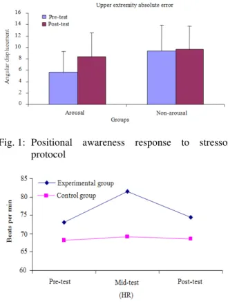
post measures in the arousal group when compared to the non-arousal group (Fig. 1). The pre-test and post-test absolute error score averages were graphed against each other for comparison (Fig. 1). The results demonstrate a statistically significant decrease in positional awareness accuracy for those individuals under increased conditions of physiological arousal. The recorded values in positional awareness revealed a +/- 2.7° change in the arousal group Vs a +/- 0.5° change in the non-arousal group.
Heart rate: Theheart rate responses indicate a state of physiological arousal. The results obtained during the testing (Fig. 2) suggest a relationship was present. Prior to data collection the heart rate was 72±13 beats per min. The mid-test heart rate was elevated to 81±13 beats per min and then decreased during post-test evaluation to 74±14 beats per min. A one-way repeated measure ANOVA was calculated comparing pre and mid-test, mid and post-test and pre and post-test. A significant difference was found (F = 22.19, p<0.017). Post hoc analysis indicated heart rate scores increased significantly from pre-test to mid-test and decreased significantly from mid-test to post-test.
Systolic blood pressure: Systolic Blood Pressure (BP) is also an indicator of physiological arousal. The systolic blood pressure means were observed in three stages of data collection: The pre, mid and post-test. The obtained means were 132±16, 135±10 and 132±10 mmHg, respectively, for the pre, mid and post test blood pressure measurements. The blood pressure response illustrates an increase in HR during the mid-test recording (Fig. 3). However, a one-way repeated measures ANOVA, indicated that there was no significant difference between pre-test, mid-test and post-test (F = 1.85, p<0.017) measures.
Diastolic blood pressure: Three diastolic blood pressure measurements were also recorded during the research sessions. A resting, mid and a post-stressor blood pressure were all recorded. Pre, mid and post stressor diastolic blood pressure means were 78±9, 88±13 and 80±7 mmHg, respectively. The blood pressure means (Fig. 3) illustrate a spike in diastolic blood pressure during the stressor protocol. The one-way repeated measures ANOVA was also used to compare the means in blood pressure at the pre-test, mid-test and post-test intervals. A significant difference was found (F = 14.26, p<0.017). Post hoc analysis revealed a significant difference between the pre-test and mid-test blood pressure means and the mid-test and post-test blood pressures.
Fig. 1: Positional awareness response to stressor protocol
Fig. 2: Heart rate response to stressor protocol
Fig. 3: Blood pressure response to stressor protocol
DISCUSSION
results of this exploratory investigation supported this hypothesis. The results revealed a significant difference between the position sense pre-test absolute error scores and the post-test absolute error scores. The increase in error scores following the stressor protocol suggests physiological arousal states do alter position sense perception in healthy young adults.
It was hypothesized heart rate and blood pressure measures would demonstrate a state of physiological arousal during the stressor protocol and the arousal state would alter upper extremity perception. The results of the study revealed a statistically significant increase in three of the four measures examined in the study (absolute error scores, diastolic blood pressure and heart rate). Heart rates increased during the stressor protocol as did both systolic and diastolic blood pressure. The absolute error also increased due to the stressor protocol. The increase in heart rate and diastolic blood pressure were both statistically significant and supports Cramer (2003), who suggested cognitive activities increase physiological arousal and more specifically, diastolic blood pressure response. The stressor utilized in this investigation used a stressor protocol comprised of cognitive activities, identifying the font color of a word and evaluating arithmetic under limited time constraints. These cognitive activities may be associated with the observed significant increase in diastolic blood pressure.
The physiological arousal response is due to an alteration in the central nervous system (Cramer, 2003). The change is, however, mostly due to changes within the autonomic system, which affects the cardiac muscle, smooth muscle and endocrine glands. The somatic system, affecting the efferent control of the skeletal system is not directly affected. The autonomic response to arousal is an increase in both systolic and diastolic blood pressure. The increase in blood pressure is due to alterations within the parasympathetic and the sympathetic systems, which modulate the heart.
The increased sympathetic activity is associated with preparing the body for strenuous Physical Activity (PA) or stressful situations. As PA is initiated, the desire to increase the flow of oxygenated blood to the area involved in PA is created. In response to the increased desire for more oxygen, the heart rate increases, the blood vessels in the area constrict increasing blood pressure and some systems, such as the digestive system and urinary system, experience minimal activity. This preparation for PA is commonly referred to as the fight-or-flight response. During the fight-or-flight response, norepinephrine and adrenaline are released, which reinforce the sympathetic response. The increase in norepinephrine and adrenaline and the
effects of these neurotransmitters on the body are indicators of physiological arousal. The increase in blood pressure and heart rate the experimental group experienced characterizes a state of physiological arousal due to a response of the autonomic nervous system, specifically the sympathetic nervous system.
Based on acquired knowledge of the effects physiological arousal has on the autonomic system, it can be suggested that the position sense response is indirectly due to alterations in the sympathetic nervous system. During an aroused state, the body undergoes numerous physical changes regulated by the sympathetic nervous system: altering heart rate, constricting blood vessels, redirecting blood flow and controlling the operation of body systems. As suggested by Calvo and Alamo (1987), as more stimuli are present the motor response to stimuli becomes more variable and less accurate. This decrease in motor response may lead to inaccuracies in positional awareness and inefficient movement patterns under these aroused conditions.
CONCLUSION
In this investigation the increase in positional inaccuracy supports the hypothesis proposed by Calvo and Alamo (1987). The body is responding to the stressor, the Stroop color-word task and the simple math problems, by activating a response similar to the fight-or-flight response. As this response is initiated, attention is diverted away from the sensory information being received from the muscle spindles, joint receptors and Golgi tendon organs and is instead accommodating for a stressful situation. With less attention on processing sensory information, the body cannot perceive upper extremity position as accurately. Although the attention designated to position perception decreases, the need for attention is stable and inaccuracy in movement results.
diseased. The vast population of older adults could benefit from an investigation distinguishing how proprioceptive ability is decreased in an effort to develop interventions and programming to assist with those changes in motor function that are seen with aging. Through the use of designed interventions to help individuals move more efficiently under stressful conditions, improvements in position sense may occur, or losses in position sense may be prevented, in any individual in which movement accuracy under stressful conditions is critical.
ACKNOWLEDGEMENT
The researchers would like to thank Melissa Parker for her assistance in the data collection and manuscript preparation of this article. Ms. Parker is currently a student in the department of Physical Therapy at the University of St. Augustine.
REFERENCES
Brown, L.A., W.H. Gage, M.A. Polych, R.J. Sleik and T.R. Winder, 2002. Central set influences on gait: Age-dependent effects of postural threat. Exp. Brain Res., 145: 286-296. DOI: 10.1007/s00221-002-1082-0
Calvo, M.G. and L. Alamo, 1987. Test anxiety and motor performance: The role of muscular and attentional demands. Int. J. Psychol., 22: 165-178. DOI: 10.1080/00207598708246775
Carello, C. and M.T. Turvey, 2004. Physics and psychology of the muscle sense. Curr. Direct. Psychol. Sci., 13: 25-28. DOI: 10.1111/j.0963-7214.2004.01301007.x
Cramer, P., 2003. Defense mechanisms and physiological reactivity to stress. J. Pers., 71: 221-244. PMID: 12693516
Helmuth, L., 2000. Neuroscience: Where the brain monitors the body. Science, 290: 1668a-1668a. PMID: 17798201
Kemeny, M.E., 2003. The psychobiology of stress. Curr. Direct. Psychol. Sci., 12: 124-129. DOI: 10.1111/1467-8721.01246
Park, S., T. Toole and S. Lee, 1999. Functional roles of the proprioceptive system in the control of goal-directed movement. Percept. Motor Skills, 88: 631-647. PMID: 10483656
Redding, G.M. and B. Wallace, 1992. Effects of pointing rate and availability of visual feedback on visual and proprioceptive components of prism adaptation. J. Motor Behav., 24: 226-237. PMID: 12736128
Swanik, C.B., Lephart, S.M. and H.E. Rubash, 2004. Proprioception, Kinesthesia and balance after total knee arthroplasty with cruciate-retaining and posterior stabilized prosthesis. J. Bone Joint Surg., 86: 328-334. PMID: 14960678
Worringham, C.J. and G.K. Kerr, 2000. Proprioception and stimulus-response compatibility. Q. J. Exp.
Psychol., 53: 69-83. DOI: