pneumoniae
Topoisomerase IV: Crystal Structure of
a Gram-Positive Quinolone Target
Ivan Laponogov1., Dennis A. Veselkov1., Maninder K. Sohi1., Xiao-Su Pan2., Aniruddha Achari1¤, Cheng Yang3, Joseph D. Ferrara3, L. Mark Fisher2*, Mark R. Sanderson1*
1Randall Division of Cell and Molecular Biophysics, School of Biomedical and Health Sciences, King’s College London, London, United Kindgom, 2Molecular Genetics Group, Molecular and Metabolic Signalling Centre, Division of Basic Medical Sciences, St. George’s, University of London, London, United Kindgom,3Rigaku Americas Corporation, The Woodlands, Texas, United States of America
The 2.7 A˚ crystal structure of the 55-kDa N-terminal breakage-reunion domain of topoisomerase (topo) IV subunit A (ParC)
from Streptococcus pneumoniae, the first for the quinolone targets from a gram-positive bacterium, has been solved and
reveals a ‘closed’ dimer similar in fold toEscherichia coli DNA gyrase subunit A (GyrA), but distinct from the ‘open’ gate structure ofEscherichia coliParC. Unlike GyrA whose DNA binding groove is largely positively charged, the DNA binding site of ParC exhibits a distinct pattern of alternating positively and negatively charged regions coincident with the predicted positions of the grooves and phosphate backbone of DNA. Based on the ParC structure, a new induced-fit model for sequence-specific recognition of the gate (G) segment by ParC has been proposed. These features may account for the unique DNA recognition and quinolone targeting properties of pneumococcal type II topoisomerases compared to their gram-negative counterparts.
Citation: Laponogov I, Veselkov DA, Sohi MK, Pan X, Achari A, et al (2007) Breakage-Reunion Domain ofStreptococcus pneumoniaeTopoisomerase IV: Crystal Structure of a Gram-Positive Quinolone Target. PLoS ONE 2(3): e301. doi:10.1371/journal.pone.0000301
INTRODUCTION
DNA topoisomerases are enzymes of vital cellular importance due to their ability to alter DNA topology. Replication of duplex DNA or recombination between DNA duplexes may cause the forma-tion of DNA supercoils, knots and catenanes. The major role of topoisomerases is to control DNA topology by transient breakage of DNA strands. This activity is achieved through the formation of covalent enzyme-DNA complexes linked via phosphotyrosine bonds. Type I topoisomerases act on one strand of DNA whereas type II enzymes pass a DNA duplex through a double-strand DNA break, resulting in changes of DNA linking number in units of 1 and 2, respectively [1–3].
Type II topoisomerases comprise a family of structurally and evolutionarily conserved enzymes common to all organisms [4] and have been implicated in a variety of intracellular processes including DNA replication, transcription and chromosome segregation [5]. Because of their importance to the cell life cycle, these enzymes have been a subject of intense drug development studies and were identified as targets for a large number of natural toxins, antimicrobial agents and anti-tumour therapeutics [6–8].
Based on sequence and structural criteria, type II topoisome-rases are divided into two subclasses. The most common subclass, type IIA, includes the bacterial enzymes topo IV and gyrase, and eukaryotic topo II. Type IIB topos are structurally and biochemically distinct, and comprise a single family member, topo VI [2,9–11]. All members of the type IIA subclass exhibit significant sequence and mechanistic similarity.
Bacterial genomes usually encode two type IIA enzymes, topo IV and DNA gyrase, each composed of two subunits (ParC and ParE in topo IV, GyrA and GyrB in gyrase) assembled into a functional heterotetrameri.e.C2E2 and A2B2, respectively [6]. Both enzymes act by passing a duplex DNA segment through a transient double-stranded DNA break in another helical region. In the currently accepted model [9], the open enzyme clamp binds a DNA segment termed the ‘gate’ or G-segment. The N-terminal ATPase domains of the ParE (GyrB) subunits dimerize upon ATP
binding capturing the DNA duplex to be transported (T-segment). The T-segment is then passed through a transient break in the G-segment (opened by the N-terminal ParC (GyrA) domains), the DNA is resealed and the T-segment released through a protein gate prior to resetting of the enzyme to the open clamp form.
Although topo IV and gyrase share a high degree of similarity, their cellular functions are different. Gyrase controls DNA super-coiling and relieves topological stress arising from the translocation of transcription and replication complexes along DNA. Gyrase is unique in its ability to introduce negative supercoiling into the DNA. By contrast, topo IV is a decatenating enzyme that resolves interlinked daughter chromosomes following DNA replication. The functional differences between gyrase and topo IV are attri-buted to the non-specific binding of the C-terminal domain of the
Academic Editor:Haiwei Song, Institute of Molecular and Cell Biology, Singapore
ReceivedJanuary 5, 2007;AcceptedFebruary 21, 2007;PublishedMarch 21, 2007 This is an open-access article distributed under the terms of the Creative Commons Public Domain declaration which stipulates that, once placed in the public domain, this work may be freely reproduced, distributed, transmitted, modified, built upon, or otherwise used by anyone for any lawful purpose.
Funding:Project Grant C16747 from the Biotechnology and Biological Sciences Research Council of the United Kingdom (to L.M. Fisher and M.R. Sanderson). I. Laponogov was supported by a scholarship from the School of Biomedical and Health Sciences and ORS Award. Xiao-Su Pan, Dennis A. Veselkov and this work were funded by Project Grant C16747 from the Biotechnology and Biological Sciences Research Council of the United Kingdom (to L.M. Fisher and M.R. Sanderson).
Competing Interests:The authors have declared that no competing interests exist.
* To whom correspondence should be addressed.E-mail: mark.sanderson@kcl. ac.uk (MS); [email protected] (LF)
.These authors contributed equally to this work.
GyrA subunit that wraps DNA presenting a neighbouring T-segment for passage through the G-gate. Removal of this domain converts gyrase into an enzyme with topo IV-like activities, i.e. ATP-dependent DNA relaxation and decatenation [12].
In addition to their unusual mechanisms, topo IV and gyrase are also targets of clinically important quinolone drugs. Quino-lones exert their bactericidal effects by interfering with DNA breakage-reunion by topo IV and gyrase leading to double-stranded DNA breaks at specific sites. Recent studies have uncovered fundamental differences in quinolone action in gram-positive vis-a`-vis gram-negative bacteria. For example, though gyrase is the universal target of quinolones in gram-negative bac-teria, topo IV is the primary (or dual) target of many quinolones in gram-positive pathogens such as Streptococcus pneumoniae, a major cause of pneumonia and other serious infections [13,14]. Second, pneumococcal topo IV is more sensitive than gyrase to quinolone inhibition in vitro, the inverse of that seen inE. coli[15]. Third, quinolones trapS. pneumoniaetopo IV (and gyrase) at DNA sites (G-segments) that are distinct in sequence from those observed forE. colitopo IV and gyrase [15]. These unique features emphasize the need to analyze the structure ofS. pneumoniaetopo IV and how it binds quinolones and DNA. Progress in these areas will be important in understanding ParC-mediated resistance to quino-lones and in developing dual targeting drugs that minimize the emergence of resistance [13,16,17].
RESULTS AND DISCUSSION
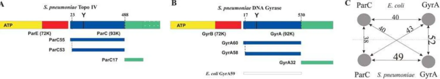
Digestion with trypsin revealed that pneumococcal ParC is organized into a 53-kDa N-terminal domain (ParC53, residues 23-487) and a compact 17-kDa C-terminal region (Figure 1A). ParC53 was enzymatically inactive whereas a recombinant ParC55 fragment (residues 1-490), when complemented with topo IV ParE, reconstituted quinolone-promoted DNA cleavage (data not shown). The DNA cleavage specificity of the ParC55-ParE complex was identical to that of pneumococcal topo IV and exhibited the same unique base recognition preferences of24G/ +8C,22A/+6T and+1G/+4C (relative to the DNA scission site between nucleotide 21 and+1). These determinants are very different from those of E. coli topo IV (preferences 21(A/G) and+1(T/A)) and E. coli gyrase (a degenerate 20-bp consensus) [15]. Sequence alignment with the N-terminal domains ofE. coli GyrA and ParC whose crystal structures have been solved [18,19] shows greater similarity ofS. pneumoniaeParC55 withE. coliGyrA andS. pneumoniaeGyrA (forms a GyrA58 (residues 17–529) tryptic fragment via GyrA60, Figure 1B) than with E. coli ParC (Figure 1C). However, these comparisons do not explain the distinctive enzyme preferences in G-segment recognition. To gain insight into the mechanism of S. pneumoniae topo IV and its
interaction with DNA and quinolones, the structure of the ParC55 fragment has been solved (Figure 2A) and the model was refined to R = 22.30% (R-free = 27.55%) at 2.7 A˚ resolution using wild-type ParC55 data (twinning fraction = 0.323, twinning operator = (k,-h,l)). The crystallization of ParC55, the quinolone target from gram-positive bacteria, and X-ray analysis were first reported by our group in Acta Cryst. (2005) A61, C176.
Crystal structure of ParC55: a closed dimer with
a distinctively charged DNA binding groove
ParC55 forms a biological dimer in a ‘closed’ conformation in this crystal form generating a ring-like structure with outer dimensions of 1006100650 A˚ and with a central hole approximately 30 A˚ in diameter, which is wide enough to pass a duplex DNA (Figure 2A). This is in contrast to the ParC structure fromE. coliwhich adopts an ‘open’ dimer conformation [18] (Figure 2B), but similar to the closed dimer of GyrA59 from E. coli [19] (Figure 2C). Each monomer of ParC55 contains two distinct regions labeled ‘head’ and ‘tail’ and exhibits an overall structural fold similar to the one observed for other topoisomerases [18–20]. The N-terminal proximal head contains the CAP-like (catabolite gene activator protein [21–23]) DNA-binding domain and the ‘tower’. The CAP-like domain is composed of a helix-turn-helix structural motif (helices a3 and a4) typical for DNA-binding proteins, with the active site Arg 117 and Tyr 118 on the neighboring ‘100-122’ loop (Figure 2A). The residues Ser 79 and Asp 83, which upon mutation give rise to quinolone-resistant pneumococci, are located primarily within the helixa4 in close proximity to the active-site tyrosine and are oriented towards the predicted position of the bound DNA. The ‘tower’ is positioned on the top of the CAP-like domain and has largelya-helical andb-sheet structural organiza-tion. High B-factor values and poor definition of several loops indicate a high level of flexibility in this region. In the closed con-formation, the ParC55 monomers form a tight contact between the a3 helices with a mean center-to-center distance of 10 A˚ resulting in the protein-protein interface with a buried surface area of 706 A˚2. This is less than in the case ofE. coliGyrA where the monomers form a protein-protein interface with a buried surface area of 1,380 A˚2. The ‘recognition’ a4 helices are further apart from each other by 5 A˚ in the case of the ParC55 dimer compared to theE. coliGyrA dimer.
The ‘tail’ has a primarilya-helical core composition and forms the so-called ‘primary’ dimer interface with a total buried surface area of 2,001 A˚2. It is very similar to equivalent domains fromE. coliGyrA andE. coliParC in terms of the overall core structure, though its outer appearance is different from the ‘tail’ present inE. coliGyrA due to a lack of extra surface loops (Figure 2A, C). Two long helices (a14 anda18) emanate from the ‘tail’ domain and join
Figure 1.Comparison of type II topoisomerases fromS. pneumoniaeandE. coli. Schematic domain organization of (A)S. pneumoniaetopoisomerase IV, (B)S. pneumoniaegyrase (with the proteolytic fragments generated by trypsin digestion superimposed) and (C) graphical representation of the sequence similarity scores for 500-residue N-terminal fragments of ParC (P72525) and GyrA (P72524) fromS. pneumoniaeand equivalent fragments of ParC (P0AFI2) and GyrA (P0AES4) fromE. colicalculated using ClustalW [49].
doi:10.1371/journal.pone.0000301.g001
it to the ‘head’ domain (a14 directly and a18 through a flexible loop and helixa19, respectively).
Superposition of the ‘closed’S. pneumoniaeParC55 and ‘open’E. coli ParC dimers reveals several significant structural differences potentially relevant to topo IV catalysis (Figure 2D). The process of changing conformation from the ‘closed’ form to an ‘open’ one will involve movement of the ‘heads’ away from each other, increasing the average distance between them by approximately 30 A˚ and rotating them by 15 degrees relative to the tail protein dimer interface and 20 degrees relative to the vertical axis of the dimer. This movement is primarily based on changes in relative orientation of the long helices (a14,a18 anda19) which join the ‘heads’ to the ‘tails’ (Figure 2D) [18]. Interestingly, in addition to several positional changes in thea-helices andb-sheets forming the CAP-like domains, perhaps the most significant difference between S. pneumoniaeandE. coliParC structures is in the fold of the 100-122 loop containing active-site Arg 117 and Tyr 118 residues (Figure 2B and Figure 3A). In the ‘closed’ S. pneumoniae ParC structure, the loop is positioned vertically and stands on the side of the DNA-binding groove orienting the active-site tyrosine towards the expected position of the DNA backbone and exposing the active-site helices (a3 and a4) to the predicted protein-DNA interface. By contrast, in the E. coli ParC, this loop forms two additional turns one of which flips the active-site tyrosine upside-down and moves it away from the dimer interface and surface of the DNA-binding groove. In addition, the loop bends into the
DNA-binding groove and covers the active-site helicesa3 anda4. It seems possible that the openE. coliParC structure corresponds to the ‘DNA-free’ form of topoisomerase IV and that the evident flexibility of the 100-122 loop is necessary to allow DNA binding to the closed enzyme conformation.
negative species Haemophilus influenzae and less so for Neisseria gonorrhoeae. It is possible that charge distribution differences relate to functional differences in DNA recognition and processing by the two enzymes. Thus,E. coli GyrA uses its C-terminal domain to bend DNA up to 180u presenting a T-segmentin cisfor passage through the G-segment and thereby introducing negative super-coils [24–28]. Unlike gyrase which has a 120–150-bp DNA footprint [29–31], topo IV lacks negative supercoiling activity and
generally binds a short region of G-segment DNA (,30 bp) [32]. The C-terminal DNA binding domain of ParC is used to guide intermolecular T segment translocation facilitating the decatena-tion of linked DNA molecules [18,32]. It remains to be determined whether differences in DNA substrate recognition and processing by topo IV and gyrase are partially facilitated by differences in charge distribution in the N-terminal DNA binding site of ParC/ GyrA.
Induced-fit model for sequence-specific DNA
binding and cleavage by ParC
In the ParC55 dimer, the catalytic Tyr 118 residues are 30 A˚ apart and are not therefore optimally placed for attack of phosphodiester bonds in the G-segment required for covalent ParC-DNA linkage and DNA breakage. This situation also applies to theE. coliGyrA dimer for which it was proposed that the G-segment is bound in a distorted (perhaps single-stranded) form placing phosphodiester bonds close enough for tyrosine attack [19]. Based on the ParC55 structure and the apparent conformational flexibility of the 100-122 loop (Figure 3), we suggest a possible alternative to the previously proposed DNA ‘‘bubble’’ mechanism [15]. In the new model, the catalytic tyrosines are moved to attack DNA as a natural consequence of G-segment recognition and binding by thea3/a4 helices and by the 100-122 loop. Based on a knowledge of the charge distribution for the DNA-binding groove of S. pneumoniaeParC, differences in folding the 100-122 loop betweenS. pneumoniaeParC andE. coliParC and the known DNA sequence preferences forS. pneumoniaeParC, we have modeled the protein-DNA binding, protein-DNA sequence recognition and cleavage process using a 24-mer duplex corresponding to the strong E binding site of topo IV [15] (Figure 5 and Movie S1). First, approximately two turns of DNA helix are captured by the middle regions of the protein heads positioning the phosphate backbone of the DNA molecule against the positively charged grooves on the surface of the CAP-like domains and neighboring towers, with the major groove of the DNA against negatively charged middle regions of the CAP-like domains. The two b-sheets with a joining loop comprising amino acids Gly 166 to Pro 179 form a tight contact with the major groove of the DNA (Figure 5A). In the next stage, the helix a4 enters the minor groove of the DNA molecule constituting the first step in DNA sequence recognition. The possibility of the helix-turn-helix motif binding the minor groove has previously been reported for human DNA repair proteinO6 -alkylguanine-DNA alkyltransferase (AGT) [33]. Alternative bind-ing of the helixa4 to either of the neighboring areas of the major Figure 3.Structural comparison between active sites of topoisomerases
from gram-positive and gram-negative bacteria. (A) Stereo view of the superposition of the active site of ParC fromS. pneumoniae(red) and ParC fromE. coli(violet). (B) Stereo view of the superposition of the active sites of ParC from S. pneumoniae (red) and GyrA fromE. coli
(blue). The active-site tyrosines and arginines are represented in CPK mode. Sites responsible for drug-resistance when mutated are represented in Licorice mode. Panels were rendered using VMD [50] and Pov-Ray.
doi:10.1371/journal.pone.0000301.g003
Figure 4.Electrostatic surface potential calculation. GRASP2 [51,52] electrostatic surface potentials within the DNA-binding grooves calculated for equivalent N-terminal fragments of: (A)S. pneumoniaeParC (ParC55), (B)E. coliParC, (C)E. coliGyrA. TheE. coliParC structure was manually brought into the ‘closed’ conformation, but the experimentally determined fold of the active site was preserved. Negatively charged surfaces are in red and positively charged surfaces are in blue.
doi:10.1371/journal.pone.0000301.g004
groove of the DNA seems less attractive as it would require a strong distortion of the active site which would physically push the active site tyrosines away from the target backbone phosphates.
Driven by the process of the helix a4 binding to the minor groove of the DNA, the helixa3 reorients itself moving into the major groove of the DNA bringing the 100-122 loop closer to the same groove. Then, the negatively charged 100-122 loop folds onto and binds to the DNA major groove providing the second step of DNA sequence recognition. As a direct consequence of DNA recognition and binding by the 100-122 loop, the active site tyrosine is moved close to the phosphate backbone of the DNA allowing DNA cleavage to occur (Figure 5B). The process of protein-DNA binding most likely involves slight opening of the protein dimer interface (by approximately 9 A˚ ) and some bending
of the DNA molecule towards the active site of the protein (an estimated radius of DNA curvature of about 70 A˚ ).
The model attractively accommodates key features of topo IV action. First, unlike the proposal for DNA-binding toE. coliGyrA, the model requires minimal distortion of protein and DNA molecules [19]. Electrostatic potential calculated for the final stage of the DNA binding process shows that the DNA backbone is properly positioned against the positively charged regions of the protein with the DNA major/minor grooves positioned against the negatively charged regions of the DNA-binding groove along the entire DNA molecule (Figure 5B). Second, it explains the mechanism of selective DNA cleavage as arising from the DNA sequence being ‘sampled’ both from the major groove (mainly by the 100-122 loop and to some extent by the helixa3) and from the Figure 5.DNA binding, recognition and cleavage byS. pneumoniaeParC. Initial (A) and final (B) stages of DNA binding and recognition are modeled and shown as orthogonal stereo representations of the protein-DNA complex (top) together with the electrostatic surface potential of the DNA binding groove (bottom). The ParC dimer is shown in cartoon mode; the helixa4 is in red, the helixa3 in cyan, the 100-122 loop in yellow and Gly 166 - Pro 179 domain in orange. The active-site tyrosines are shown in blue color using CPK mode and indicated by asterisks (magenta for unbound and yellow for bound to the DNA states). The target 59-end phosphates of the DNA molecule are indicated by circles in magenta. The DNA molecule is represented as a molecular surface model; the backbone is in pink, the cleavage points in purple, the base pairs involved in sequence specific recognition in green and non-specific base pairs in silver. (C) Stereo view of the active site of the modelled protein-DNA complex ofS. pneumoniae
ParC after cleavage and separation of the DNA fragments in cartoon (left) and surface (right) representation. Topo IV cleaves DNA with a 4-base stagger and the active site tyrosines are covalently attached to the 59-ends of the DNA. Ser 79 and Asp 83 which upon mutation lead to quinolone resistance in bacteria are shown in purple, the helixa4 is in cyan, the helixa3 in yellow, the 100-122 loop with active site tyrosine in red and Gly 166-Pro 179 domain in orange. DNA molecule backbone is shown in yellow and side chains are in blue. The position of the covalent bond between the active-site tyrosine and the target 59-end DNA backbone phosphate is indicated by a yellow asterisk in a magenta circle. Panels were rendered using VMD [50], Pov-Ray and PyMOL [53].
minor groove (by the helixa4). Nucleotides+4,+6 and+8 (+1,22 and 24 on the opposite strand) are placed close to the 100-122 loop and the helicesa3/a4 (Figure 5C) consistent with the strong DNA cleavage determinants observed at these positions for pneumococcal topo IV [15]. Involvement of the 100-122 loop in DNA sequence recognition may also help explain the divergence of the DNA sequence preferences between ParC and GyrA from different organisms despite very high conservation of the residues within helices a3 and a4 which are involved in protein-DNA contact formation (Figure S2).
Finally, the ParC55 structure and modeling of DNA recognition are relevant to quinolone action. At the final stages of DNA bind-ing and cleavage (Figure 5C), the quinolone resistance sites (Ser 79 and Asp 83, which are located within the helixa4 bound to the minor groove of the DNA molecule) will be positioned next to the 39-end of the cleaved DNA strand with Ser 79 being closest to the point of cleavage (Figure 5C). It is known that quinolones bind strongly to topoisomerase-DNA complexes stabilizing the cleaved form, but very weakly to the protein or duplex DNA alone [34,35]. Moreover, quinolones preferentially bind single-stranded DNA [34]. These features indicate that the 39-end of the cleaved DNA (-1 position) is a potential site for quinolone binding due to likely distortion of the double-stranded DNA at the cleavage point, as supported by clerocidin labeling experiments [36]. Sequence homology (Figure 1C) and similarity in the disposition of muta-tional hotspots in ParC and GyrA (Figure 3B) suggest that selective quinolone targeting of ParC inS. pneumoniaeand GyrA inE. coli arises from structural conservation of high affinity drug binding sites. TheS. pneumoniaeParC55 structure presented here provides a basis for approaching key aspects of DNA recognition and quinolone action in gram-positive bacteria.
MATERIALS AND METHODS
Trypsin digestion of
S. pneumoniae
ParC and GyrA
ParC protein (10mg) was incubated for 1 hour at 37uC with bovine pancreatic trypsin (0.25 to 4mg) in reaction buffer containing 50 mM Tris-HCl, pH 7.5, 55 mM KCl, 2 mM MgCl2, 5 mM DTT and 10% glycerol (total volume of 20ml). Reactions were stopped by addition of SDS to a final concentration of 1% followed by boiling for 5 minutes. Protein products were separated in a NuPAGETM4–12% Bis-Tris gradient gel run at 100 Volts for 2.5 hours and stained with Coomassie blue. Proteins were electro-blotted onto polyvinylidene fluoride membrane and their N-terminal sequences were determined using the Edman procedure developed by Dr. Arthur Moir, University of Sheffield.
For tryptic digestion ofS. pneumoniaeGyrA protein, a time course was performed. The reaction mixture contained 90mg of GyrA and 2.25mg of trypsin in the same digestion buffer used for ParC (total volume of 150ml). At 0, 0.5, 1, 2, 5, 10 and 30 min, a 20ml aliquot was removed and the reaction was stopped. Proteins were analysed by electrophoresis and sequenced as described for ParC. Recombinant ParC, ParE, GyrA and GyrB were prepared as described previously [8].
Expression of
S. pneumoniae
ParC55
A 1.4-kb fragment of theparCgene ofS. pneumoniaestrain 7785 was amplified by PCR using forward primer N6894, 59 -TGGGC-TTTGTATCATATGTCTAAC (NdeI site overlapping the initi-ation ATG codon is underlined) and reverse primer VPC4 (59
-TAGCTGTATCAATCTCGAGT-GCTTTCGCAG, parC
nu-cleotide position 1482 to 1452, engineered XhoI site underlined). The NdeI-XhoI digested PCR product was ligated into pET29a, yielding an expression plasmid that inE. coliinducibly expressed
the 55.5 kDa N-terminal ParC domain (ParC55). The protein constitutes residues 1-490 of S. pneumoniae ParC bearing a C-terminal hexahistidine tag and a single conserved amino acid substitution (I489L) necessary for plasmid construction. Seleno-methionine-substituted ParC55 was prepared by induction inE. coli strain B834(DE3)pLysS (Novagen). To produce ParC55 Cys426, theparCArg426 codon of the expression plasmid was altered using the Quikchange site-directed mutagenesis kit (Stratagene). Mutant proteins were overexpressed inE. coliand purified by Ni2+
-NTA chromatography using the same conditions as for wild-type ParC55.
Crystallization, data collection, structure solution
and refinement
The first crystallization of ParC55, the quinolone target from gram-positive bacteria, and preliminary X-ray data analysis were reported by our group (in Acta Cryst. (2005) A61, C176).
The purified ParC55 protein was dialyzed against 20 mM Tris-HCl, pH 7.0, 200 mM NaCl, 10% glycerol, 1 mM b -mercap-toethanol, 0.05% NaN3 then concentrated to 3-4 mg/ml using PEG 35,000 at 4uC. ParC55 was crystallized by hanging drop vapor diffusion in 24-well Limbro plates using 0.5 ml reservoir volumes. Hanging drops were formed by mixing equal volumes of protein and crystallization solutions. Crystals grew at ambient temperature within 3 to 10 days and diffracted to 2.7 A˚ . The best diffracting wild-type ParC55 crystals were grown from 100 mM Tris-HCl, pH 5.5, 200 mM NaCl, 1 mM b-mercaptoethanol, 0.05% NaN3and 10% of 1:1 ethanol-isopropanol as precipitant. Cys426 mutant was crystallized from 100 mM Tris-HCl, pH 6.5, 200 mM NaCl, 1 mMb-mercaptoethanol, 0.05% NaN3using 4– 8% PEG 400 as a precipitant and 1 mM of hexamine cobalt chloride as an additive.
The crystals were soaked for 3 to 5 seconds in cryoprotectant solutions composed of either the crystallization solutions plus 25% glycerol or 30% MPD, 150 mM NaCl, 0.05% NaN3, 20 mM Tris-HCl, pH 7.0 and immediately frozen in a nitrogen cryo-stream.
Data sets were collected at ESRF (Grenoble, France), SRS (Daresbury Laboratory, UK) and at Rigaku Americas Corporation (The Woodlands, Texas, USA), processed and integrated by HKL2000 [37] (ParC Cys426) and XDS [38] (wild-type ParC55) software. The cell dimensions for the best diffracting ParC crystal were:a= 136.92 A˚ ,b= 137.85 A˚ ,c= 326.02 A˚ ,a=b=c= 90.0u. Relatively high Rcryst(7–20% for different crystals) together with high mosaicity (0.8u–1.4u) and peculiarities of the diffraction patterns indicated that the crystal lattice was twinned. The space group was established to be I222 with non-crystallographic sym-metry operators mimicking the symsym-metry of the I4 space group. In addition, most of the crystals were twinned with the twinning operator (k,-h,l) equivalent to one of the symmetry operators within I4. Data sets were tested for twinning using the UCLA Merohedral Crystal Twinning Server [39] and CNS [40]. The twinning fraction varied from 0.32 to 0.43 for different data sets. The only data set which did not show signs of twinning was the one collected on the Cys426 mutant using cobalt hexamine chloride as an additive. The resolution of this data was 3.25 A˚ and the model could not be refined to an R factor of lower than 30%, which is probably due to an unaccounted contribution from cobalt.
close to the NCS operator. The structure was solved by Molecular Replacement. The best two data sets in terms of resolution (wild-type ParC55) and completeness (Cys426 mutant ParC55) were used for the structure solution and refinement (Table 1).
The initial model was built using 3D-JIGSAW [41–43]. Due to a higher sequence similarity, the GyrA from E. coli was picked automatically as a prototype [19]. The rotational search per-formed in CNS using theE. coliGyrA-based model gave the same rotational solutions for different data sets and the results were confirmed by AMoRe (CCP4) [44,45]. The initial translational searches in I222 space group were not successful either in CNS or in AMoRe. The data set collected on Cys426 mutant with chromium wavelength (l= 2.29 A˚ ) was integrated in P1 space group and used for a thorough translational search to overcome this problem. The monomers were introduced into the trans-lational search in P1 space group one-by-one in different orienta-tions derived from the previous cross-rotational search and I222 space group symmetry operators. One sequential search solution resulted in formation of a reasonable biological dimer and its results were transferred back into the I222 space group giving a half of the asymmetric unit content. The asymmetric unit was completed in I222 space group using NCS-related dimer in-troduced into the translational search. Finally, the asymmetric unit was established to be composed of two biological dimers related to each other by an approximately 90 degree rotation around thec axis.
The structure was refined in CNS through several cycles of simulated annealing/positional/B-factor refinement using strict NCS operators in the initial cycles and restrained NCS operators in the final ones. Manual backbone and side chain refitting were performed using Coot [46]. The model was refined to R = 22.30% (R-free = 27.55%) at 2.7 A˚ resolution using wild-type ParC55 data (twinning fraction = 0.323).
The model was cross-checked using the anomalous data collected on the chromium edge for the Cys426 mutant. An anomalous difference Fourier map was generated in CCP4 [44]
Table 1.Summary of the crystallographic analysis
. . . .
Data set Wild type ParC55 ParC Cys426
Radiation source ESRF Grenoble, Beam Line BM 30A
Rigaku MicroMax-007HF generator fitted with a chromium anode and a R-AXIS IV++detector
Wavelength (A˚ ) 0.91694 2.29090
Resolution (A˚) 2.67 3.25
Space group I222 I222
Cell dimensions
a, b, c(A˚) 136.92,137.85, 326.02 136.39, 137.64, 328.52
a,b,c(u) 90, 90, 90 90, 90, 90 Completeness (%) 97.2 96.0
Redundancy 5.4 8.2
Rsym(%)* 9.3 (21.2) 7.3 (34.4)
No of sulfur sites (per monomer) 16 17
Resolution (A˚) 2.7 3.25
R/Rfree(%)** 22.30/27.55 31.14/34.46
r.m.s. bond lengths (A˚) r.m.s. bond angles
0.013 1.72
*R
sym=SSj|,I.-Ij|/S,I.. **R/R
free=S||Fobs|-|Fcalc||/|Fobs|, where the working and free R-factors are calculated using the working and free reflection sets, respectively. The free reflections were held
aside throughout refinement. doi:10.1371/journal.pone.0000301.t001
..
...
....
...
...
....
...
...
....
...
...
....
...
...
....
...
...
....
...
...
....
...
...
....
.
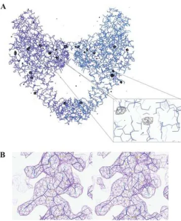
Figure 6.Electron density maps confirming the molecular replacement solution. (A) Superposition of the anomalous difference Fourier map calculated using Cys426 data collected on the chromium edge contoured at 3.5swith the final refined model of ParC55. Panels were rendered using Coot [46]. (B) Stereo view of a region of the 2Fobs-Fcalc electron density map from wild-type ParC55 data contoured at 1.5s with the final refined model superimposed.
using the ParC Cys426 data set collected at the chromium wavelength and phases from the refined wild-type ParC55 model. The map showed clear anomalous peaks around sulfur atoms of corresponding methionines as well as cysteine 426, confirming the molecular replacement solution (Figure 6A).
Close inspection of the 2Fobs-Fcalcelectron density maps built using both wild-type ParC55 and Cys426 mutant data sets have shown a very good coverage for most of the backbone and side-chains (ure 6B). The overall B-factors of the structure are relatively high with the average value of 67.42 A˚2and are similar to those shown for GyrA from E. coli, which indicates a high level of flexibility of the molecules [19]. The terminal residues (1 to 27 and 483 to 496) as well as the residues in flexible regions:
Chain A: 172, 283–285, 291–295, 301–304, 408; Chain B: 172, 299, 408;
Chain C: 306, 408–409;
Chain D: 110, 171–174, 253, 282–284, 316–318, 408–409 were omitted from the final structure because no convincing electron density was observed for them.
The final Ramachandran plot indicated that most of the residues are in the favored regions: 83.2% most favored, 13.8% allowed, 2.3% generously allowed, 0.7% disallowed according to PROCHECK [47,48].
Data deposition: The atomic coordinates and structure factors have been deposited with the Protein Data Bank, www.rcsb.org (PDB ID code 2NOV).
SUPPORTING INFORMATION
Figure S1 Electrostatic surface potential modeling. GRASP2 [51,52] electrostatic surface potentials within the DNA-binding grooves calculated for DNA-binding domains of topoisomerases IIA from different organisms using models generated by 3D-JIGSAW [41–43] on the base of the known structure of E. coli GyrA (1AB4).
Found at: doi:10.1371/journal.pone.0000301.s001 (3.64 MB PDF)
Figure S2 Modeling of the DNA sequence recognition by
topoisomerase IV. (A) Amino acid sequence alignment for the regions of GyrA and ParC from E. coli and S. pneumoniae com-prising helices a3 and a4 and the 100-122 loop. The residues within thea-helices which are likely to interact directly with the incoming DNA helix are indicated by color (red fora3 and blue for a4) and asterisks. Active site tyrosines and arginines are in green. (B) Model of the bound state for the protein-DNA complex betweenS. pneumoniaeParC and the DNA E site [15]. The DNA is in Licorice mode and the protein is in cartoon mode. The positions on the DNA helix are given by numbers in yellow circles. The nucleotides are indicated by square boxes. Amino acids of the
100-122 loop are indicated by ovals with corresponding names. Helixa4 is in cyan and helixa3 is in yellow. Active site tyrosine and arginine are in CPK mode and the point of the DNA cleavage is indicated by yellow asterisk in red circle. Ser 79 and Asp 83 are shown using VDW representation and are in yellow and red respectively. The panel was generated using VMD [50] and Pov-Ray.
Found at: doi:10.1371/journal.pone.0000301.s002 (3.14 MB PDF)
Movie S1 A schematic process of DNA binding and recognition byS. pneumoniaeParC. The process is shown both in cartoon and electrostatic potential surface modes. The positions of the active-site tyrosines are indicated by arrows when the tyrosines approach the target DNA backbone phosphates. The DNA backbone is in rose, the base pairs involved in sequence specific DNA recognition are in green, the nucleotides adjacent to the cleavage points are in purple in Licorice mode and the non-specific base pairs are in silver. The active-site tyrosines of the ParC dimer are represented in CPK mode in blue. The helixa3 is in cyan, the helixa4 is in red, the 100-122 loop is in yellow and the Gly 166-Pro 179 region is in orange. The movie was generated using VMD [50], Pov-Ray and PyMOL [53].
Found at: doi:10.1371/journal.pone.0000301.s003 (4.77 MB MPG)
ACKNOWLEDGMENTS
We thank Drs. Pierre Rizkallah, Rob Kehoe, James Nicholson, Miroslav Papiz, Michele Cianci and Mark Ellis at the SRS, Daresbury laboratory for their invaluable assistance whilst collecting a number of data associated with this project on several beamlines. We also thank Drs. Andrew Thompson and Gordon Lennard for their help in collecting the Se-Met MAD data at ESRF beamlines ID-14 and ID-29, although due to twinning problems this could not ultimately be used in the structure solution. We acknowledge Dr. Juan Fontecilla-Camps and Dr. Michel Roth for giving us access to beamline ESRF BM 30A for data collection. We are grateful to Drs Edward Morris and Paula da Fonseca of the Structural Electron Microscopy Team from Imperial College and the Institute of Cancer Research for their help in collecting electron microscopy data, though due to low resolution the data could not assist us in finding the structure solution. Mark R. Sanderson would like to thank Prof. Nigel Grindley F.R.S. of the Department of Molecular Biophysics and Biochemistry, Yale University, for hosting his sabbatical during which part of this manuscript was written. Dr. Jim Warwicker is thanked for discussion on electrostatics software.
Author Contributions
Conceived and designed the experiments: MS LF. Performed the experiments: MS IL DV MS XP. Analyzed the data: MS IL DV LF AA. Contributed reagents/materials/analysis tools: MS XP LF CY JF. Wrote the paper: MS IL DV MS LF.
REFERENCES
1. Berger JM (1998) Structure of DNA topoisomerases. Biochim Biophys Acta 1400: 3–18.
2. Champoux JJ (2001) DNA topoisomerases: structure, function, and mechanism. Annu Rev Biochem 70: 369–413.
3. Wang JC (2002) Cellular roles of DNA topoisomerases: a molecular perspective. Nat Rev Mol Cell Biol 3: 430–440.
4. Huang WM (1994) Type II DNA topoisomerase genes. Adv Pharmacol 29A: 201–225.
5. Maxwell A (1996) Protein gates in DNA topoisomerase II. Nat Struct Biol 3: 109–112.
6. Drlica K, Zhao X (1997) DNA gyrase, topoisomerase IV, and the 4-quinolones. Microbiol Mol Biol Rev 61: 377–392.
7. Maxwell A (1997) DNA gyrase as a drug target. Trends Microbiol 5: 102–109.
8. Pan XS, Fisher LM (1999)Streptococcus pneumoniaeDNA gyrase and topoisomerase IV: overexpression, purification, and differential inhibition by fluoroquinolones. Antimicrob Agents Chemother 43: 1129–1136.
9. Corbett KD, Berger JM (2004) Structure, molecular mechanisms, and evolutionary relationships in DNA topoisomerases. Annu Rev Biophys Biomol Struct 33: 95–118.
10. Gadelle D, Filee J, Buhler C, Forterre P (2003) Phylogenomics of type II DNA topoisomerases. Bioessays 25: 232–242.
11. Wang JC (1998) Moving one DNA double helix through another by a type II DNA topoisomerase: the story of a simple molecular machine. Q Rev Biophys 31: 107–144.
12. Kampranis SC, Maxwell A (1996) Conversion of DNA gyrase into a conven-tional type II topoisomerase. Proc Natl Acad Sci U S A 93: 14416–14421.
13. Pan XS, Fisher LM (1997) Targeting of DNA gyrase inStreptococcus pneumoniaeby sparfloxacin: selective targeting of gyrase or topoisomerase IV by quinolones. Antimicrob Agents Chemother 41: 471–474.
14. Pan XS, Fisher LM (1998) DNA gyrase and topoisomerase IV are dual targets of clinafloxacin action inStreptococcus pneumoniae. Antimicrob Agents Chemother 42: 2810–2816.
15. Leo E, Gould KA, Pan XS, Capranico G, Sanderson MR, et al. (2005) Novel symmetric and asymmetric DNA scission determinants forStreptococcus pneumoniae
topoisomerase IV and gyrase are clustered at the DNA breakage site. J Biol Chem 280: 14252–14263.
16. CBSN: Canadian Bacterial Surveillance Network (2006),http://microbiology. mtsinai.on.ca/data/sp/indexOld.shtml..
17. Bellon S, Parsons JD, Wei Y, Hayakawa K, Swenson LL, et al. (2004) Crystal structures of Escherichia coli topoisomerase IV ParE subunit (24 and 43 kilodaltons): a single residue dictates differences in novobiocin potency against topoisomerase IV and DNA gyrase. Antimicrob Agents Chemother 48: 1856–1864.
18. Corbett KD, Schoeffler AJ, Thomsen ND, Berger JM (2005) The structural basis for substrate specificity in DNA topoisomerase IV. J Mol Biol 351: 545–561. 19. Morais-Cabral JH, Jackson AP, Smith CV, Shikotra N, Maxwell A, et al. (1997)
Crystal structure of the breakage-reunion domain of DNA gyrase. Nature 388: 903–906.
20. Berger JM, Gamblin SJ, Harrison SC, Wang JC (1996) Structure and mechanism of DNA topoisomerase II. Nature 379: 225–232.
21. McKay DB, Steitz TA (1981) Structure of catabolite gene activator protein at 2.9 A˚ resolution suggests binding to left-handed B-DNA. Nature 290: 744–749. 22. Passner JM, Steitz TA (1997) The structure of a CAP-DNA complex having two cAMP molecules bound to each monomer. Proc Natl Acad Sci U S A 94: 2843–2847.
23. Schultz SC, Shields GC, Steitz TA (1991) Crystal structure of a CAP-DNA complex: the DNA is bent by 90 degrees. Science 253: 1001–1007. 24. Brown PO, Cozzarelli NR (1979) A sign inversion mechanism for enzymatic
supercoiling of DNA. Science 206: 1081–1083.
25. Mizuuchi K, Fisher LM, O’Dea MH, Gellert M (1980) DNA gyrase action involves the introduction of transient double-strand breaks into DNA. Proc Natl Acad Sci U S A 77: 1847–1851.
26. Corbett KD, Shultzaberger RK, Berger JM (2004) The C-terminal domain of DNA gyrase A adopts a DNA-bendingb-pinwheel fold. Proc Natl Acad Sci U S A 101: 7293–7298.
27. Reece RJ, Maxwell A (1991) The C-terminal domain of theEscherichia coliDNA gyrase A subunit is a DNA-binding protein. Nucleic Acids Res 19: 1399–1405. 28. Ruthenburg AJ, Graybosch DM, Huetsch JC, Verdine GL (2005) A superhelical spiral in the Escherichia coli DNA gyrase A C-terminal domain imparts unidirectional supercoiling bias. J Biol Chem 280: 26177–26184.
29. Morrison A, Cozzarelli NR (1981) Contacts between DNA gyrase and its binding site on DNA: features of symmetry and asymmetry revealed by protection from nucleases. Proc Natl Acad Sci U S A 78: 1416–1420. 30. Fisher LM, Mizuuchi K, O’Dea MH, Ohmori H, Gellert M (1981) Site-specific
interaction of DNA gyrase with DNA. Proc Natl Acad Sci U S A 78: 4165–4169. 31. Kirkegaard K, Wang JC (1981) Mapping the topography of DNA wrapped around gyrase by nucleolytic and chemical probing of complexes of unique DNA sequences. Cell 23: 721–729.
32. Peng H, Marians KJ (1995) The interaction ofEscherichia colitopoisomerase IV with DNA. J Biol Chem 270: 25286–25290.
33. Daniels DS, Woo TT, Luu KX, Noll DM, Clarke ND, et al. (2004) DNA binding and nucleotide flipping by the human DNA repair protein AGT. Nat Struct Mol Biol 11: 714–720.
34. Shen LL, Baranowski J, Pernet AG (1989) Mechanism of inhibition of DNA gyrase by quinolone antibacterials: specificity and cooperativity of drug binding to DNA. Biochemistry 28: 3879–3885.
35. Willmott CJ, Maxwell A (1993) A single point mutation in the DNA gyrase A protein greatly reduces binding of fluoroquinolones to the gyrase-DNA complex. Antimicrob Agents Chemother 37: 126–127.
36. Richter SN, Leo E, Giaretta G, Gatto B, Fisher LM, et al. (2006) Clerocidin interacts with the cleavage complex ofStreptococcus pneumoniaetopoisomerase IV to induce selective irreversible DNA damage. Nucleic Acids Res 34: 1982–1991. 37. Otwinowski Z, Minor W (1997) Processing of X-ray diffraction data collected in
oscillation mode. Method Enzymol: 307–325.
38. Kabsch W (1993) Automatic processing of rotation diffraction data from crystals of initially unknown symmetry and cell constants. J Appl Crystallogr 26: 795. 39. Yeates TO (1997) Detecting and overcoming crystal twinning. Method
Enzymol: 344–360.
40. Brunger AT, Adams PD, Clore GM, DeLano WL, Gros P, et al. (1998) Crystallography & NMR system: A new software suite for macromolecular structure determination. Acta Crystallogr D Biol Crystallogr 54: 905–921. 41. Bates PA, Kelley LA, MacCallum RM, Sternberg MJE (2001) Enhancement of
protein modeling by human intervention in applying the automatic programs 3D-JIGSAW and 3D-PSSM. Proteins 45: 39–46.
42. Bates PA, Sternberg MJE (1999) Model building by comparison at CASP3: using expert knowledge and computer automation. Proteins 37: 47–54.
43. Contreras-Moreira B, Bates PA (2002) Domain Fishing: a first step in protein comparative modelling. Bioinformatics 18: 1141–1142.
44. The CCP4 suite: programs for protein crystallography. Acta Crystallogr D Biol Crystallogr 50: 760–763.
45. Navaza J (2001) Implementation of molecular replacement in AMoRe. Acta Crystallogr D Biol Crystallogr 57: 1367–1372.
46. Emsley P, Cowtan K (2004) Coot: model-building tools for molecular graphics. Acta Crystallogr D Biol Crystallogr 60: 2126–2132.
47. Laskowski RA, MacArthur MW, Moss DS, Thornton JM (1993) PROCHECK: a program to check the stereochemical quality of protein structures. J Appl Crystallogr 26: 283.
48. Morris AL, MacArthur MW, Hutchinson EG, Thornton JM (1992) Stereo-chemical quality of protein structure coordinates. Proteins 12: 345–364. 49. Chenna R, Sugawara H, Koike T, Lopez R, Gibson TJ, et al. (2003) Multiple
sequence alignment with the Clustal series of programs. Nucleic Acids Res 31: 3497–3500.
50. Humphrey W, Dalke A, Schulten K (1996) VMD: visual molecular dynamics. J Mol Graph 14: 27–38.
51. Petrey D, Honig B (2003) GRASP2: visualization, surface properties, and electrostatics of macromolecular structures and sequences. Method Enzymol 374: 492–509.
52. Warwicker J, Watson HC (1982) Calculation of the electric potential in the active site cleft due toa–helix dipoles. J Mol Biol 157: 671–679.
![Figure 4. Electrostatic surface potential calculation. GRASP2 [51,52] electrostatic surface potentials within the DNA-binding grooves calculated for equivalent N-terminal fragments of: (A) S](https://thumb-eu.123doks.com/thumbv2/123dok_br/17101215.237460/4.918.89.446.83.519/electrostatic-potential-calculation-electrostatic-potentials-calculated-equivalent-fragments.webp)