Review Article
Chagas Disease: Still Many Unsolved Issues
José M. Álvarez,
1Raissa Fonseca,
1Henrique Borges da Silva,
1Cláudio R. F. Marinho,
2Karina R. Bortoluci,
3Luiz R. Sardinha,
4Sabrina Epiphanio,
5and Maria Regina D’Império Lima
11Department of Immunology, Biomedical Sciences Institute, University of S˜ao Paulo, 05508-000 S˜ao Paulo, SP, Brazil
2Department of Parasitology, Biomedical Sciences Institute, University of S˜ao Paulo, 05508-000 S˜ao Paulo, SP, Brazil
3Department of Biological Sciences, UNIFESP (Campus Diadema), 09972-270 Diadema,SP, Brazil
4Hospital Israelita Albert Einstein, 05652-000 S˜ao Paulo, SP, Brazil
5Department of Clinical and Toxicological Analyses, Faculty of Pharmaceutical Sciences, University of S˜ao Paulo,
05508-000 S˜ao Paulo, SP, Brazil
Correspondence should be addressed to Jos´e M. ´Alvarez; [email protected]
Received 22 April 2014; Accepted 15 June 2014; Published 29 June 2014
Academic Editor: Edecio Cunha-Neto
Copyright © 2014 Jos´e M. ´Alvarez et al. his is an open access article distributed under the Creative Commons Attribution License,
which permits unrestricted use, distribution, and reproduction in any medium, provided the original work is properly cited.
Over the past 20 years, the immune efector mechanisms involved in the control ofTrypanosoma cruzi, as well as the receptors
participating in parasite recognition by cells of the innate immune system, have been largely described. However, the main questions on the physiopathology of Chagas disease remain unanswered: “Why does the host immune system fail to provide sterile immunity?” and “Why do only a proportion of infected individuals develop chronic pathology?” In this review, we describe the mechanisms proposed to explain the inability of the immune system to eradicate the parasite and the elements that allow the development of chronic heart disease. Moreover, we discuss the possibility that the inability of infected cardiomyocytes to sense
intracellularT. cruzicontributes to parasite persistence in the heart and the development of chronic pathology.
1. A Brief Overview of Chagas Disease
Chagas disease is caused by Trypanosoma cruzi and rep-resents an important health problem in Latin America, with approximately 8 million chronically infected people [1]. Recently, as a consequence of human migrations, Chagas dis-ease has become a potential public health issue in developed countries, and signiicant increases in conirmed cases have been reported in the USA, Canada, Europe, Japan, and Aus-tralia [2]. he invasion of the human host frequently occurs by the passage through damaged skin or intact mucosa of metacyclic trypomastigotes released with the feces of infected triatomines ater their blood meal. Alternatively, infection through other routes, such as oral, congenital, and blood transfusion/organ transplantation, also occurs. Because it is an obligate intracellular parasite, T. cruzi can be found in the vertebrate host as amastigotes, the intracellular replicative form, and as extracellular trypomastigotes circulating freely in the blood and tissues. he infection has a self-limiting
acute phase, with patent (or subpatent) parasitemia, which goes unnoticed in many infected individuals. At this stage, the parasites actively replicate in many diferent cell types, such as macrophages; smooth, striated, and cardiac muscle cells; adipocytes; and cells of the central nervous system [3].
While a small proportion of patients succumbs to the acute phase of the disease, the development of the adaptive immune response typically provides control of theT. cruzi
infection, albeit nonsterile control. Failing to completely eradicate the parasite, individuals remain infected for life and establish a dynamic equilibrium with the parasite that results in diferent clinical outcomes. hus, while many chronically infected individuals remain in the asymptomatic indeterminate phase, a signiicant proportion (30–35%) of patients develop the cardiac or digestive manifestations of chronic disease: cardiomyopathy that may lead to congestive heart failure, arrhythmia and, eventually, patient death, and esophageal or colonic megasyndromes. hese are irreversible pathological changes that occur despite parasite scarcity. Volume 2014, Article ID 912965, 9 pages
Recapitulating human chagasic myocarditis, mice surviving long-term infection by certain stocks of T. cruzi develop chronic myocardial lesions [4,5].
2. The Host Immune Response against
T. cruzi
he immune system is well equipped to detect and control
T. cruzi parasites through the combined efect of diverse branches of the immune response. CD4+and CD8+T cells, as well as B cells, contribute to control the parasite through cytokine secretion, cellular cytotoxicity, and speciic antibody production [6–8]. From the end of the acute phase and throughout chronic infection,T. cruzi-speciic IgG antibodies actively participate in the removal of extracellular parasites released from ruptured tissue nests, an efect that presumably occurs by promoting parasite phagocytosis by macrophages and neutrophils. In addition, speciic IgG antibodies mediate the removal of blood trypomastigotes, a clearance process in which complement and mononuclear phagocytes from the liver, spleen, and lungs appear to be involved [9–11]. he IFN-� produced by activated CD4+ and CD8+ T cells, NK cells, and CD4−CD8−��T cells plays a crucial role in parasite elimination [12,13]. IFN-�potentiates the efector activity of macrophages by inducing the transcription of the inducible nitric oxide synthase (iNOS) gene, notably increasing the production of nitric oxide, which has a potent efect on
T. cruzi killing [14, 15]. In addition, IFN-� promotes the immunoglobulin switch to IgG subclasses with high opsoniz-ing and complement-activatopsoniz-ing potential. Lastly, cytotoxic CD8+T cells also contribute toT. cruzicontrol through the recognition and destruction of cells that harbor intracellular forms of the parasite [16].
3. Limitations to the Innate and Acquired
Immune Responses That Contribute to
Parasite Persistence
One of the most intriguing questions of human and experi-mentalT. cruziinfection is why the immune system fails to totally eradicate the parasite. At irst glance, the inability of the infected host to attain sterility suggests that the immune efector activity directed against the parasite is insuicient or inappropriate due to defective activation of the speciic immune response or excessive regulation of this response.
In this context, we outline in this section the diferent escape mechanisms employed byT. cruziparasites and dis-cuss the hypothesis generated to explain an immune system failure. At the beginning of the infection (before development of the parasite-speciic response),T. cruzi trypomastigotes escape lysis by the complement system, an evasion strategy that results from the presence of complement-regulatory molecules on the parasite surface [17]. In addition, internal-ized parasites of diverseT. cruzistrains escape the phagocytic vacuole of unprimed resident macrophages [18], a strategy that relies on a variety of molecules with antioxidant prop-erties [19, 20]. Nevertheless, as infection progresses, these two evasion strategies are largely circumvented by the devel-opment of the speciic humoral response and the induction
of macrophage activation by IFN-� and other cytokines. BecauseT. cruzistrains display diferent levels of antioxidant activity that directly correlate with strain virulence [21], it remains unclear whether IFN-�confers efective macrophage protection against anyT. cruziparasite or results in diferent degrees of intracellular parasite destruction for diferent isolates.
Pattern recognition receptors (PRRs), such as toll-like receptors (TLRs) 2, 4, 7, and 9, nucleotide-binding oligomer-ization domain-like receptor (NOD) 1 and NATCH, LRR, and PyD domains-containing protein 3 (NLRP3) have been shown to participate inT. cruzidetection by macrophages and dendritic cells [22–27]. However, a deicient innate immune response due to poor PRR signaling by pathogen-associated molecular patterns (PAMPs) has been proposed as a mecha-nism involved in parasite escape. his hypothesis is supported by data showing that, compared with mice infected by wild-typeT. cruziparasites, mice infected with transgenicT. cruzi
expressing Salmonella typhimurium lagellin (liC) display an increased innate response mediated by macrophages and dendritic cells and notably enhanced adaptive immunity [28]. More importantly, in the chronic phase, mice infected with liC-transgenicT. cruzidisplay notably reduced parasite levels in relation to those infected with wild-type parasites.
Parasite persistence has also been attributed to the rel-atively slow development ofT. cruzi-speciic CD8+T efec-tor cells [29], a phenomenon explained by diverse facefec-tors, including the postulated poor PAMP activity ofT. cruzi[28] and the induction of a strong Fas expression on T. cruzi -speciic CD8+ T cells [30]. Another possible reason for the failure to acquire sterile immunity is the clonal dominance of the lymphocyte response to T. cruzi infection. his is illustrated by the observation that the CD8+T cell response to the abundantly expressedT. cruziantigen amastigote surface protein-2 (ASP2) is restricted to a small number of clones [31]. Narrowing the scope of parasite peptides that are recognized by CD8+ T cells may impair complete parasite eradication during chronic infection and the control of reinfection, which frequently occurs in endemic areas.
0
0 20 40 60 80 100
0 20 40 60 80 100
0 20 40 60 80 100
103 104 105 0 103 104 105 0 103 104 105
CD4+ CD8+
PD-L1 PD-L1
PD-L1
Control Chronic
Control Chronic
Control Chronic
Monocytes
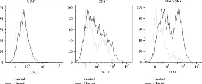
Figure 1: Expression of PD-L1 by iniltrating leukocytes in a chronically infected heart. C3H/HePAS mice were infected with 4×10⁵Sylvio
X10/4 trypomastigotes obtained from LLCMK2 cultures. At day 400 postinfection, mice were sacriiced, and the heart tissue was digested with collagenase to isolate the iniltrating leukocytes. PD-L1 expression was analyzed by low cytometry. Heart leukocytes from a pool of age-matched noninfected mice were also included.
elimination and permitting the perpetuation of lesions in the chronic phase remain to be determined. In this respect, our data showing the intense expression of PD-L1 in the heart-iniltrating leukocytes of mice chronically infected by Sylvio X10/4T. cruziparasites suggest the involvement of this regulatory circuit in parasite persistence (Figure 1).
he mechanisms of parasite escape discussed above refer to limitations of the innate or acquired protective immune response toT. cruzi that appear to operate in all infected individuals. However, in addition to these gen-eral elements, we must consider that individuals difer in the intensity/efectiveness of their anti-T. cruzi humoral and cellular responses, a consequence of polymorphisms in genes associated with the immune response [38]; these polymorphisms inluence the intensity of the anti-T. cruzi
efector activity, yielding diferent levels of residual parasites and undesired tissue lesions in chronically infected human patients. In addition, these polymorphisms inluence the residual parasite distribution in diferent tissues/organs, an element closely connected to the development of the diferent forms of the disease.
he isogenic strains of mice also difer in the immune response toT. cruzi. hese diferences are oten critical for acute phase survival [39] and most likely determine the par-asite load in the chronic phase. However, not a single mouse strain has been reported to promote the complete elimination of the parasite. herefore, regardless of the mouse/parasite strain combination, the inevitable outcome of murine infec-tion byT. cruziappears to be parasite persistence, a result analogous to that observed in human patients.
4. Only a Proportion of
T. cruzi
-Infected
Individuals Show Chronic Pathologies
A signiicant proportion of chronically infected individuals develop the cardiac and digestive forms of the disease,but the largest fraction present the indeterminate form. Importantly, although many indeterminate patients remain asymptomatic for the rest of their lives, it is estimated that, each year, 2.5% of infected individuals evolve from the indeterminate to the clinical forms [40]. Chronic chagasic cardiomyopathy (CCC) represents the main cause of death in T. cruzi-infected patients. Moreover, this clinical form represents an important social burden in terms of lost labor hours and hospital costs. he situation of CCC patients is worrisome because the speciic anti-T. cruzidrugs, currently limited to benznidazol and nifurtimox, show limited eicacy in chronically infected patients.
Because of parasite scarcity in the inlamed heart, CCC was long considered an autoimmune disease directed against self-epitopes showing cross-reactivity with parasite antigens [41]. According to this view, lesions were thought to occur as a result of T cell reactivity against myosin and other heart-derived proteins [42] as well as humoral reactivity to the beta-1-adrenergic and M2 cholinergic receptors, leading to autonomic nervous system imbalance [43]. However, in the last 20 years, cumulative evidence has promoted a change in our understanding of this process. First, immunohis-tochemistry data showed that, in patients with CCC, the level ofT. cruziantigen correlated with the intensity of the inlammatory iniltrate [44]. Furthermore,T. cruziDNA was found in the heart of diseased CCC patients but not in the heart of patients with the indeterminate form [45]. In contrast, patients with megaesophagus, one of the digestive forms of the disease, displayed positive PCR for T. cruzi
(a) (b)
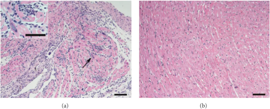
Figure 2: Parasite persistence is not the necessary outcome of heart infection byT. cruziparasites. C57BL/6 mice were infected with 103T.
cruziblood trypomastigotes of the Y strain. At days 18 (a) and 180 (b) postinfection, mice were sacriiced, and the heart tissue was
formalin-ixed, included in parain, and stained with hematoxylin/eosin. Arrow in the igure shows an amastigote nest, which is magniied at the insert.
Bars in igures correspond to 200�m and to 100�m at the insert.
agreed that the inlammatory iniltrate in CCC is caused by a response directed toward locally persisting parasites [48]. Still, because Chagas disease is a highly heterogeneous process inluenced by host genetics, we cannot discard that the immune response in the heart of CCC patients could include, besides leukocyte reactivity towards locally persist-ing parasites, diferent degrees of autoreactivity. Furthermore, it is possible that a proportion of CCC patients shows cardiac pathology in the absence of locally persisting infection. his last possibility is illustrated by a recent report using bioluminescence imaging, in which mice chronically infected with luciferase expressingT. cruziparasites display mild heart inlammation in the absence of local parasitism [49]. Further studies on the contribution of autoimmunity are required for a full comprehension of CCC physiopathology. hese studies may eventually reveal alternative therapeutic approaches to attenuate heart tissue inlammation in chagasic patients.
Nonetheless, because most data suggest the association of chronic cardiac pathology with local parasite persistence, CCC is presently understood as an expression of the host’s incapacity to totally eliminate the heart parasitism, a particu-larity of the host’s failure to eradicateT. cruzifrom the organ-ism. his leads us to question why heart parasitism occurs in only a fraction ofT. cruzichronically infected individuals.
5. Where Does
T. cruzi
Persist in
the Chronically Infected Host?
he tissue distribution of T. cruzi parasites varies among chronically infected individuals. As indicated above, patients with cardiomyopathy and megaesophagus harbor parasites in the heart and esophagus, respectively. However, are there in these patients, as well as in those with the indeterminate form, other locations where the parasite is relatively safe from com-plete elimination by the immune system?A priori, because parasite persistence is the rule, it is reasonable to hypothesize that immune-privilegedT. cruzireservoirs must exist in all chronically infected hosts. One of these parasite niches could be the central adrenal vein, which, in autopsy studies of patients with chronic Chagas disease, has been found to
frequently harbor amastigote nests; further, the presence of these nests shows a close correlation with heart pathology parameters such as the intensity of leukocyte iniltration and myocardial ibrosis [50]. Additionally, both brown and white adipose tissues, as well as the colon and stomach, have been described as locations where T. cruzi parasites chronically persist [49,51,52]. Moreover, because of parasite persistence in the cardiac tissue of CCC patients, the heart could be one of these niches for a fraction of the infected population.
Another issue is whether parasite dissemination through the blood or by spreading from neighboring tissues occurs in the chronic host. Are theT. cruzifound in the hearts of CCC patients the consequence of chronic phase dispersion from other tissues or do they result from the perpetuation of heart colonization during the acute phase? his is an important question because chronic phase reinvasion could readdress the problem of heart parasite persistence outside the cardiac tissue.
On the other hand, it is important to note the observation made by diferent research groups that mice from certain strains can eliminate T. cruzi from the heart. his occurs in C57BL/6 mice infected with a sublethal dose of Y strain parasites; these mice exhibit strong leukocyte iniltration with the presence of amastigote nests in the heart in the late acute phase but no signs of infection or pathology in this organ in the chronic phase (Figure 2). his observation indicates that parasite persistence is not the necessary outcome of heart infection. Moreover, these results open the possibility that the indeterminate group of human patients might include patients in whom the cardiac infection was resolved in addi-tion to individuals in whom the heart was never colonized by the parasite.
6. Elements Involved in Parasite
Persistence in the Heart or Other
Tissues: Parasite Tropism
correlations with the sylvatic or peridomestic forms of trans-mission as well as with the occurrence (or lack thereof) of diferent chronic pathologies. Mixed infections by multiple
T. cruziisolates are frequently observed in chronic chagasic patients.
Parasite tropism was originally deined as the preferential invasion of a cell type by aT. cruziclone/isolate. Nevertheless, because the infected host exhibits considerable variability in its tissue responses to the parasite, tropism has to be redeined as the outcome of the interaction of a deined
T. cruzi clone/isolate in a particular individual, a process largely dependent on the parasite and host genetics. he importance of the host in the development of chronic heart pathology by a singleT. cruzi parasite is clearly illustrated in the murine model of infection by parasites of theT. cruzi
clone Sylvio X10/4, which results in chronic cardiomyopathy in C3H/HePAS mice but not in C57BL/6 or A/J mice [47].
herefore, while (at least for mice and men) the lack of a spontaneous cure and, consequently, the persistence of the parasite are general problems associated with T. cruzi
infection, independent of parasite and host diversity, the development of chronic heart disease appears to be limited to particular host-parasite combinations.
7. Is Parasite Persistence Merely the Result of
a Deficit in the Local Immune Response?
Diferent research groups have analyzed the heart-iniltrating leukocytes of CCC patients and mice with chronic car-diomyopathy [54–58]. hese studies have yielded valuable data regarding the distribution of leukocyte populations, surface marker expression, and the production of cytokines, chemokines, and other mediators. Nonetheless, the gathered information did not help to determine whether a local immune deicit exists because the characteristics of an efec-tive local immune response are undeined. his is because in those chronic settings in which an efective response could eventually occur, such as in chronically infected mice with no cardiac pathology or in patients with the indeterminate form, there are by deinition no heart iniltrates to dissect. herefore, if there is a defect in the local immune response in the heart of CCC patients, it has yet to be found.
Immune response analysis of the blood of patients with the cardiac and indeterminate forms has been used as an indirect means of searching for the presumed local immune defect, aiming to reveal a special immune signature that would explain why the parasite persists in the heart of CCC patients. Remarkably, these studies have revealed that the production of IFN-�and other proinlammatory cytokines is higher in CCC patients than in indeterminate form patients [59–61] and that asymptomatic patients exhibit augmented TREG numbers and IL-10 levels [59, 62]. Because a
proin-lammatory response is considered the appropriate approach to eliminateT. cruziparasites, these results conlict with the hypothesis that parasite evasion is the result of a deicient immune response. Furthermore, while systemic studies do not suggest a local deicit in the anti-T. cruzi immune response of CCC patients, they do indicate that these patients
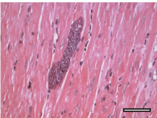
Figure 3: Lack of sensing of amastigote nests could contribute to parasite perpetuation and pathology in the chronically infected heart. Lack of sensing could occur independently of the origin of cardiomyocyte-invading trypomastigotes, that is, from a local ruptured nest or metastasis from a distant niche. he heart picture shown corresponds to a C3H/HePAS mouse infected for more than
200 days with 106Sylvio X10/4 trypomastigotes. he tissue section
was stained with hematoxylin-eosin. Bar corresponds to 100�m
(reproduced from C. R. F. Marinho,Microbes and Infection[64] with
permission from Elsevier).
display an aggressive deregulated response that may explain the presence of tissue damage in the afected heart [63].
TNF-� and IL-1� [66]. A way to reconcile these in vitro
and in vivo indings is that the failure of cardiomyocyte signaling in vivo could be an adaptive trait that develops during the course of infection in only certain parasite-host combinations. he possibility thatT. cruziinterferes with the physiology of infected structural cells is supported by other studies. In this manner, cruzipain, an enzyme abundantly found inT. cruziparasites, has been shown to interfere with cardiomyocyte apoptosis through activation of the NF�B and PI3K/Akt and MEK1/ERK1/2 pathways in the host cell, which lead to increased expression of antiapoptotic Bcl-2 molecules and increased arginase expression [67]. It is therefore conceivable that the crosstalk of the intracellular parasite with the signaling pathways of these structural cells might negatively impact the production of chemotactic molecules.
A deicit in intracellular parasite sensing, although important, is insuicient to guaranteeT. cruzievasion in the chronically infected heart. his is the case given that, sooner or later, an undetected amastigote nest will spontaneously dis-rupt, releasing extracellular parasites that, ater detection by antibodies, will cause complement and resident macrophages to generate mediators for leukocyte recruitment. hus, for long-term parasite perpetuation to occur, it is predictable that a small fraction of nest-released trypomastigotes will reinvade relatively distant cardiomyocytes, where they may remain unnoticed and out of the reach of cytotoxic CD8+T cells in the newly formed iniltrate.
As stated above, defective parasite sensing by cardiomy-ocytes could be an adaptive process of the heart tissue that develops with the length of infection. his process most likely exhibits a great degree of variability, relecting the genetics of the host and the parasite. Moreover, its occurrence in the cardiac tissue is not surprising because the heart is a vital organ that must have special mechanisms designed to protect its integrity.
8. Local Parasite Destruction
versus Immunopathology
heoretically, if two hosts are unable to control tissue par-asites, the one with a greater local inlammatory response will pay a higher price by provoking greater damage of the infected tissue [68]. herefore, if defective local sensing occurs in CCC patients, strong local immune responses would clearly represent a detrimental factor in the induc-tion of pathology, thus explaining the reported associa-tions between high levels of cardiac dysfunction and geno-types associated with high reactivity [69,70]. Furthermore, the inverse correlation observed in CCC patients between the intensity of electrocardiogram abnormalities and IL-10 plasma levels [59] reinforces this view. hat is, to respond strongly when there is a gap in localT. cruzicontrol clearly represents a deleterious manner of dealing with the parasite. Entering into a persistent cycle of an intense local efector response with no resolution is, to a certain extent, a form of autoaggression, considering the high price paid by the organism in terms of tissue damage. It is not, however,
an aggression speciically directed against self-antigens but the unwanted price for unceasingly attempting to completely eliminate a small number of parasites that persist in a fraction of chronically infected individuals. Paradoxically, to protect heart integrity, cardiomyocytes may allow parasite persistence, which indirectly results in tissue damage every time a nest breaks open and new leukocyte iniltrates are formed.
A deicit in the interaction of T. cruzi parasites with tissue structural cells could also be involved in parasite persistence at locations other than the heart. his could occur in any infected patient, independent of whether the infection is cardiac, digestive, or indeterminate. In contrast to the heart, however, in many of these locations, the bystander tissue damage resulting from the persistent immune reaction against parasites might not be suicient to compromise the function of the infected tissue.
Future Perspectives
While extensive research has deciphered the local and sys-temic immune responses of chronically T. cruzi-infected hosts in the last two decades, future studies will need to focus on thein vivointeraction of parasites with structural cells, in both the heart and other tissues. Although these studies currently face great technical challenges, they will be of great importance to improve our knowledge about Chagas disease pathology.
Disclosure
Figure 3 is reprinted with permission from Elsevier from Microbes and Infection [64].
Conflict of Interests
he authors declare that there is no conlict of interests regarding the publication of this paper.
Acknowledgments
he authors extend their thanks to Rog´erio Silva do Nasci-mento and Bernardo Paulo Albe for providing technical support. Financial support was provided by Grants FAPESP 2013/08199-0 and CNPq 303269/2010-3.
References
[1] OMS, “Chagas disease (American trypanosomiasis),” Fact Sheet
340, 2013, http://www.who.int/mediacentre/factsheets/fs340/
en/index.html.
[2] J. R. Coura and P. A. V˜ıas, “Chagas disease: a new worldwide
challenge,”Nature, vol. 465, no. 7301, pp. S6–S7, 2010.
[3] A. L. Bombeiro, L. A. Gonc¸alves, C. Penha-Gonc¸alves et al., “IL-12p40 deiciency leads to uncontrolled Trypanosoma cruzi dissemination in the spinal cord resulting in neuronal death and
motor dysfunction,”PLoS ONE, vol. 7, no. 11, Article ID e49022,
[4] S. G. Andrade and Z. A. Andrade, “Pathology of prolonged
experimental Chagas’ disease,”Revista do Instituto de Medicina
Tropical de Sao Paulo, vol. 10, no. 3, pp. 180–187, 1968.
[5] R. P. Laguens, P. C. Meckert, and R. J. Gelpi, “Chronic Chagas disease in the mouse. I. Electrocardiographic and
morpholog-ical patterns of the cardiopathy.,”Medicina B, vol. 41, no. 1, pp.
35–39, 1981.
[6] F. G. Araujo, “Development of resistance toTrypanosoma cruzi
in mice depends on a viable population of L3T4+ (CD4+) T
lymphocytes,”Infection and Immunity, vol. 57, no. 7, pp. 2246–
2248, 1989.
[7] R. L. Tarleton, B. H. Koller, A. Latour, and M. Postan,
“Suscepti-bility of�2-microglobulin-deicient mice toTrypanosoma cruzi
infection,”Nature, vol. 356, no. 6367, pp. 338–340, 1992.
[8] A. M. Rodriguez, F. Santoro, D. Afchain, H. Bazin, and A. Capron, “Trypanosoma cruzi infection in B-cell-deicient rats,” Infection and Immunity, vol. 31, no. 2, pp. 524–529, 1981. [9] L. F. Umekita, H. A. Takehara, and I. Mota, “Role of the antibody
Fc in the immune clearance ofTrypanosoma cruzi,”Immunology
Letters, vol. 17, no. 1, pp. 85–89, 1988.
[10] I. Mota and L. F. Umekita, “he efect of C3 depletion on the
clearance ofTrypanosoma cruzi induced by IgG antibodies,”
Immunology Letters, vol. 21, no. 3, pp. 223–226, 1989.
[11] L. R. Sardinha, T. Mosca, R. M. Elias et al., “he liver plays a major role in clearance and destruction of blood
trypomastig-otes in Trypanosoma cruzichronically infected mice,” PLoS
Neglected Tropical Diseases, vol. 4, no. 1, article e578, 2010. [12] F. Cardillo, J. C. Voltarelli, S. G. Reed, and J. S. Silva, “Regulation
of Trypanosoma cruzi infection in mice by gamma interferon
and interleukin 10: role of NK cells,”Infection and Immunity,
vol. 64, no. 1, pp. 128–134, 1996.
[13] L. R. Sardinha, R. M. Elias, T. Mosca et al., “Contribution of
NK, NK T, �� T, and �� T cells to the gamma interferon
response required for liver protection against Trypanosoma
cruzi,”Infection and Immunity, vol. 74, no. 4, pp. 2031–2042,
2006.
[14] R. E. McCabe, S. G. Meagher, and B. T. Mullins, “Endogenous
interferon-�, macrophage activation, and murine host defense
against acute infection with Trypanosoma cruzi,”Journal of
Infectious Diseases, vol. 163, no. 4, pp. 912–915, 1991.
[15] R. T. Gazzinelli, I. P. Oswald, S. Hieny, S. L. James, and
A. Sher, “he microbicidal activity of interferon-�-treated
macrophages against Trypanosoma cruzi involves an L-arginine-dependent, nitrogen oxide-mediated mechanism inhibitable by interleukin-10 and transforming growth
factor-�,” European Journal of Immunology, vol. 22, no. 10, pp.
2501–2506, 1992.
[16] H. P. Low, M. A. M. Santos, B. Wizel, and R. L. Tarleton,
“Amastigote surface proteins ofTrypanosoma cruziare targets
for CD8+CTL,”Journal of Immunology, vol. 160, no. 4, pp. 1817–
1823, 1998.
[17] K. A. Joiner, W. D. daSilva, M. T. Rimoldi, C. H. Hammer, A. Sher, and T. L. Kipnis, “Biochemical characterization of
a factor produced by trypomastigotes ofTrypanosoma cruzi
that accelerates the decay of complement C3 convertases,”he
Journal of Biological Chemistry, vol. 263, no. 23, pp. 11327–11335, 1988.
[18] N. Nogueira and Z. Cohn, “Trypanosoma cruzi: mechanism
of entry and intracellular fate in mammalian cells,”Journal of
Experimental Medicine, vol. 143, no. 6, pp. 1402–1420, 1976.
[19] H. Castro and A. M. Tom´as, “Peroxidases of trypanosomatids,” Antioxidants and Redox Signaling, vol. 10, no. 9, pp. 1593–1606, 2008.
[20] L. Piacenza, M. P. Zago, G. Pelufo, M. N. Alvarez, M. A. Basombrio, and R. Radi, “Enzymes of the antioxidant network
as novel determiners of Trypanosoma cruzi virulence,”
Inter-national Journal for Parasitology, vol. 39, no. 13, pp. 1455–1464, 2009.
[21] L. Piacenza, M. N. Alvarez, G. Pelufo, and R. Radi, “Fighting the oxidative assault: the Trypanosoma cruzi journey to infection,” Current Opinion in Microbiology, vol. 12, no. 4, pp. 415–421, 2009.
[22] M. A. S. Campos, I. C. Almeida, O. Takeuchi et al., “Activation of toll-like receptor-2 by glycosylphosphatidylinositol anchors
from a protozoan parasite,”Journal of Immunology, vol. 167, no.
1, pp. 416–423, 2001.
[23] A. Oliveira, J. R. Peixoto, L. B. de Arrada et al., “Expression of functional TLR4 confers proinlammatory responsiveness to Trypanosoma cruzi glycoinositolphospholipids and higher
resistance to infection with T. cruzi,”Journal of Immunology, vol.
173, no. 9, pp. 5688–5696, 2004.
[24] B. C. Caetano, B. B. Carmo, M. B. Melo et al., “Requirement of UNC93B1 reveals a critical role for TLR7 in host resistance
to primary infection with Trypanosoma cruzi,” Journal of
Immunology, vol. 187, no. 4, pp. 1903–1911, 2011.
[25] G. K. Silva, F. R. S. Gutierrez, P. M. M. Guedes et al., “Cutting edge: nucleotide-binding oligomerization domain 1-dependent responses account for murine resistance against Trypanosoma
cruzi infection,”Journal of Immunology, vol. 184, no. 3, pp. 1148–
1152, 2010.
[26] G. K. Silva, R. S. Costa, T. N. Silveira et al., “Apoptosis-associated speck-like protein containing a caspase recruitment domain
inlammasomes mediate IL-1�response and host resistance to
Trypanosoma cruziinfection,”he Journal of Immunology, vol.
191, no. 6, pp. 3373–3383, 2013.
[27] V. M. Gon�alves, K. C. Matteucci, C. L. Buzzo et al., “NLRP3
controls Trypanosoma cruzi infection through a
caspase-1-dependent IL-1R-incaspase-1-dependent NO production,”PLoS Neglected
Tropical Diseases, vol. 7, no. 10, article e2469, 2013.
[28] S. P. Kurup and R. L. Tarleton, “Perpetual expression of PAMPs necessary for optimal immune control and clearance of a
persistent pathogen,” Nature Communications, vol. 4, article
2616, 2013.
[29] F. Tzelepis, B. C. G. De Alencar, M. L. O. Penido, R. T. Gazzinelli, P. M. Persechini, and M. M. Rodrigues, “Distinct
kinetics of efector CD8+cytotoxic T cells ater infection with
Trypanosoma cruzi in na¨ıve or vaccinated mice,”Infection and
Immunity, vol. 74, no. 4, pp. 2477–2481, 2006.
[30] J. R. Vasconcelos, O. Bru˜na-Romero, A. F. Ara´ujo et al., “Pathogen-induced proapoptotic phenotype and high CD95 (Fas) expression accompany a suboptimal CD8+ T-cell
response: reversal by adenoviral vaccine,”PLoS Pathogens, vol.
8, no. 5, Article ID e1002699, 2012.
[31] F. Tzelepis, B. C. G. de Alencar, M. L. O. Penido et al., “Infection with Trypanosoma cruzi restricts the repertoire of
parasite-speciic CD8+ T cells leading to immunodominance,”Journal
of Immunology, vol. 180, no. 3, pp. 1737–1748, 2008.
[32] M. C. Albareda, G. C. Olivera, S. A. Laucella et al., “Chronic
human infection withTrypanosoma cruzidrives CD4+T cells
to immune senescence,”he Journal of Immunology, vol. 183, no.
[33] J. K. Leavey and R. L. Tarleton, “Cutting edge: dysfunctional CD8+ T cells reside in nonlymphoid tissues during chronic
Trypanosoma cruzi infection,”Journal of Immunology, vol. 170,
no. 5, pp. 2264–2268, 2003.
[34] D. L. Martin, D. B. Weatherly, S. A. Laucella et al., “CD8+ T-cell
responses toTrypanosoma cruziare highly focused on
strain-variant trans-sialidase epitopes,”PLoS Pathogens, vol. 2, no. 8,
pp. 731–740, 2006.
[35] D. L. Martin, M. Postan, P. Lucas, R. Gress, and R. L. Tarleton,
“TGF-�regulates pathology but not tissue CD8+ T cell
dys-function during experimental Trypanosoma cruzi infection,” European Journal of Immunology, vol. 37, no. 10, pp. 2764–2771, 2007.
[36] M. C. Albareda, A. M. de Rissio, G. Tomas et al., “Polyfunc-tional T cell responses in children in early stages of chronic
Trypanosoma cruzi infection contrast with monofunctional
responses of long-term infected adults,”PLoS Neglected Tropical
Diseases, vol. 7, no. 12, article e2575, 2013.
[37] F. R. Gutierrez, F. S. Mariano, C. J. Oliveira et al., “Regulation of Trypanosoma cruzi-induced myocarditis by programmed death
cell receptor 1,”Infection and Immunity, vol. 79, no. 5, pp. 1873–
1881, 2011.
[38] C. M. Ayo, M. M. Dalalio, J. E. Visentainer et al., “Genetic susceptibility to Chagas disease: an overview about the infection and about the association between disease and the immune
response genes,”BioMed Research International, vol. 2013,
Arti-cle ID 284729, 13 pages, 2013.
[39] C. Sanoja, S. Carbajosa, M. Fresno, and N. Giron`es, “Analysis
of the dynamics of iniltrating CD4+T cell subsets in the heart
during experimentalTrypanosoma cruziinfection,”PLoS ONE,
vol. 8, no. 6, Article ID e65820, 2013.
[40] J. C. Pinto Dias, “he indeterminate form of human chronic
Chagas’ disease. A clinical epidemiological review,”Revista da
Sociedade Brasileira de Medicina Tropical, vol. 22, no. 3, pp. 147– 156, 1989.
[41] R. Ribeiro-Dos-Santos, J. O. Mengel, E. Postol et al., “A heart-speciic CD4+ T-cell line obtained from a chronic chagasic mouse induces carditis in heart-immunized mice and rejection
of normal heart transplants in the absence of Trypanosoma
cruzi,”Parasite Immunology, vol. 23, no. 2, pp. 93–101, 2001.
[42] E. Cunha-Neto, M. Duranti, A. Gruber et al., “Autoimmunity in Chagas disease cardiopathy: biological relevance of a car-diac myosin-speciic epitope crossreactive to an
immunodom-inant Trypanosoma cruzi antigen,”Proceedings of the National
Academy of Sciences of the United States of America, vol. 92, no. 8, pp. 3541–3545, 1995.
[43] L. Sterin-Borda and E. Borda, “Role of neurotransmitter autoantibodies in the pathogenesis of chagasic peripheral
dysautonomia,”Annals of the New York Academy of Sciences, vol.
917, pp. 273–280, 2000.
[44] M. D. L. Higuchi, M. M. Reis, V. D. Aiello et al., “Association of
an increase in CD8+ T cells with the presence ofTrypanosoma
cruziantigens in chronic, human, chagasic myocarditis,”
Amer-ican Journal of Tropical Medicine and Hygiene, vol. 56, no. 5, pp. 485–489, 1997.
[45] E. M. Jones, D. G. Colley, S. Tostes, E. R. Lopes, C. L. Vnencak-Jones, and T. L. McCurley, “Ampliication of a Trypanosoma cruzi DNA sequence from inlammatory lesions in human
cha-gasic cardiomyopathy,”American Journal of Tropical Medicine
and Hygiene, vol. 48, no. 3, pp. 348–357, 1993.
[46] A. R. Vago, A. M. Macedo, S. J. Adad, D. D’Avila Reis, and R.
Correa-Oliveira, “PCR detection ofTrypanosoma cruziDNA in
oesophageal tissues of patients with chronic digestive Chagas’
disease,”he Lancet, vol. 348, no. 9031, pp. 891–892, 1996.
[47] C. R. F. Marinho, D. Z. Bucci, M. L. Z. Dagli et al., “Pathol-ogy afects diferent organs in two mouse strains chronically
infected by aTrypanosoma cruziclone: a model for genetic
studies of Chagas'disease,”Infection and Immunity, vol. 72, no.
4, pp. 2350–2357, 2004.
[48] R. L. Tarleton, “Parasite persistence in the aetiology of Chagas
disease,”International Journal for Parasitology, vol. 31, no. 5-6,
pp. 550–554, 2001.
[49] M. D. Lewis, A. F. Francisco, M. C. Taylor et al.,
“Biolu-minescence imaging of chronicTrypanosoma cruziinfections
reveals tissue-speciic parasite dynamics and heart disease in the
absence of locally persistent infection,”Cellular Microbiology,
2014.
[50] V. de Paula Antunes Teixeira, V. Hial, R. A. da Silva Gomes et al., “Correlation between adrenal central vein parasitism and heart
ibrosis in chronic chagasic myocarditis,”American Journal of
Tropical Medicine and Hygiene, vol. 56, no. 2, pp. 177–180, 1997. [51] T. P. Combs, S. Mukherjee, C. J. G. De Almeida et al., “he
adipocyte as an important target cell forTrypanosoma cruzi
infection,”he Journal of Biological Chemistry, vol. 280, no. 25,
pp. 24085–24094, 2005.
[52] A. V. Ferreira, M. Segatto, Z. Menezes et al., “Evidence for
Trypanosoma cruziin adipose tissue in human chronic Chagas
disease,”Microbes and Infection, vol. 13, no. 12-13, pp. 1002–1005,
2011.
[53] B. Zingales, S. G. Andrade, M. R. S. Briones et al., “A new
consensus forTrypanosoma cruziintraspeciic nomenclature:
second revision meeting recommends TcI to TcVI,”Memorias
do Instituto Oswaldo Cruz, vol. 104, no. 7, pp. 1051–1054, 2009. [54] L. G. Nogueira, R. H. B. Santos, B. M. Ianni et al., “Myocardial
chemokine expression and intensity of myocarditis in Chagas
cardiomyopathy are controlled by polymorphisms inCXCL9
andCXCL10,”PLoS Neglected Tropical Diseases, vol. 6, no. 10,
Article ID e1867, 2012.
[55] D. B. Rocha Rodrigues, M. A. dos Reis, A. Romano et al., “In situ expression of regulatory cytokines by heart inlammatory
cells in Chagas’ disease patients with heart failure,”Clinical
and Developmental Immunology, vol. 2012, Article ID 361730, 7 pages, 2012.
[56] E. Cunha-Neto, V. J. Dzau, P. D. Allen et al., “Cardiac gene expression proiling provides evidence for cytokinopathy as a
molecular mechanism in Chagas'disease cardiomyopathy,”he
American Journal of Pathology, vol. 167, no. 2, pp. 305–313, 2005. [57] M. M. Reis, M. D. L. Higuchi, L. A. Benvenuti et al., “An in situ quantitative immunohistochemical study of cytokines and IL-2R+ in chronic human chagasic myocarditis: correlation
with the presence of myocardialTrypanosoma cruziantigens,”
Clinical Immunology and Immunopathology, vol. 83, no. 2, pp. 165–172, 1997.
[58] J. C. Silverio, I. R. Pereira, M. D. C. Cipitelli et al., “CD8+ T-cells expressing interferon gamma or perforin play antagonistic roles in heart injury in experimentalTrypanosoma cruzi-elicited
cardiomyopathy,” PLoS Pathogens, vol. 8, no. 4, Article ID
e1002645, 2012.
[59] G. R. Sousa, J. A. Gomes, R. C. Fares et al., “Plasma cytokine expression is associated with cardiac morbidity in chagas
disease,”PLoS ONE, vol. 9, no. 3, Article ID e87082, 2014.
that development of severe cardiomyopathy in human Chagas’
disease is due to a h1-speciic immune response,”Infection and
Immunity, vol. 71, no. 3, pp. 1185–1193, 2003.
[61] C. Poveda, M. Fresno, N. Giron´es et al., “Cytokine proiling in Chagas disease: towards understanding the association with
infectingTrypanosoma cruzidiscrete typing units (A BENEFIT
TRIAL SubStudy),”PLoS ONE, vol. 9, no. 3, Article ID e0091154,
2014.
[62] F. F. de Ara´ujo, R. Corrˆea-Oliveira, M. O. C. Rocha et al.,
“Foxp3+CD25high CD4+regulatory T cells from indeterminate
patients with Chagas disease can suppress the efector cells and cytokines and reveal altered correlations with disease severity,” Immunobiology, vol. 217, no. 8, pp. 768–777, 2012.
[63] W. O. Dutra, C. A. S. Menezes, F. N. A. Villani et al., “Cel-lular and genetic mechanisms involved in the generation of protective and pathogenic immune responses in human Chagas
disease,”Memorias do Instituto Oswaldo Cruz, vol. 104, no. 1, pp.
208–218, 2009.
[64] C. R. F. Marinho, L. N. Nu˜nez-Apaza, K. R. Bortoluci et al., “Infection by the Sylvio X10/4 clone of Trypanosoma cruzi:
relevance of a low-virulence model of Chagas’ disease,”Microbes
and Infection, vol. 11, no. 13, pp. 1037–1045, 2009.
[65] G. Vianna, “Contribuic¸˜ao para o estudo da Anatomia Patol´ogica
da Molestia de Carlos Chagas,”Memorias do Instituto Oswaldo
Cruz, vol. 3, p. 276, 1911.
[66] F. S. Machado, G. A. Martins, J. C. S. Aliberti, F. L. A. C. Mestriner, F. Q. Cunha, and J. S. Silva, “Trypanosoma cruzi-infected cardiomyocytes produce chemokines and cytokines that trigger potent nitric oxide-dependent trypanocidal
activ-ity,”Circulation, vol. 102, no. 24, pp. 3003–3008, 2000.
[67] M. D. P. Aoki, R. C. Cano, A. V. Pellegrini et al., “Diferent signaling pathways are involved in cardiomyocyte survival
induced by aTrypanosoma cruziglycoprotein,”Microbes and
Infection, vol. 8, no. 7, pp. 1723–1731, 2006.
[68] L. R˚aberg, D. Sim, and A. F. Read, “Disentangling genetic variation for resistance and tolerance to infectious diseases in
animals,”Science, vol. 318, no. 5851, pp. 812–814, 2007.
[69] R. Ramasawmy, K. C. Fae, E. Cunha-Neto et al.,
“Polymor-phisms in the gene for lymphotoxin-�predispose to chronic
chagas cardiomyopathy,”Journal of Infectious Diseases, vol. 196,
no. 12, pp. 1836–1843, 2007.
[70] G. Zafra, C. Morillo, J. Mart´ın, A. Gonz´alez, and C. I. Gonz´alez,
“Polymorphism in the 3�UTR of the IL12B gene is associated
with Chagas’ disease cardiomyopathy,”Microbes and Infection,
Submit your manuscripts at
http://www.hindawi.com
Stem Cells
International
Hindawi Publishing Corporationhttp://www.hindawi.com Volume 2014
Hindawi Publishing Corporation
http://www.hindawi.com Volume 2014
INFLAMMATION
Hindawi Publishing Corporation
http://www.hindawi.com Volume 2014
Behavioural
Neurology
Endocrinology
International Journal ofHindawi Publishing Corporation
http://www.hindawi.com Volume 2014 Hindawi Publishing Corporation
http://www.hindawi.com Volume 2014
Disease Markers
Hindawi Publishing Corporation
http://www.hindawi.com Volume 2014
BioMed
Research International
Oncology
Journal ofHindawi Publishing Corporation
http://www.hindawi.com Volume 2014
Hindawi Publishing Corporation
http://www.hindawi.com Volume 2014 Oxidative Medicine and Cellular Longevity Hindawi Publishing Corporation
http://www.hindawi.com Volume 2014
PPAR Research
The Scientiic
World Journal
Hindawi Publishing Corporation
http://www.hindawi.com Volume 2014
Immunology Research
Hindawi Publishing Corporation
http://www.hindawi.com Volume 2014
Journal of
Obesity
Journal ofHindawi Publishing Corporation
http://www.hindawi.com Volume 2014
Hindawi Publishing Corporation
http://www.hindawi.com Volume 2014
Computational and Mathematical Methods in Medicine
Ophthalmology
Journal ofHindawi Publishing Corporation
http://www.hindawi.com Volume 2014
Diabetes Research
Journal ofHindawi Publishing Corporation
http://www.hindawi.com Volume 2014
Hindawi Publishing Corporation
http://www.hindawi.com Volume 2014
Research and Treatment
AIDS
Hindawi Publishing Corporation
http://www.hindawi.com Volume 2014
Gastroenterology Research and Practice
Hindawi Publishing Corporation
http://www.hindawi.com Volume 2014
Parkinson’s
Disease
Evidence-Based Complementary and Alternative Medicine
Volume 2014 Hindawi Publishing Corporation