Oral & Maxillofacial Pathology Journal [ OMPJ ] V ol 2 No 1 Jan- Jun 2011 ISSN 0976-1225
TAURODONTISM OF MULTIPLE TE E TH – A CASE RE PORT
Joshy V.R1, Maji Jose2, Rajeesh Mohammed3
1
Professor, Dept of Oral Pathology, PSM Dental College, A k k ik k avu, Thrissur, Kerala
2
Professor, 3
A ssistant Professor [Former], Dept of Oral Pathology, Yenepoya Dental College, Karnatak a, India Corresponding Author: Dr. Joshy V. R, email: joshyhi@ rediffmail.com
Abstract
Taurodontism is a rare dental anomaly affecting primarily the molars and are usually found in association with other anomalies or as a part of syndrome. This anomaly which was considered as a feature of primitive man is also reported in modern man with less prevalence rate. In this article we are reporting rare case of a fifteen year old male patient presented with taurodontism involving all the developed molars of all four quadrants, which was not associated with any other anomalies or syndromes.
Key words – Taurodontism, bull’s tooth, permanent molars, diagnosis
Introduction
Taurodontism is considered as a variation in tooth morphology that occurs more often in molars and occasionally in premolars. This anomaly is characterized by enlarged pulp chamber with more apical positioning of floor of pulp chamber and furcation of root. Witkop defined Taurodontism as “teeth with large pulp chambers in which the
bifurcation or trifurcation are displaced apically, so that the chamber has greater apico-occlusal height than in normal teeth and lacks the constriction at the level of cemento-enamel junction (CE J). The distance from the trifurcation or bifurcation of the root to the CE J is greater than the occluso-cervical distance”1.
This anomaly was first reported in the remnants of prehistoric hominids by de Terra in 1903 and by Gorjanovic – Kramberger and Aldoff in 1907². Pickerill in 19093 noted this in modern man. However
the term “taurodontism” was first used by Sir Arthur Keith in 19134 to describe the teeth of prehistoric
people, the Neanderthals and Heidelberg. He coined this term from the Latin Word tauro (for bull) and Greek term dont (for tooth) because of the
morphological resemblance of affected tooth to the tooth of ungulates, especially bulls.
Taurodontism has been reported by several authors to be a primitive pattern. Witkop suggested that, this anomaly is more often found in populations in which the teeth are used as tools1. Contradicting
Witkop’s observation Mena had suggested that this anomaly cannot be considered as a racial trait because it has been found in different races, and in widely separated areas5. Although few, the number of reports
in the literature show that taurodontism is no more to be considered as a feature observed in Neanderthal man, rather also seen in present day man. Reports of taurodontism involving permanent dentition6,7,8,
deciduous dentition9 or both 10,5 are found in the
literature.
E tiology of taurodontism is diverse commonly attributed to the failure of invagination of the epithelial root sheath sufficiently early to form the cynodont. Although the exact mode of genetic
transmission is not understood, the hereditary tendency of taurodontism is well established. Shaw (1928)11
claimed that the trait is inherited as an autosomal recessive disorder. Dominant inheritance was suggested by the 2-generation pedigrees reported by Goldstein and Gottlieb(1973)12 and Gramer and Zusman (1967).13
Witkop and Rao(1971)14 found no affected parents in 8
cases they investigated. Jaspers and Witkop(1980)15 and
J. Varrela et al (1990)16 pointed out association of
taurodontism with X-chromosome aneuploidy. Blumberg & co workers17 studied the trait and ascribed
taurodontism to a polygenic system and described the anomaly as a continuous trait without discrete mode of expression. Reichart and Quast18 had reported a case
Oral & Maxillofacial Pathology Journal [ OMPJ ] V ol 2 No 1 Jan- Jun 2011 ISSN 0976-1225
and suggested that the influence of external factors also should be considered.
Taurodontism often occur with other anomalies although many isolated cases have been reported. It has been found to occur as a part of several well known syndromes such as Klienfelter’s syndrome19-21 Down’s syndrome22-24, trichodento-
Osseous syndromes25, orofacial digital syndrome or
Mohr Syndrome26, ectodermal dysplasia,27,28 and many
other less common syndromes. Taurodontism has also been reported associated with Dwarfism29, Cleft
palate30 and other dental anomalies such as
hypodontia31-33, microdontia and dens invaginatus34,
amelogenesis imperfecta 35-37etc.
The incidence of taurodontism has been reported to be highly variable in modern man. The prevalence of taurodontism was reported to be 0.57– 3.2% of white Americans17,21, 8% of Jordanian,38 and
46.4% of young adult Chinese39, 5.6% in Israeli
people40, 9.9% in normal Dutch31and 33–41% of
certain Africans.41
Taurodontism primarily affect the molar teeth and rarely premolars42,43. Only few cases are reported,
where multiple teeth are affected by taurodontism44, 45.
In this article, we are presenting a rare case in which patient presented with multiple taurodontism involving the molars of all four quadrants. In contrast to most of the reports found in literature, this patient did not have positive family history or any associated disorders or features of syndromes.
Case report
A 15 year old boy reported to the dental clinic for treatment of painful ulcer on left cheek mucosa. On examination it was observed that patient was
undergoing orthodontic treatment to correct the malaligned teeth. An ulcer was found on the left buccal mucosa measuring 0.5X 0.5cm with irregular margins, opposite to upper first molar. The molar band of fixed orthodontic appliance was found to be impinging on the ulcer. Clinically the lesion was diagnosed as traumatic ulcer.
To evaluate the status of orthodontic treatment and periodontal condition, panoramic radiograph was taken. The radiograph (Fig.1) revealed that permanent first and second molars of all four quadrants were with enlarged pulp chambers without cervical constriction and short root, suggestive of taurodontism.
The old dental records of the patient submitted, also had Intraoral periapical radiographs of maxillary and mandibular posterior region which were taken before starting orthodontic treatment. Those radiographs (Fig 2(A&B) and 3(A&B) clearly revealed that the floor of pulp chamber of all molar teeth was displaced apically. To confirm the diagnosis of Taurodontism, we have applied the mathematical criteria put forward by Shiffman and Chanannel40(Fig.
4).
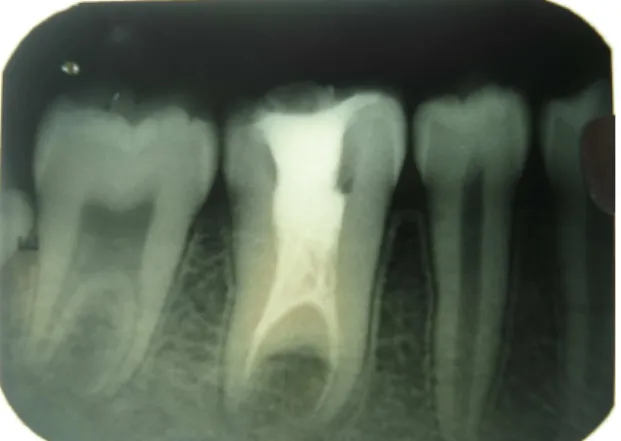
The premolars also showed widened pulp chamber and root canal. The status of third molars could not be assessed because they were in the developing stages. IOPA radiograph of mandibular right region also revealed root canal treated mandibular first molar.
No other remarkable dental findings were observed clinically or radiographically. Past medical history was non contributory. General physical examination did not reveal any significant findings. Patient’s physical and mental development was within normal rates for his age.
Topical anesthetic, antiseptic gel was prescribed to reduce the symptoms and to promote healing of ulcer on buccal mucosa.
Fig. 1- Panoramic radiograph showing multiple Taurodonts (first and second permanent molars of all four quadrants).
Oral & Maxillofacial Pathology Journal [ OMPJ ] V ol 2 No 1 Jan- Jun 2011 ISSN 0976-1225
pulp chamber and apically shifted floor of pulp chamber.
Fig 3: IOPA showing first and second molars with typical features of Taurodontism and widened pulp canals of Premolars.
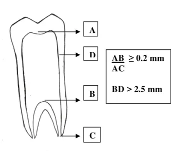
Fig 4 – Illustration showing Shiffman and Chanannel’scriteria for diagnosing Taurodontism. Discussion
Taurodontism is an anomaly of multi rooted teeth with enlargement of pulp chamber at the expense of root. A taurodont does not exhibit any unique morphologic clinical characteristics which may aid in its recognition. Diagnosis of taurodont is usually made in a radiograph. In a radiograph taurodont shows a long rectangular body with short roots, the pulp chamber is elongated in the apico- occlusal direction and lacks
constriction at the cervix. The apico-occlusal height of the pulp chamber varies depending upon the type of taurodontism.
Various diagnostic approaches were adopted by authors who have reported cases of taurodontism. In 1928 Shaw41 classified taurodontism arbitrarily, based
on relative degree of apical displacement of floor of pulp chamber into hypotaurodontism, meso and hypertaurodontism. The pulp chamber may extend to the apex of the tooth in hypertaurodontism and in that case the pulpal floor appears as a shelf in close proximity to the apices. The apical extension may be mild to moderate in hypo- or mesotaurodontism.
A more effective approach for assessing taurodontism was put forward by Feichtinger and Rossiwall46. They suggested that a tooth can be
considered as a taurodont only if the distance from the furcation of the root to the CE J is greater than the cervico-occlusal distance. Later in 1978 Shiffman and Chanannel40 established mathematical criteria which are
adopted by various authors for assessing their cases. According to this criteria, a tooth is considered as a taurodont if the distance from the lowest point of roof of the pulp chamber (A) to the highest point of the floor (B), divided by the distance from A to the root apex (C) is equal to or greater than 0.2 mm, and when the distance from B to the CE J (D) is greater than 2.5 mm. (Fig 4)
Only little information is available about the clinical significance of taurodontism. According to Widerman and Serene47 unusual shape of the root
canals in taurodonts may cause difficulty in endodontic treatment. Sathyanarayana and Carounanidy reported a case of Taurodontism involving mandibular left first and second molars in which they have performed conventional endodontic treatment. According to them endodontic treatment in taurodont was challenging. E ven in our case we have observed an endodontically treated first molar which was clinically and
radiographically symptom free.
Since the available data on prevalence of Taurodontism is limited, we opine that detailed larger scale studies have to be carried out to assess its prevalence in the general population and to compare it with other ethnic groups.
Reference:
1. Witk op CJ Jr:Manifestations of genetic diseses s in Human Pulp. Oral Surg 1971;32:278-283.
2. Gorjanovic-Kramberger K. Uber prismatische molarwurzeln rezenter und diluvialer Menschem. A nat Anz 1908;32:401-430.
AB
≥ 0.2 mm
AC
BD > 2.5 mm
A
D
B
Oral & Maxillofacial Pathology Journal [ OMPJ ] V ol 2 No 1 Jan- Jun 2011 ISSN 0976-1225
3. Mark T, Jaspers MT. Taurodontism in Down’s Syndrome. Oral Surg. 1981;51:632-636.
4.Keith A . Problems relating to the teeth of the earlier forms of prehistoric man. Proc R Soc Med. 1913;6:103-110.
5. Mena CA . Taurodontism Oral Surg Oral Med Oral Pathol 1971;32:812-823.
6.Mangron JJ. Two cases of taurodontism in modern human jaws. Br Dent J 1962;113:309-312.
7. Tik u A , Damle SG, Nadk arni UM. Kalask ar RR. Hypertaurodontism in molars and premolars: Management of two rare cases. J Indian Soc Pedo Prev Dent 2003;21:131-134.
8. Sathyanarayana R, Carounanidy U. Taurodontism – Review and an endodontic case report. E ndodontology. 2001;13: 8-10. 9. Bhat SS, Sargod S, Mohammed SV . Taurodontism in deciduous molars – A case report. J Indian Soc Pedo Prev Dent. 2004;22(4):193-196
10. Rao A , Arathi R. Taurodontism of deciduous and permanent molars: Report of two cases. J Indian Soc Pedod Prev Dent 2006;24:42-44
11. Shaw, J. C. M. Taurodont teeth in South A frican races. J. A nat. 1928; 62: 476-498.
12. Goldstein, E .; Gottlieb, M. A .Taurodontism: familial tendencies demonstrated in eleven of fourteen case reports. Oral Surg. Oral Med. Oral Path. 1973; 36: 131-144.
13. Gamer, S.; Zusman, S. H.Taurodontism in a 15-year-old boy and his mother. J. S. Calif. Dent. A ssoc. 1967; 35: 441-444.
14. Witk op, C. J.; Rao, S. Inherited defects in tooth structure.
Birth Defects Orig. Art. Ser. 1971;7 (7): 153-184.
15.Jaspers, M. T.; Witk op, C. J., Jr.Taurodontism, an isolated trait associated with syndromes and X-chromosomal aneuploidy.
A m. J. Hum. Genet. 1980; 32: 396-41.
16. V arella J, A lvesalo L , Mayhall J. Taurodontism in 45,X Females. J Dent Res, 1990; 69(2):494-495.
17. Blumberg JE , Hylander WL , Goepp RA . Taurodontism: a biometric study. A m J Phys A nthropol. 1971; 34:243–255. 18. Reichart P, Quast U. Mandibular infection as a possible etiological factor in Taurodontism. J Dent 1975; 3: 198- 202. 19. Keeler C. Taurodont malar and shovel incisors in Klinefelter’s Syndrome. J Hered 1973;64: 234-236. 20. Gardner DG, Girgis SS. Taurodontism, shovel shaped incisors and the Klinefelter’s Syndrome. J Can Dent A ssoc 1978; 44:372-373.
21.Yeh SC, Hsu TY. E ndodontic treatment in taurodontism with Klinefelter's syndrome: a case report. Oral Surg Oral Med Oral Pathol Oral Radiol E ndod. 1999; 88:612–615. 22. Jaspers MT. Taurodontism in Down’s syndrome. Oral surg Oral med Oral Pathol. 1981;51:632-636.
23. Bell J, Civil CR, Townsend GC, Brown RH. The prevalence of Taurodontism in Down’s syndrome. J ment Defic Res 1989; 33:467-76.
24. A lpoz A R, E ronat C. Taurodontism in children associated with trisomy 21 syndrome. J Clin Pediatr Dent. 1997; 22:137–39.
25. L ichetenstein JR, Warson RJorgenson R, Dorst JP ,
McKusick V A , The Tricho- Dento-Osseous Syndrome. A m J Hum Genet. 1972;24:569-582.
26. Witk op CJ. Clinical aspects of dental anomalies. Int Dent J. 1976; 26:378–390.
27. Crawford PJ, A lfred HJ, Clark e A . Clinical and
radiographic dental findings in X- link ed hypohydrotic ectodermal dysplasia. J med Genet. 1991;28:181-185.
28. Stenvick A , Zachrisson BU, Svatun B. Taurodontism. Oral Surg Oral Med Oral Pathol1972;33:841-845. 29. Gardner DG, Girgis SS. Taurodontism, short roots and external resorption associated with short stature and a small head. Oral Surg Oral Med Oral Pathol 1977;44:271-73. 30. L aatik ainen T, Ranta R. Taurodontism in twins with cleft lip and/ or palate. E ur J Oral Sci. 1996; 104: (2; part 1). 82– 86.
31. Schalk – van der Weide Y, Steen WH, Bosman F. Taurodontism and length of teeth in patients with oligodontia. J Oral Rehábil. 1993;20(4):401-412
32. Kuchler C, E ryca, Risso A , Patricia, Costa DC, Marcelo, Modesto, A driana, V ieira R, A lexandre. A ssessing the proposed association between tooth agenesis and taurodontism in 975 paediatric subjects. Int J Paed Dent.2008; 18(3): 231-234.
33. Stenvik A , Zachrisson BU, Svatun B. Taurodontism and concomitant hypodontia in siblings. Oral Surg Oral Med Oral Pathol. 1972; 33:841–845.
34. Ireland E J, Black JP, Scures CC. Short root , Taurodontia and multiple dens invaginatus. J Pedod 1987;11:164-175. 35. E lzay RP, Chamberlain DH. Differential diagnosis of enlarged dental pulp chambers: a case report of amelogenesis imperfection with taurodontism. J Dent Child 1986; 53: 388 - 390.
36. A lfred MJ, CrawfordPJ. V ariable expression in A melogenesis Imperfecta with Taurodontism. J Oral Pathol1988;17:327-333.
37.Crawford PJ, E vans RD, A ldred MJ. A melogenesis imperfecta: autosomal dominant hypomaturation- hypoplasia type with Taurodontism. Br. Dent J 1988;164:71-73.
38. Darwazeh A M, Hamasha AA , Pillai K. Prevalence of taurodontism in Jordanian dental patients. Dentomaxillofac Radiol. 1998; 27:3163–3165.
39. MacDonald-Jank owsk i DS, L i TT. Taurodontism in young adult Chinese population. Dentomaxillofac Radiol. 1993; 22:3140–3144.
40. Schiffman A , Chanannel I. Prevalence of taurodontism found in radiographic dental examination of 1200 young adult Israeli patients. Community Dent oral E pidemiol 1978; 6: 200-203.
41.Shaw JCM. Taurodont teeth in South A frican races. J A nat 1928;62: 476-498.
42.Madeira MC, L eite HF, Filho WDN, Simoes S. Prevalence of taurodontism in premolars. Oral Surg Oral Med Oral Pathol 1986;61: 158-162.
Oral & Maxillofacial Pathology Journal [ OMPJ ] V ol 2 No 1 Jan- Jun 2011 ISSN 0976-1225
45. Sert S
, Bayırlı G.
Taurodontism in Six Molars: A Case Report. Journal of E ndodontics. 2004; 30(8):601– 602. 46. Feichtinger C, Rosiwall B. Taurodontism in human sex chromosome aneuploidy. A rch Oral Biol 1977; 22:327-329. 47. Widerman FH, Serene TP. E ndodontic therapy involving a taurodont tooth. Oral Surg. 1971; 32:618-620.Source of Support: Nil, Conflict of Interest: None