Mini-implants: Mechanical resource
for molars uprighting
Susiane Allgayer1, Deborah Platcheck2, Ivana Ardenghi Vargas3, Raphael Carlos Drumond Loro4
How to cite this article: Allgayer S, Platcheck D, Vargas IA, Loro RCD. Mini-implants: Mechanical resource for molars uprighting. Dental Press J Orthod. 2013 Jan-Feb; 18(1):134-42.
Submitted: March 16, 2011 - Revised and accepted: August 31, 2011.
» Patients displayed in this article previously approved the use of their facial and intraoral photographs.
Contact address: Susiane Allgayer
PUCRS – Pontifícia Universidade Católica do Rio Grande do Sul Av. Ipiranga, 6681 – Prédio 06 – Sala 209
CEP: 90619-900 – Porto Alegre / RS, Brazil E-mail: [email protected]
Introduction:The early orthodontic treatment allows correction of skeletal discrepancies by growth control, and the elimination of deleterious habits, which are risk factors for the development of malocclusions, favoring for the correction of tooth positioning later in a second treatment stage. During development of teeth and occlusion, the mandibular second molars commonly erupt in the oral cavity ater all other teeth of the anterior region. In their eruptive process there may be a condition known as tooth impaction, which precludes its complete eruption and requires proper uprighting treat-ment. The temporary anchorage devices allow disimpaction and movement of these teeth directly to their inal position, without the need of patient compliance or reaction movements in other parts of the arch. Objective:This paper aims at describing a case report of the treatment of a patient with Angle Class II malocclusion, performed in two phases, in which mini-implants were used for uprighting the impacted mandibular second molars.
Keywords: Corrective orthodontics. Orthodontic anchorage procedures. Impacted tooth.
1 PhD student of Orthodontics and Facial Orthopedics, PUC-RS. 2 PhD and Professor of Orthodontics, ABO/RS.
3 PhD in Dentistry, ULBRA.
4 PhD in Oral and Maxillofacial Surgery and Professor at Graduation and
Post-graduation courses, PUCRS.
» The author reports no commercial, proprietary or inancial interest in the prod-ucts or companies described in this article.
Introdução:o tratamento ortodôntico precoce permite a correção das discrepâncias esqueléticas por meio do controle de crescimento e a eliminação de hábitos deletérios, que são fatores de risco para o desenvolvimento de más oclusões, que favorecem a correção do posicionamento dentário mais tardiamente, em uma segunda fase do tratamento. Durante o desenvolvimento da dentição e da oclusão, normalmente o segundo molar inferior chega à cavidade bucal após todos os dentes posicionados anteriormente a ele. Durante seu processo eruptivo, pode ocorrer uma condição chamada “impacção dentária”, em que sua erupção completa é interrompida, exigindo tratamento apropriado para verticalização. Os dispo-sitivos temporários de ancoragem permitem a desimpacção e a movimentação desses dentes diretamente às suas posições inais, sem a necessidade de cooperação do paciente e sem movimento de reação nas outras unidades da arcada. Objeti-vo: descrever o tratamento de um caso de má oclusão Classe II de Angle, realizado em duas fases, durante o qual foram utilizados mini-implantes para verticalização dos segundos molares inferiores impactados.
1,000.3,4,5,6 The most probable causes of impaction of second molars seem to be related to the excessive size of these teeth, deficient mandibular growth, in-adequate length of the mandibular arch or only due to an abnormal eruption pathway.7,8
The treatment options may involve or not sur-gery for exposure of the molar crown and utilization of fixed and/or removable appliances.7-10
The utilization of temporary implants for orth-odontic anchorage allowed a new perspective in the orthodontic treatment, especially in the perma-nent dentition. Mini-implants, palatal implants and mini-plates are currently used in several clinical sit-uations, as the treatment of open bites and upright-ing of molars without adverse effects on the adjacent teeth.11-18
Class II is the severe malocclusion most frequent-ly found and is characterized by “distal positioning” of the mandibular teeth compared to the maxillary teeth, which may be caused by bone dysplasia or for-ward positioning of the alveolar process or maxillary dental arch, or even by the combination of skeletal and dental factors.9
This case report describes the orthodontic treat-ment of a patient with Angle Class II malocclusion with impaction of the mandibular second molars, in which two mini-implants were used as an anchorage aid for uprighting of the teeth, which were impacted on the distal aspect of mandibular first molars.
CASE REPORT
Caucasian patient of female gender sought for ini-tial orthodontic treatment at the age of 9 years and 5 months. The general health status was good and there was no history of severe diseases or traumas. On the clinical examination, it was observed that the patient was in the intermediate mixed dentition and presented tongue thrusting and speech disorder.
patient presented a favorable mandibular growth pat-tern in both horizontal and vertical directions. Con-cerning the dental relationships, the maxillary and mandibular incisors were slightly buccally tipped and protruded (1-NA 6 mm, 1-NA 25o, 1-NB 6 mm, 1-NB 25o, and IMPA 95o). The patient presented overjet of 3.5 mm, anterior open bite, negative tooth size discrepancy of 4 mm in the maxillary arch and
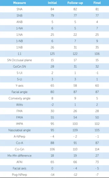
Table 1 - Cephalometric measurements.
Measure Initial Follow-up Final
SNA 84 82 81
SNB 79 77 77
ANB 5 5 4
1-NA 6 5 7
1.NA 25 22 25
1-NB 6 7 9
1.NB 26 31 35
1.1 125 122 106
SN.Occlusal plane 15 17 15
GoGn.SN 28 31 32
S-Ul 2 1 -1
S-Ll 3 3 1
Y-axis 65 58 60
Facial angle 80 87 87
Convexity angle 8 9 5
Witts -2 1 2
FMA 30 26 28
FMIA 55 54 50
IMPA 95 100 102
Nasolabial angle 95 109 105
A-NPerp - 4 - 2 - 1
Co-A 88 91 87
Co-Gn 106 110 114
Mx-Mn diference 18 19 27
AFAI 65 66 73
Facial axis 0 - 4 - 5
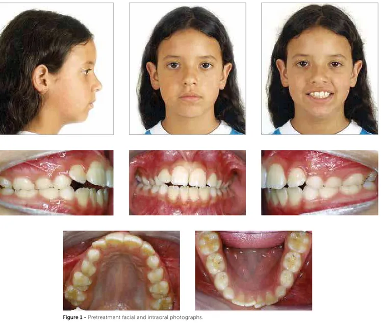
Figure 1 - Pretreatment facial and intraoral photographs.
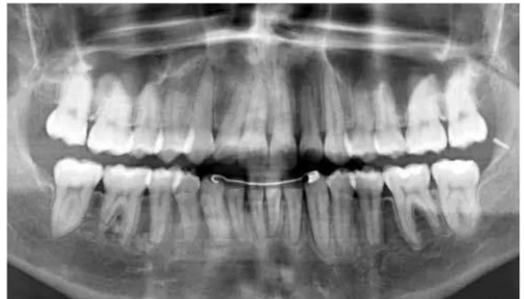
During the eruption process, the mandibular second molars presented a marked mesial eruption pathway (Fig 6), leading to impaction on the irst molars. Surgi-cal removal of third molars was indicated to enhance the uprighting of second molars. The same procedure com-prised surgical exposure and bonding of a bracket on the cusp of these teeth and placement of mini-implants (Ortoimplante Conexão, 2.0 x 9.0, medium transmu-cous proile) for anchorage on the retromolar region, distal and occlusal to the second molars, using a muco-periosteal lap11. This site was selected to allow support for orthodontic eruption of the impacted second molars in distal and occlusal direction (Fig 7A).
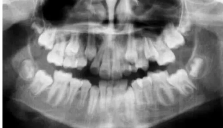
Figure 3 - Initial panoramic radiograph.
The treatment objectives were to correct the skel-etal and dental Class II malocclusion and intercept the malocclusion, providing conditions for adequate growth of the bone bases, thus improving the dentoal-veolar morphology. Therefore, it was necessary to con-trol the maxillary protrusion by redirecting growth, to eliminate oral habits, to re-establish normal lip and tongue functions to correct the overbite, and to con-trol the eruption of mandibular second molars, which presented a mesial eruption pathway.
TREATMENT PROGRESS
Initially, a maxillary appliance with tongue crib was used for six months, for tongue reeducation. Af-ter achievement of adequate overbite (Fig 3), correc-tion of the skeletal discrepancy was initiated by redi-recting maxillary growth with an extraoral traction appliance, during 10 months (Fig 5).
The fixed orthodontic appliance was placed us-ing standard edgewise brackets, and the alignment and leveling stage was initiated.
Figure 5 - Intermediate lateral cephalometric radiograph and tracing. Figure 6 - Intermediate panoramic radiograph.
The affected teeth were gradually moved using chain elastics, improving their positioning (Fig 7B). The period required for uprighting was 18 months. The mini-implants did not present mobility and the patient hygiene was excellent.
The maxillary second molars did not erupt (Fig 7B). After surgical removal of fibrosis, they erupted with palatal tipping and the mandibular second mo-lars were in buccoversion, which caused a crossbite that was treated by placement of a contracted lin-gual archwire, besides expansion of the maxillary arch. Simple cross vertical elastics were also used on brackets bonded on the palatal aspects of maxillary right and left second molars and buccal aspect of the mandibular right and left second molars. After 18
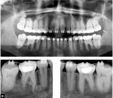
months, they presented good inclination in the bone base (Fig 7C), were included in the archwire, the lin-gual arch and mini-implants were removed and cor-rective fixed appliances were placed for treatment fi-nalization. There was resorption of the distal root of the mandibular right first molar, which was followed up radiographically (Figs 7C and D).
The total treatment time with the fixed applianc-es was eight years, due to the period elapsed to wait for eruption of the maxillary and mandibular second molars. After orthodontic finishing and improve-ment of occlusion, the appliance was removed and a wraparound removable orthodontic retainer was placed in the maxillary arch, as well as a mandibular 3x3 bonded lingual retainer.
Figure 7B - Intermediate radiograph and photographs.
Figure 7A - Intermediate radiograph and photographs.
Figure 7C - Intermediate radiograph and photograph. Figure 7D - Intermediate radiographs.
Figure 8 - Final facial and intraoral photographs.
Figure 9 - Final lateral cephalometric radiograph and tracing. Figure 10 - Final panoramic radiograph.
Figure 11 - Superimposition of cephalometric tracings at pretreatment (black) and posttreatment (red), with parallel Sella-Nasion lines and register at Sella for growth analysis. The redirection of maxillary growth combined to the fa-vorable mandibular horizontal growth corrected the malocclusion.
Both mini-plates and endosseous implants are costly and difficult to be removed.13,18,25
However, skeletal anchorage has surely been an important tool in Orthodontics. These treatments require minimum patient compliance and a good oral hygiene can be more easily maintained. Even in patients who do not need prosthetic rehabilitation, recent studies have used the retromolar, palatal and alveolar regions for the placement of implants only for orthodontic purposes, for induced movement of teeth or segments.16
TREATMENT RESULTS
At treatment completion, the facial outcome was excellent. The intraoral analysis revealed Class I molar and canine relationship, adequate overjet and overbite, coincident midlines and adequate intercuspation be-tween the dental arches, including the second molars. The panoramic radiograph evidenced correct parallel-ism between the roots, and the cephalometric tracing and superimposition on the cephalogram revealed the dental and skeletal changes achieved at treatment com-pletion (Figs 8 - 11, Tab 1).
DISCUSSION
Tooth impaction may be caused by factors as he-redity, malposition of the tooth germ, overretention of deciduous teeth, localized pathological lesions, re-duced arch length and deficient growth of the man-dibular ramus.9 The mandibular second molars may be impacted or severely malpositioned and are often blocked under the distal convexity of permanent first molars. Their early repositioning is usually advanta-geous during active root development.9,10,19
several clinical situations, they are preferred as a skel-etal anchorage method25.
The site for fixation of the orthodontic mini-implant should present sufficient quantity of cortical bone tissue to assure immediate mechanical stability, minimum discomfort to the patient, safety to ana-tomical structures, as well as to allow the application of adequate biomechanics. The retromolar region21,22 is indicated to promote the uprighting of molars be-cause it increases the distal force component. When mini-implants are placed at this region, it is also pos-sible to achieve extrusion forces with a distal com-ponent, another reason for selecting this treatment.26
One of the goals of orthodontic treatment is to achieve a harmonious arch shape. Transverse problems should be corrected soon after diagnosis to prevent bone deficiencies.27 When a tooth presents crossbite, it rarely presents alterations only in its axial inclina-tion. The opposing tooth in the other arch is often also malpositioned. Thus, the mandibular tooth may be in buccoversion and the maxillary tooth in palato-version. In these cases, both should be corrected and simple cross elastics are indicated. In the present case, brackets bonded on the surfaces of molars were used to position the elastics to correct the crossbite. The technique is relatively easy, but requires patient com-pliance.28 Combined to elastics, a contracted lingual archwire soldered to the bands on the mandibular right and left second molars was placed, because they presented with marked buccoversion.
Similar to the aforementioned problem, deleterious habits should be interrupted as early as possible, to re-establish the normal lip and tongue functions in order to allow a correct overbite. In the present case, a max-illary removable appliance fabricated with clear acrylic and a tongue crib fabricated with 0.7mm stainless steel wire was used for tongue reeducation. This appliance — which is easy to be made and is ixed in the oral
the force is removed, there is the repair process and reestablishment of the periodontal ligament.30 In the present case, resorption of the mandibular right irst molar observed during treatment was controlled by ra-diographic follow-up and force control. Considering the large movement of the second molar, repair of the periodontal ligament of the mandibular right irst mo-lar and reestablishment of the normal functions of the two teeth, resorption of the irst molar was clinically acceptable (Figs 7C and D).
Ultimately, the mini-implants provide biome-chanical advantages that allow an easier and more effective treatment, without the need of patient compliance.17,26 The preference of clinicians for a certain treatment modality must not be necessar-ily followed. After deciding that the utilization of mini-implants is safe and necessary for the treat-ment, the area of insertion should be selected con-sidering the accessibility, conditions of soft and hard tissues, biomechanical utility in orthodontics, comfort to the patient and possibility of irritation of the adjacent oral tissues.26
CONCLUSION
1. Hitchin AD, Durh MDS, Edin RCS. The impacted maxillary canine. Br Dent J. 1956;100(1):1-14.
2. Lindauer SJ, Rubenstein LK. Canine impaction identiied early with
panoramic radiographs. J Am Dent Assoc. 1992;123(3):91-7.
3. Grover PS, Lorton L. The incidence of unerupted permanent teeth and
related clinical cases.Oral Surg Oral Med Oral Pathol. 1985;59(4):420-5. 4. Varpio M, Wellfelt B. Disturbed eruption of the lower second molar:
clinical appearance, prevalence, and etiology. ASDC J Dent Child. 1988;55(2):114-8.
5. Montelius GA. Impacted teeth: a comparative study of chinese and
canadian dentitions. J Dent Res. 1932;12(6):931-8.
6. Johnsen DC. Prevalence of delayed eruption of permanent teeth as a
result of local factors. J Am Dent Assoc. 1977;94(1):100-6.
7. Lima CEO, Henriques JFC, Janson GRP, Freitas MR. Segundo molar
inferior impactado: revisão e apresentação de um caso clínico. Rev Clín Orthod Dental Press. 2004;2(6):68-75.
8. Teixeira RG, Vidal BC, Bastos EPS. Reposicionamento cirúrgico de um
segundo molar inferior direito impactado com cárie: relato de caso. J Bras Ortodon Ortop Facial. 2000;5(30):76-81.
9. Moyers RE. Ortodontia. 4ª ed. Rio de Janeiro: Guanabara Koogan; 1991.
10. Pogrel MA. The surgical uprighting of mandibular second molars. Am J Orthod Dentofacial Orthop. 1995;108(2):180-3.
11. Faber J, Velasque F. Titanium miniplate as anchorage to close a premolar space by means of mesial movement of the maxillary molars. Am J Orthod Dentofacial Orthop. 2009;136(4):587-95.
12. Cope JB. Temporary anchorage devices in orthodontics: a paradigm shift. Semin Orthod. 2005;11(1):3-9.
13. Faber J, Morum TFA, Leal S, Berto PM, Carvalho CKS. Miniplacas permitem tratamento eiciente e eicaz da mordida aberta anterior. Rev Dental Press Ortod Ortop Facial. 2008;13(5):144-57.
14. Faber J. Ancoragem esquelética com miniplacas. In: Lima Filho RMA, Bolognese AM. Ortodontia: arte e ciência. Maringá: Dental Press; 2007. p. 449-73.
15. Kuroda S, Katayama A, Takano-Yamamoto T. Severe anterior open-bite case treated using titanium screw anchorage. Angle Orthod. 2004;74(4):558-67.
16. Erverdi N, Keles A, Nanda R. The use of skeletal anchorage in open bite treatment: a cephalometric evaluation. Angle Orthod. 2004;74(3):381-90.
REFERENCES
17. Sugawara J, Kanzaki R, Takahashi I, Nagasaka H, Nanda R. Distal movement of maxillary molars in nongrowing patients with the skeletal anchorage system. Am J Orthod Dentofacial Orthop. 2006;129(6):723-33. 18. Sugawara J, Daimaruya T, Umemori M, Nagasaka H, Takahashi I,
Kawamura H, et al. Distal movement of mandibular molars in adult patients with skeletal anchorage system. Am J Orthod Dentofacial Orthop. 2004;125(2):130-8.
19. Vedtofte H, Andreasen JO, Kjaer I. Arrested eruption of the permanent lower second molar. Eur J Orthod. 1999;21(1):31-40.
20. Rindler A. Efects on lower third molars after extraction of second molars. Angle Orthod. 1977;47(1):55-8.
21. Higuchi KW, Slack JM. The use of titanium ixtures for intraoral anchorage to facilitate orthodontic tooth movement. Int J Oral Maxillofac Implants. 1991;6(3):338-44.
22. Roberts WE, Marshal KJ, Mozsary PG. Rigid endosseous implant utilized as anchorage to protract molars and close an atrophic extraction site. Angle Orthod. 1990;60(2):135-52.
23. Lee JS, Kim DH, Park YC, Kyung SH, Kim Tk, The eicient use of midpalatal miniscrew implants. Angle Othod. 2004;74(5):711-4. 24. Wehrbein H, Merz BR, Diedrich P, Glatzmaier J. The use of palatal
implants for orthodontic anchorage. Design and clinical application of the orthosystem. Clin Oral Implants Res. 1996;7(4):410-6.
25. Joseph S. Petrey, Marnie M. Saunders, G. Thomas Kluemper, Larry L. Cunningham, and Cynthia S. Beeman. Temporary anchorage device insertion variables: efects on retention. Angle Orthod. 2010;80(4):634-41.
26. Lee JS, Kim DH, Park YC, Vanardall RL. Aplicação dos mini-implantes ortodônticos. São Paulo: Quintessence; 2009.
27. Joondeph DR. Mysteries of asymmetries. Am J Orthod Dentofacial Orthop. 2000;117(5):577-9.
28. Assed S. Odontopediatria: bases cientíicas para a prática clínica. São Paulo: Artes Médicas; 2005.
29. Tanaka OM, Amorin LH, Shintcovsk RL, Hirata TM. Treatment of patient with severaly shortened maxillary central incisor roots. J Clin Orthod. 2008;42(12):729-31.