Analysis of the rotational position of the
maxillary first permanent molar in normal
occlusion and Class II, division 1 malocclusion
Marisa Helena Zingaretti Junqueira*, Karyna Martins Valle-Corotti**, Daniela Gamba Garib**, Ricardo Brandão Vieira***, Flavio Vellini Ferreira****
Objective: The purpose of this study was to evaluate and compare the rotational position of maxillary first permanent molars (U6) in subjects in the permanent dentition presenting normal occlusion and Class II, division 1 malocclusion. Methods: Casts of 60 subjects with normal oc-clusion (Group 1, mean age 15.1 years) and 120 with untreated Class II, division 1 malococ-clusion (Group 2, mean age 15.5 years) were evaluated. The maxillary dental casts were scanned and the position of maxillary molars was analyzed using three angular measurements and one linear measurement, named indicators 1, 2, 3 and 4, respectively. The mesiopalatal rotation of maxillary first molars was evidenced by an increase in the values of indicators 1 and 4 and decrease in the indicators 2 and 3. Comparisons between groups were performed using Student´s t test for mea-surements with normal distribution and by the Mann-Whitney test for non-normal distribution, at p<0.05. Results: Statistically significant differences were found between Groups I and II for all indicators, on both right and left sides. Conclusion: It was concluded that individuals with Class II, division 1 malocclusion present greater mesiopalatal rotation of the maxillary first molars.
Abstract
Keywords: Molar rotation. Normal occlusion. Class II, Division 1. Orthodontics.
* MSc in Orthodontics, São Paulo City University.
** MSc, PhD, Professor of Orthodontics, São Paulo City University.
*** Professor of Orthodontics, Bauru Dental School, and Craniofacial Anomalies Rehabilitation Hospital, São Paulo University. **** Chairman, Masters Course in Orthodontics, São Paulo City University.
intROduCtiOn
Good treatment finishing requires knowl-edge on the characteristics of an ideal occlu-sion. Andrews1 reported the six keys to normal occlusion to describe the ideal relationship be-tween the dental arches, contact points bebe-tween maxillary and mandibular teeth, individual po-sitioning of teeth concerning their tipping and angulation, tooth rotation and curve of Spee. It should be highlighted that an ideal occlusion
should present all these characteristics de-scribed by Andrews.
Rotations of first molars are observed in most cases of Angle Class II division 1 malocclusion, which increases the space occupied by the teeth in the dental arch.3,6 Despite the good positioning of premolars and canines, in some cases, rotation of the first molar occurs due to incorrect posi-tioning induced by local etiologic factors, such as proximal caries, during development of the mixed or permanent dentition.7
Several authors6,9,11,13,17 specifically addressed the rotation of the permanent maxillary first mo-lar. In addition to a thorough literature review, they have conducted measurements on maxillary dental casts and concluded that the position of the first molar is very important for the orthodontic practice. Three positions of this tooth are signifi-cant: positioning in the maxilla, axial inclination and rotation in the long axis. A guideline for clini-cal evaluation of the position of maxillary first molars on the occlusal view has been proposed19 using a line traced through the tips of distobuccal and mesiopalatal cusps of the permanent maxil-lary first molar. It was observed that, in normal occlusion, this line should pass through the distal third of the canine on the opposite side, and that this was a good guideline to analyze the mesio-palatal rotations characteristic of mesial displace-ment of first molars in malocclusions. Cetlin and Ten Hoeve4 described that, when the two maxil-lary first molars are well positioned, their buccal aspects are parallel to each other.
In another study,10 the rotations of maxillary first molars were quantified by measuring the an-gle formed by the intersection of two lines, name-ly one formed by the union of two points corre-sponding to the tips of buccal cusps and the other to the midpalatal suture. The means observed for this angle were 14.08º and 12.76º for the right and left maxillary molars, respectively, in patients with acceptable occlusions.
Based on data in the literature, this study evaluated and compared the rotation of the maxillary first molar in patients with permanent
dentition with normal occlusion and Class II di-vision 1 malocclusion.
MAtERiAL And MEtHOdS
This study was approved by the Institutional Review Board of University Cidade de São Paulo, under protocol number 13180098. The study was conducted on sixty pairs of dental casts with nor-mal occlusion not submitted to orthodontic treat-ment, in the age range 11 years 11 months to 31 years 10 months, with mean age 15 years 1 month, being 32 females and 28 males, from the files of the Department of Orthodontics of Bauru Dental School, University of São Paulo, which comprised the sample in Group 1 (Control). The study also included 120 pairs of dental casts before cor-rective treatment, presenting Class II division 1 malocclusion, being 70 females and 50 males, in the age range 10 years to 35 years 1 month, with mean age 15 years 6 months (Group 2), from the files of the Training and Specialization Courses in Orthodontics of Prev Odonto Study and Research Center at the city of Rio de Janeiro, state of Rio de Janeiro, which constituted the sample in Group 2.
The sample in Group 2 met the following inclu-sion criteria: permanent dentition with all present teeth (regardless of the presence of second and third molars); trimmed dental casts with defined occlu-sion; bilateral molar Class II division 1 relationship; no congenital malformations; no surgical skeletal dysplasias; no teeth with ectopic eruption; no abnor-malities of shape and size of the molars, premolars and canines; no unilateral or bilateral posterior cross-bite; absence of anterior and posterior open cross-bite; no hypodontia, tooth extractions and supernumerary teeth; no fractures or severe wear of the cusps of molars and premolars; and no prosthetic restorations on the molars, premolars and canines.
A
1
B
R P1
P2
MV
DV ML
RP1
RP2 ML DV
MV P2 P1 R
A
C
D B
E
3 R
B
2
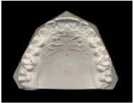
Fourteen points were marked on each digi-tized image, being 2 on the midpalatal suture and 12 divided between the right and left sides of the maxillary dental arch (Figs 1 and 2), which were later measured on the Radiocef software.18
The following points were used:
» RP1: most anterior region of the midpalatal suture, close to the first or second premolars. » RP2: posterior region of the midpalatal
su-ture, close to the first or second molars. » MV: mesiobuccal cusp tip of maxillary first
molar.
» DV: distobuccal cusp tip of maxillary first molar.
» ML: mesiolingual cusp tip of maxillary first molar.
» P1: most external point tangent to the buccal aspect of the first premolar, marked on the occlusal view.
» P2: most external point tangent to the buc-cal aspect of the second premolar, marked on the occlusal view.
» R: most distal point on the distal aspect of the maxillary canine.
The lines defined for the analysis were gener-ated by the union of marked points, being one central and the others on the right and left sides of the dental arch (Figs 3, 4, 5 and 6).
» Line A: union of points RP1 and RP2. » Line B: union of points MV and DV. » Line C: union of points MV and ML. » Line D: union of points P1 and P2. » Line E: union of points DV and ML.
Each line was traced on the right and left sides of the dental arch, except for line A, which were then named Bd, Be, Cd, Ce, Dd, De, Ed and Ee.
The angles defined for the analysis were gen-erated by the intersection of the aforementioned lines, bilaterally (Figs 3, 4 and 5).
» Angle 1: intersection of line A with line B. » Angle 2: intersection of line A with line C. » Angle 3: intersection of line B with line D. Angle 1 indicated mesial rotation when in-creased, while angles 2 and 3 indicated rotation
FIGURE 1 - Points marked on the digital images of dental casts.
FIGURE 4 - Angle 2 (Line A.Line C). FIGURE 6 - Linear measurement (R). FIGURE 2 - Scanned image of dental cast on the
computer screen with the points marked.
FIGURE 5 - Angle 3 (Line B.Line D).
when decreased in relation to the values found for normal occlusion.
A linear measurement was defined for the analysis (Fig 6), for both sides of the dental arch, as the distance perpendicular to point R at one side to the line generated by points DV and ML on the opposite side. These were named Rd (for the right side) and Re (for the left side). Thus, the distance Rd is the distance from point Rd to line Ee and vice-versa. This measurement indicates that, the greater the distance found, the greater is the mesial rotation of the molar.
StAtiStiCAL AnALYSiS
The groups of normal occlusion and Class II malocclusion were analyzed by descrip-tive statistics, with achievement of arithmetic means, medians, standard deviations, mini-mum and maximini-mum values of measurements obtained for each indicator of the rotational position of the first molar, between the com-ponents in the two groups.
The results of measurements of each indi-cator obtained on the right and left sides were compared by the Student t test for indicators with normal distribution and by the non-para-metric Mann-Whitney test for the others.
To determine the proper statistical test to be applied, the Kolmogorov-Smirnov test was initially applied to each set of measurements for analysis of their distribution. It was estab-lished that measurements presenting coefficient of variation above 30% would be submitted to non-parametric statistical analysis.
intRA-EXAMinER ERROR
The method error was evaluated by re-analy-sis of 20 dental casts randomly selected from the two groups. This allowed the achievement of data on the same dental casts at two moments, T1 and T2, indicating the first and second analy-ses, respectively, obtained at a 30-day interval. Based on the data in T1 and T2, the Dahlberg
formula (1940) was applied to estimate the ca-sual errors. Thereafter, the measurements were compared by the paired t test and Wilcoxon test for evaluation of systematic errors, at a signifi-cance level of 5% (p<0.05).
To compare the results of measurements of each indicator obtained on the right and left sides, Student´s t test was applied for indicators whose measurements presented normal distri-bution and the non-parametric Mann-Whitney test for the others.
In order to compare the rotation of first mo-lars between groups with normal occlusion and Class II malocclusion, comparisons were per-formed between the measurements obtained for each indicator. Student´s t test was applied for in-dicators with normal distribution, while indica-tors with wide variation in the values were ana-lyzed by the non-parametric Mann-Whitney test. The characteristics of distribution of values were determined by application of the Kolmogorov-Smirnov test. The arithmetic mean and standard deviation were used to identify the group with highest value for indicators with normal distribu-tion, while the median, minimum and maximum values were used for the others.
A significance level of 5% (p<0.05) was con-sidered for all statistical analyses.
RESuLtS
Evaluation of intra-examiner error
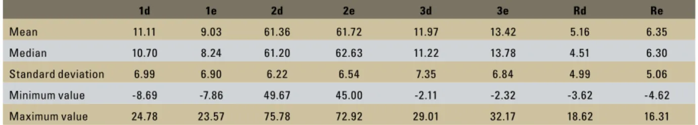
TABLE 1 - Arithmetic means, medians, standard deviations, minimum and maximum values of the rotational position of maxillary first molars in the normal occlusion sample.
TABLE 2 - Arithmetic means, medians, standard deviations, minimum and maximum values of the rotational position of maxillary first molars in the Angle Class II division 1 sample.
Table 2 presents the arithmetic means, medians, standard deviations, minimum and maximum val-ues of indicators related to the rotational position of maxillary first molars in the group with normal occlusion. Considering the means between the right and left sides, the values of the indicators were, re-spectively, 10.07, 61.54, 12.69 and 5.75, with stdard deviations ranging from 6.22º to 7.35º for an-gular measurements and 5 mm for indicator 4.
Table 3 demonstrates the results of the ro-tational position of maxillary first molars in the group with Angle Class II, division 1 malocclusion (Group 2). In this group the measurements exhib-ited different values compared to group 1, with means of 14.97, 57.44, 5.52 and 11.38. However, the standard deviation values were similar, ranging from 6.55º to 8.72º for the indicators 1, 2 and 3, and 5 mm for the linear measurement.
Table 4 presents the comparison between groups according to the results of Student´s t test and Mann-Whitney test, which revealed statis-tically significant differences between the two study groups for all indicators analyzed (p<0.01).
diSCuSSiOn
Rotational position of maxillary irst molar in normal occlusion
The rotational position of the maxillary first molar was evaluated in a sample of Brazilian Cau-casoid individuals with normal occlusion. No dif-ference was observed between the right and left sides for the four indicators used to evaluate the position of the molar (Table 5). These indicators have been previously used by some authors for clinical evaluation or in studies, in both normal occlusion and Class II samples.4,6,9,10-13,19
The mean between measurements on the right and left sides for the angle 1 was 10.07º. Considering the normality values of means and standard deviations, minimum and maximum values of the angles 1d and 1e, demonstrated in Table 3, it was observed that in dental casts with normal occlusion the guideline on the parallelism between the buccal aspects suggested by Cetlin and Ten Hoeve4 was confirmed in only a few cases. This evidence is based on the fact that the line used in this study for angle 1 and the line of Cetlin and Ten Hoeve4 are nearly parallel to each other.
1d 1e 2d 2e 3d 3e Rd Re
Mean 11.11 9.03 61.36 61.72 11.97 13.42 5.16 6.35
Median 10.70 8.24 61.20 62.63 11.22 13.78 4.51 6.30
Standard deviation 6.99 6.90 6.22 6.54 7.35 6.84 4.99 5.06
Minimum value -8.69 -7.86 49.67 45.00 -2.11 -2.32 -3.62 -4.62
Maximum value 24.78 23.57 75.78 72.92 29.01 32.17 18.62 16.31
1d 1e 2d 2e 3d 3e Rd Re
Mean 14.98 14.97 58.20 56.69 5.11 5.93 10.89 11.88
Median 15.00 14.83 58.68 56.38 6.08 6.66 10.89 11.49
Standard deviation 6.79 6.55 6.85 7.15 8.50 8.72 5.38 5.21
Minimum value -2.65 -1.43 39.88 39.88 -16.24 -18.03 -5.97 -6.63
Indicator Test P
Group 1 Group 2
Mean or Median
Mean or Median
Angle 1d U = 3.18 0.001** 10.70 15.00
Angle 1e U = 5.25 0.000** 8.24 14.83 Angle 2d t = 3.01 0.003** 61.36 58.20
Angle 2e t = 4.57 0.000** 61.72 56.69
Angle 3d U = 4.86 0.000** 11.22 6.08
Angle 3e U = 5.43 0.000** 13.78 6.66 Distance Rd U = 6.39 0.000** 4.51 10.89
Distance Re U = 6.00 0.000** 6.30 11.49 TABLE 3 - Unpaired t test (t) and Mann-Whitney test (U), significance levels (p), arithmetic means and medians for each indicator analyzed, for groups 1 and 2.
** significant at 1% level.
TABLE 4 - Paired t and Mann-Whitney tests and significance levels (p) for each indicator analyzed, between the right and left sides for dental casts of groups 1 and 2.
ns = non significant.
Group 1 Group 2
Indicator Test p Test P
Angle 1 U = 1.64 0.104ns U = 1.25 0.211ns
Angle 2 t = -0.31 0.758ns t = 1.66 0.098ns
Angle 3 U = 1.11 0.269ns U = 0.74 0.461ns
Distance R U = 1.30 0.197ns U = 1.46 0.144ns
TABLE 5 - Comparison of means of indicators on the right and left sides for normal occlusion in this study and results of other authors.
TABLE 6 - Comparison of means of indicators on the right and left sides for Angle Class II division 1 malocclusion in this study and results of other authors.
Indicator 1 Indicator 2 Indicator 3 Indicator R
Junqueira et al. (present paper) 10.07o 61.54o 12.69o 5.75 mm
Henry11 (1956) 11.15o
Friel9 (1959) 58.25o
Lamons and Holmes13 (1961) 61o
Orton17 (1966)* 10o *
Ricketts19 (1969) 0 to -2.6 mm
Dahlquist, Gebauer and Ingerwall6 (1996) 11.11o 61.5o 9.9o * 7.4 mm
Hansen et al.10 (1997) 13.42o 64o
Nery and Barbosa16 (2003) 12.2o 63.2o
Indicator 1 Indicator 2 Indicator 3 Indicator R
Junqueira et al.
(present paper) 14.98
o 57.45o 12.69o 5.75 mm
Henry11
(1956) 18.8
o
Nery and Barbosa16 (2003)
15.5o 55.79o
Kanomi et al.12
(2004) 63.5
Angle 2 presented a mean value between sides of 61.54º. Values reported in the literature6,9,10,13 range from 58.2º to 64º, similar to the present re-sults (Table 6). The same was observed for indica-tor 3 (mean 12.69º), which presented similar val-ues as those described in the literature16,17, nearly 10º. These different results observed in different studies may have occurred due to slight differ-ences in the methodology employed to evaluate the rotation of the molar.
The linear measurement in the present study (indicator 4) exhibited a mean value of 5.75 mm. This measurement was suggested by Ricketts19 in 1969, characterizing the normal positions of max-illary first molars. The author considered that the first molar was well positioned when a line passing through the tips of distobuccal and mesiopalatal cusps intersected the distal third of the canine on the opposite side. Therefore, considering that a maxillary canine presents an average mesiodistal dimension of 8 mm,8 this line should fall within a range between the distal aspect of the canine and a point up to 2.6 mm mesially to this tooth. The val-ues obtained in this study ranged from -4.62 mm (line passing 4.62 mm mesially to the distal aspect of the canine) to 18.62 mm (line passing distally to the distal aspect of the canine), being that all cases presented Angle Class I molar relationship, according to key I of Andrews1. Therefore, these results differ from the values demonstrated in pre-vious studies.6,19 Analysis of the sample distribu-tion revealed that only 10% of the cases followed the guideline proposed by Ricketts,19 confirming the findings of a previous study10. The wide varia-tion in the values of this measurement indicates that it may impair the definition of the molar po-sitioning in relation to the malocclusion.
Rotational position of maxillary irst molar in Angle Class ii division 1 malocclusion
The same indicators of the rotational position-ing of the first molar in normal occlusions were ap-plied for evaluation in the Class II malocclusion.
The angle 1, with mean of 14.98º, was slightly lower than the values observed in previous stud-ies11,16, which reveal values ranging between 15.5º and 20º. Only one study12 described values higher than 60º, probably because the study sample in-cluded 148 pretreatment dental casts of patients with different malocclusions, thus not limited to Class II division 1 malocclusion.
The angle 2 exhibited mean value of 57.45º, close to the values reported in the literature (63.5°, 55.79°),12,16 confirming that the maxil-lary first molar presents a different positioning in the Class II malocclusion compared to nor-mal occlusion.
The indicator 3 presented the wider variation, on both right and left sides. The reason for such variation in this angle was that different bucco-lingual positions of the premolars were observed, which caused variations in the position of points P1 and P2 bilaterally. Therefore, there were also variations in the inclination of line D and con-sequently of angle 3. By the practical analysis of dental casts, it was thus considered that this was the least reliable among all indicators for evalu-ation of molar rotevalu-ation, because it depends on unstable reference points located on teeth that may be malpositioned.
The angle 3 and linear measurement R exhib-ited mean values of 5.52° and 11.38 mm which differed from the group with normal occlusion yet no data in the literature are available for com-parison, as displayed in Table 6.
Comparison of rotational position of maxillary irst molar in Class II division 1 malocclusion and normal occlusion
The molar rotation in mesiopalatal direction implies higher values for the angle 1 and for the linear measurement R, with lower values for an-gles 2 and 3 compared to the values of the same indicators in the normal occlusion.
for 2 and 3, compared to the values observed for the group with normal occlusion (p<0.001). These results were compatible with the values expected for molars with mesiopalatal rota-tions, which evidences a greater tendency to rotation in the Class II division 1 malocclusion. The groups presented statistically significant differences for all indicators.
The literature unanimously states that the mesiopalatal rotations of molars are common characteristics in malocclusions, especially in the Class II, division 1 malocclusion.1,3,6,10,13,15 Liu and Melsen14 reported a rate of 85% of molars with mesiopalatal rotations in a sample with Class II, division 1 malocclusion.
Considering the values of arithmetic means, standard deviations, minimum and maximum val-ues of indicators 2d and 2e in both groups, it is observed that there is a range of measurement val-ues that is common to both groups, even though their means exhibited statistically significant dif-ferences (p<0.003). This observation occurred due to the wide variation in the measurements obtained and their ranges of standard deviation. Thus, analysis of the magnitude of a single indica-tor does not allow to state if it is a case of normal occlusion or Class II malocclusion, or if it refers to a molar with or without rotation. Probably there
are other factors that, associated to the indicators, are determinant for the diagnosis of molar rota-tion, such as the dental arch shape, anatomical shape of the first molar crown, early loss of de-ciduous molars and proximal caries in the decidu-ous and mixed dentition, which may be addressed in future studies.
Based on the present results, the greater me-siopalatal rotation of maxillary first molars in the Class II division 1 malocclusion was confirmed. This highlights the need of careful analysis of den-tal casts and investigation of the involvement of molar positioning in the malocclusion.
When the rotation of molars is diagnosed, the treatment plan should be directed to this prob-lem, which may be solved by the utilization of dif-ferent appliances as the headgear, transpalatal bar or even distalizers that may correct the rotation of molars and reach the Class I molar relationship desired in the orthodontic treatment.
COnCLuSiOnS
1. Andrews LF. The six keys to normal occlusion. Am J Orthod. 1972;62(3):296-309.
2. Angle EH. Classiication of malocclusion. Dent Cosmos. 1899; 41(3):248-64.
3. Braun S, Kusnoto B, Evans CA.The effect of maxillary irst molar
derotation on arch length. Am J Orthod Dentofacial Orthop.
1997 Nov;112(5):538-44.
4. Cetlin NM, Ten Hoeve A. Nonextraction treatment. J Clin
Orthod. 1983 Jun;17(6):396-413.
5. Dahlberg G. Statistical methods for medical and biological students. New York: Interscience; 1940.
6. Dahlquist A, Gebauer U, Ingervall B. The effect of a transpalatal
arch for the correction of irst molar rotation. Eur J Orthod. 1996 Jun;18(3):257-67.
7. Dale JG. Interceptive guidance of occlusion with emphasis on diagnosis. In: Graber TM, editor. Orthodontics: current principles and techniques. 2ª ed. Rio de Janeiro: Guanabara Koogan; 1996. p. 264-346.
8. Della Serra O, Vellini FV. Anatomia dental. São Paulo: Artes
Médicas; 1970.
9. Friel S. Determination of the angle of rotation of the upper irst
molar to the median raphe of the palate in different types of
malocclusion. Dental Practitioner. 1959;9:72-8.
10. Hansen GK, Caruso JM, West V, Andreiko CA, Farrage JR, Jeiroudi MT. The rotation of maxillary irst molars, mandibular irst molars, and maxillary irst premolars in acceptable
occlusions. Aust Orthod J. 1997 Mar;14(4):242-6. REfEREnCES
11. Henry RG. Relationship of the maxilary irst molar in normal occlusion and malocclusion. Am J Orthod. 1956;42:288-306.
12. Kanomi R, Hidaka O, Yamada C, Takada K. Asymmetry in
the condylar long axis and irst molar rotation. J Dent Res. 2004;83(2):109-14.
13. Lamons FF, Homes CW. The problem of the rotated maxillary
irst permanent molar. Am J Orthod. 1961;47(4):246-72.
14. Liu D, Melsen B. Reappraisal of Class II molar relationships diagnosed from the lingual side. Clin Orthod Res. 2001;4(2):97-104.
15. McNamara JA, Brudon WL. Tratamiento ortodòncico y ortopédico en la dentición mixta. 1ª ed. Ann Arbor: Needham Press; 1995.
16. Nery PCB, Barbosa JA. Rotação de primeiros molares superiores na oclusão normal e má oclusão de Classe II divisão 1 de Angle. Rev Dental Press Ortod Ortop Facial. 2003
set-out;8(5):101-12.
17. Orton HS. An evaluation of ive methods of de-rotating upper molar teeth. Dent Pract Dent Rec. 1966 Mar;16(7):279-86. 18. Radiomemory. Manual do programa Radiocef versão 4.17. Belo
Horizonte; 2004.
19. Ricketts RM. Occlusion - the medium of dentistry. J Prosthet Dent. 1969;21(1):39-60.
Contact address
Karyna Martins do Valle-Corotti Rua Dr. Almeida Cintra 6 -16
CEP: 17.012-480 - Vila Universitária - Bauru / SP E-mail: [email protected]