1271
FIRST CASE OF VISCERAL LEISHMANIOSIS/HIV COINFECTION IN NIŠ – SOUTHEASTERN SERBIA
G. MARJANOVIĆ1,4, NATAŠA MILADINOVIĆ TASIĆ1,2, SIMONA GABRIELLI3,
SUZANA OTAŠEVIĆ1,2, LIDIJA POPOVIĆ DRAGONJIĆ1,4, BRANISLAVA KOCIĆ1,2,
VALENTINA ARSIĆ ARSENIJEVIĆ5, LJILJANA TADIĆ6 and GABRIELLA CANCRINI3
1 University of Niš, Medical Faculty of Niš, 18000Niš, Serbia 2 Institute of Public Health Niš, 18000Niš, Serbia 3 “Sapienza” University of Rome, Medical Faculty, Rome Italy
4 Clinical Center Niš, 18000Niš, Serbia
5 University of Belgrade, Medical Faculty, 11000 Belgrade, Serbia 6 Military Hospital, 18000Niš, Serbia
Abstract - Visceral leishmaniosis (VL) has emerged as an important opportunistic parasitosis associated with human im-munodeiciency virus (HIV) infection. he aim of this paper is to report the irst case of Leishmania/HIV coinfection in a patient from Niš (Southeastern Serbia). Microscopical examination of Giemsa-stained bone marrow (BM) smears show the presence of Leishmania spp. amastigotes based on their morphological characteristics. In spite of the parasitologi-cal inding, the serologiparasitologi-cal test applied gave negative results. Molecular analyses conirmed the infection and allowed us to identify the leishmania species as Leishmania infantum (100% identity). VL/HIV coinfection has important clinical, diagnostic and epidemiological implications. In fact, the failure of serological tests is expected in this condition, and the application of molecular diagnostics to the blood may ofer, apart from an easy and non-invasive diagnostic opportunity, the possibility of warning about the risk of possible nosocomial infections.
Key words:Visceral leishmaniosis, HIV co-infection, molecular diagnosis
INTRODUCTION
he irst report of HIV and VL coinfection appeared in 1985 and since then the number of cases has in-creased rapidly in southern Europe. Countries from other parts of Europe have also reported this coinfec-tion (Alvar et al., 2008).
Visceral leishmaniosis has emerged as an im-portant opportunistic parasitosis associated with HIV infection. In southern Europe, it was ob-served that up to 70% of cases of VL in adults were associated with HIV infection (Alvar et al., 1997,
Lopez-Velez et al., 1998). Of the first 1700 cases of coinfection reported to the WHO by 1998, 1440 were from southwestern Europe (Guerin et al., 2002).
A recent study (del Giudice et al., 2002) revealed that the incidence of VL in HIV-infected patients decreased from 11.6 ± 1.2 per 10 000 persons in the years before 1996 to 6.3 ± 0.7 per 10 000 persons af-ter 1996, the year when highly active antiretroviral therapy (HAART) was initiated in France. Similar data have been reported from Spain (de La Rosa et al., 2002). However, at present the beneits of HAART are only available to 5% or less of HIV-infected patients in the world, so a decrease in this coinfection can be expected only in developing countries (UNAIDS: AIDS Epidemic Update 2002. www.unaids. org.).
In Serbia, from the beginning of this century to date, 22 cases of visceral leishmaniosis have been de-scribed, but no case of HIV/leishmania coinfection has been reported (Dakic et al., 2009). To report the irst case of this opportunistic parasitosis in AIDS patient from Niš (Southeastern Serbia) is the aim of this paper.
MATERIALS AND METHODS
Case History
A 58 year old patient, permanently resident in the Nišava district (in a village near the Niš municipality) who had travelled as a construction worker in Iraq in 1992 and Ukraine in 2002, was hospitalized at the Clinic of Hematology in March of 2011 with severe malaise, weight loss (40 kg weight loss over a period of 12 months), anemia, hypergammaglobulinemia, pancytopenia, splenomegalia and asthenia.
Ater a bone marrow (BM) aspiration, intracellu-lar and extracelluintracellu-lar parasites (amastigotes of
Leish-mania spp.) were detected using a conventional
mi-croscopic examination of the BM smears. A sample of the BM and serum were sent to the Department of Parasitology, Faculty of Medicine, Niš.
Diagnosis of VL was conirmed by a double test of the microscopic examination of Giemsa-stained BM smears that was carried out by a clinical doc-tor and parasitologist to check for the repeatability of the morphological identiication. he serologic
test showed negative results. Leishmaniosis was con-irmed by molecular methods and the identiication of parasites was completed. Due to suspected HIV infection, a serological diagnosis was performed and HIV positivity was established by an enzyme-linked immunosorbent assay (ELISA) and conirmed by Western Blot (WB) analysis.
Ater being transferred to the Clinic for Infec-tious Diseases, the patient underwent treatment for leishmaniosis (liposomal amphotericin B, 4 mg/kg bodyweight/day (he lived until receiving irst three doses, ater days 1, 5 and 10). In addition to antileish-manial chemotherapy, the patient started to receive HAART and prophylactic therapy for other oppor-tunistic infectious diseases, according to the oicial protocols for HIV&AIDS treatment, as the level of his CD4 and CD8 cells was 21 cells/µl and 118 cells/ µl, respectively. During the course of therapy, adult respiratory distress syndrome (ARDS) occurred, which was the immediate cause of death.
For hematological investigations, BM and pe-ripheral blood were taken. BM smears were stained by the Giemsa method and were microscopically ex-amined (500-1000X magniication). Blood was used to prepare a serum sample; blood was also submitted to the Rapid Dipstick rK39 test (DiaSys Europe Ltd, Wokingham, UK). his is a qualitative membrane-based immunoassay using the recombinant antigen K39, which is part of the Leishmania chagasi kinesin-related protein and is speciic for all members of the
Leishmania donovani complex (Burns et al., 1993).
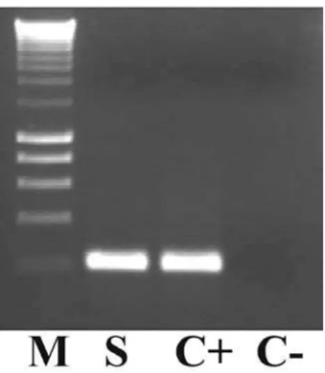
DNA was extracted from the remaining blood (200 µL) and subjected to polymerase chain reaction with speciic primers LEI 1-2 (Rogers et al., 1990), which amplify a fragment of 116 bp of the kinetoplast DNA, followed by sequencing of the amplicon and submis-sion of the sequences obtained to a BLAST Identity Search to give the most likely identiication of leish-mania species involved in the infection.
RESULTS
amastigotes based on their morphological character-istics, and allowed us to diagnose the visceral form of leishmaniosis (Fig. 1).
In spite of the parasitological inding, the sero-logical test applied gave a negative result. Molecular analyses conirmed the infection and allowed us to identify the leishmania species as Leishmania infan-tum (100% identity) (Fig. 2).
DISCUSSION
An increasing spread of Leishmania infantum, which causes zoonotic visceral and cutaneous leishmanio-sis in humans and dogs (the reservoir host), and L. Fig. 2. PCR with speciic primers LEI 1-2
tropica, which causes anthroponotic cutaneous leish-maniasis, has been evidenced in recent years in Euro-pean countries. he high prevalence of asymptomat-ic human carriers of L. infantum in southern Europe (Moral et al., 2002, Martín-Sánchez et al., 2004, Prat-long et al., 2004, Marty et al., 2007) suggests that this latent parasitosis is a great public health problem, as demonstrated by the increase of HIV and leishmania
coinfection prevalence (Alvar et al., 1997).
Leishmaniosis is becoming the third most fre-quent opportunistic parasitic disease in HIV patients ater toxoplasmosis and cryptosporidiosis (Desjeux et al., 2003). he HIV/AIDS pandemic has modiied the natural history of leishmaniosis (Alvar et al., 1997). HIV infection increases the risk of developing VL by 100 to 2,320 times in areas of endemicity: it reduces the likelihood of a therapeutic response, and greatly increases the probability of relapse (Lopez-Velez et al., 1998). It is thought that the parasitic infection found concurrently with HIV induces chronic immune activation and therefore an increased HIV load and accelerated progression of AIDS (Alvar et al., 2008), whereas immunological disturbances caused by HIV are particularly favorable for the uncontrolled multi-plication of the parasite (Alvar et al., 2008).
he patient with HIV/leishmaniosis coinfection reported here is the irst diagnosed case in Serbia. It is very diicult to say if this leishmaniosis was an im-ported or an autochthonous infection. he patient’s travel history suggests he could have been infected in Iraq where he was working; in this country some endemic areas for L. infantum have been determined (WHO, 2010). However, people can be infected in southern Serbia as well.
he irst autochthonous cases of VL in Serbia were recorded in 1945 in Niš and the Dobrič district, and this was where the patient was resident (Simić, 1957). Moreover, in the period from 1946 to 1948 in the territory of southern, eastern and western Serbia, more than 350 cases of kala-azar (VL) were recorded (Simić, 1957). he studies performed at that time established that the type of kala-azar was similar to that observed in the Mediterranean basin. As for the
reservoirs of infection, the presence of Leishmania spp. was proven in dogs (most commonly in asymp-tomatic infection) in each region where kala-azar was identiied in humans. he studies performed in Niš in 1955 showed that over 2% of dogs in the area had an asymptomatic infection (Simić, 1957). Rare autochthonic cases were reported in the Niš munici-pality in 1968 and 1969 when the presence of vectors, such as Phlebotomus major, P. simici and P. perfiliewi, was also reported (Petrović, 1980).
Epidemiologic data show that in the period from 1991 to 2000 there were 39 cases of VL reported in Serbia and Montenegro, with only one case of im-ported leishmaniosis (Dakic et al., 2009).
A recent retrospective epidemiologic and diag-nostic study of VL in Serbia for the period 2001-2007 has demonstrated a visit to the Montenegrin coast to be a predominant risk factor in the 22 individu-als diagnosed with VL, apart from one case of VL that occurred in southern Serbia and which probably represented a dormant focus of infection (Dakic et al., 2009).
he WHO concluded that more extensive and eicient surveillance is necessary in Europe to as-sess the emergence of leishmaniosis (WHO, 2009). Increased awareness of leishmaniosis is mandatory even in areas where it is not endemic.
In view of the growing epidemiologic problem of VL spread and especially VL/HIV coinfection, the surveillance of leishmaniosis is imperative, both in human and dog populations in Serbia, paying special attention to the southern regions of the country. Epi-demiologic surveillance would certainly prevent an epidemic outburst of the infections.
coin-fected patients have a positive Leishmania serology (Montalban et al., 1990, Gari-Toussaint et al., 1994). In fact, anti-Leishmania antibodies in AIDS patients are 50 times lower than in those with an intact im-mune system (Mary et al., 1992). herefore, using se-rological methods many false-negative results should be expected in HIV-infected individuals. Ideally, at least two diferent serological tests should be used for each patient, and the leishmanial antigens employed should be freshly prepared, to increase their sensitiv-ity (Desjeux et al., 2003).
Polymerase chain reaction (PCR) revolutionized the possibility of diagnosing the etiologic agents of infectious diseases. In the past decade, PCR-based techniques have been progressively more applied to diagnosis leishmaniosis, but its use is, to date, lim-ited to tertiary health centers. To avoid invasive pro-cedures, peripheral blood is oten used, and the re-ported sensitivity of PCR on blood ranges from 70% to 96% (Takagi et al., 2009).
CONCLUSION
VL/HIV coinfection has important clinical, diagnos-tic and epidemiological implications. In this condi-tion, the failure of serological tests is to be expected and, apart from being an easy and non-invasive agnostic approach, the application of molecular di-agnostics to the blood may give a warning about the risk of possible nosocomial infections.
Acknowledgments - his work was inancially supported by
the Serbian Ministry of Education and Science, Grants No 41018 and No 175034.
REFERENCES
Alvar, J., Aparicio, P., Asefa, A., Den Boer, M., Cañavate, C., De-det, J.P., Gradoni, L., Ter Horst, R., López Vélez, R., and J. Moreno (2008). he Relationship between Leishmaniasis and AIDS: the Second 10 Years Clin. Microbiolo. Review.
21 (2), 334- 359.
Alvar, J., Cañavate, C., Gutiérrez-Solar, B., Jiménez, M., Laguna, F., López Vélez, R., Molina, R., and J. Moreno (1997). Leish-mania and human immunodeiciency virus co-infection: the irst 10 years. Clin Microbiol Rev. 10 (2), 298-319.
Burns, J.,M., Shreler, W.,G., Benson, D.,R., Ghalib, H.,W. and R.S.G Badaró (1993). Molecular characterization of a ki-nesin-related antigen of Leishmania chagasi that detects speciic antibody in African and American visceral leish-maniasis. Proc Natl Acad Sci USA. 90, 775-779.
Dakić, Z.D., Pelemis, M.R., Stevanović, G.D., Poluga, J.L., Lavadinović, L.S., Milošević, I.S., Indjić, N.K., Ofori-Belić, I.V., and M.D. Pavlović (2009). Epidemiology and diag-nostics of visceral leishmaniasis in Serbia. Clin Microbiol Infect. 15, 1173-1176.
de La Rosa, R., Pineda, J.A., Delgado, J., Macias, J., Morillas, F., Mira, J.A., et al. (2002). Incidence and risk factors for symptomatic visceral leishmaniasis among human immu-nodeiciency virus type 1-infected patients from Spain in the era of highly active antiretroviral therapy. J Clin Micro-biol. 40, 762-7.
del Giudice, P., Mary-Krause, M., Pradier, C., Grabar, S., Della-monica, P., Marty, P. et al. (2002). Impact of highly active antiretroviral therapy on the incidence of visceral leishma-niasis in a French cohort of patients infected with human immunodeiciency virus. J Infect Dis.186, 1366-70.
Desjeux, P (2001). he increase in risk factors for leishmaniasis worldwide. Trans R Soc Trop Med Hyg. 95 (3), 239-43.
Desjeux, P., and J. Alvar (2003). Leishmania/HIV co-infections: epidemiology in Europe. Ann Trop Med and Parasitol.97
(1), S3-S15.
Gari-Toussaint, M., Lelievre, A., Marty, P., and Y. Le Fichoux (1994). Contribution of serological tests to the diagnosis of visceral leishmaniasis in patients infected with the hu-man immunodeiciency virus. Trans R Soc Trop Med Hyg.
88, 301-2.
Guerin, P.J., Olliaro, P., Sundar, S., Boelaert, M., Crot, S.L., Desjeux, P., et al. (2002). Visceral leishmaniasis: current status of control, diagnosis, and treatment, and a proposed research and development agenda. Lancet Infect Dis. 2, 494-501.
Leishmaniasis: background information. A brief history of the disease. WHO. 2009. Available from: www.who.int/leish-maniasis/en
Lopez-Velez, R., Perez-Molina, J.A., Guerrero, A., Baquero F., Vil-larrubia J., Escribano, L., Bellas, C., Perez-Corral, F., and J. Alvar (1998). Clinicoepidemiologic characteristics, prog-nostic factors, and survival analysis of patients coinfected with human immunodeiciency virus and Leishmania in an area of Madrid, Spain. Am. J. Trop. Med. Hyg., 58, 436–443.
the canine reservoir and phlebotomine vectors. Trop Med Int Health. 13(2), 256-64.
Martín-Sánchez, J., Pineda, J.A., Morillas-Márquez, F., García-García, J.A., Acedo, C. and J. Macías (2004). Detection of Leishmania infantum kinetoplast DNA in peripheral blood from asymptomatic individuals at risk for paren-terally transmitted infections: relationship between poly-merase chain reaction results and other Leishmania infec-tion markers. Am J Trop Med Hyg. 70(5), 545-8.
Marty, P., Izri, A., Ozon, C., Haas, P., Rosenthal, E., Del Giudice, P., et al. (2007). A century of leishmaniasis in Alpes-Mari-times, France. Ann Trop Med Parasitol. 101(7), 563-74.
Mary, C., Lamouroux, D., Dunan, S., and M Quilici (1992). West-ern blot analysis of antibodies to Leishmania infantum antigens: potential of the 14-KD and 16-KD antigens for diagnosis and epidemiologic purposes. Am J Trop Med Hyg. 47, 764-71.
Milovanovic, M., аnd D. Popovic (1960). Addition to research of kala-azar epidemic in NR Serbia. Voice Hyg Inst. 9, 23–27.
Montalban, C., Calleja, J.,L., Erice, A., Laguna, F., Clotet, B., Podzamczer, D., et al. (1990). Visceral leishmaniasis in pa-tients infected with human immunodeiciency virus. Co-operative Group for the Study of Leishmaniasis in AIDS. J Infect. 21, 261-70.
Moral, L., Rubio, E.M., and M. Moya (2002). A leishmanin skin test survey in the human population of l’Alacantí region (Spain): implications for the epidemiology of Leishmania infantum infection in southern Europe. Trans R Soc Trop Med Hyg. 96(2), 129-32.
Petrović, Z. (1980). Epidemiology of kala-azar in Serbia. Bel-grade: Institute for Medical Research (Serbian).
Pratlong, F., Rioux, J.A., Marty, P., Faraut-Gambarelli, F., Dereure, J., Lanotte, G., et al. (2004). Isoenzymatic analysis of 712
strains of Leishmania infantum in the south of France and relationship of enzymatic polymorphism to clinical and epidemiological features. J Clin Microbiol. 42 (9), 4077-82.
Ready, P. D. (2010). Leishmaniasis emergence in Europe. Euro-surveillance. 15(10), 1-11.
Report of a meeting of the WHO Expert Committee on the Con-trol of Leishmaniases, WHO technical report series; no. 949, Geneva, 22-26 March 2010. Available from: whqlib-doc.who.int/trs/WHO_TRS_949_eng.pdf
Rogers, B. B., Alpert, L.C., Hine E. A. S., and G. J. Bufone (1990). Analysis of DNA in Fresh and Fixed Tissue by the Poly-merase Chain Reaction. American Journal of Pathology.
136(3), 541-548.
Simić, Č. (1957). Protozoan parasites of man and domestic ani-mals. Belgrade, 1957.
Sundar, S. (2001). Drug resistance in Indian visceral leishmania-sis. Trop Med Int Health. 6, 849-54.
Sundar, S., Jha, T.K., hakur, C.P., Engel, J., Sindermann, H., Fis-cher, C., et al. (2002) Oral Miltefosine for Indian Visceral Leishmaniasis. N Engl J Med. 347, 1739-6.
Takagi, H., Makoto, I., Mohammad, Z.I., Abdur, R., Saifuddin Ekram, E.R.M., Yoshihisa, H., Eisei, N., and E. Kimura (2009). Sensitive, Speciic, and Rapid Detection of Leish-mania donovani DNA by Loop-Mediated Isothermal Am-pliication. Am. J. Trop. Med. Hyg. 81 (4), 578–582.
UNAIDS: AIDS Epidemic Update 2002. www.unaids.org.
World Health Organization (WHO). Regional Oice for Europe. Vectorborne and rodentborne diseases. WHO. 29 Septem-ber 2009.