889 889 889 889 889 Mem Inst Oswaldo Cruz, Rio de Janeiro, Vol. 99(8): 889-893, D ecem ber 2004
Subclinical Form of the American Visceral Leishmaniasis
M ônica Elinor Alves Gama/+, Jackson M aurício Lopes Costa* , Cláudia M aria Castro Gomes* * , Carlos Eduardo Pereira Corbett* *
Departamento de Medicina III (Pediatria) *Departamento de Patologia, Universidade Federal do Maranhão, Praça Gonçalves Dias 21 ILA, 65020-270 São Luis, MA, Brasil **Departamento de Patologia, Universidade de São Paulo, São Paulo, Brasil
The subclinical form of visceral leishmaniasis (VL) shows nonspecific clinical manifestations, with difficulties being frequently met in its clinical characterization and diagnostic confirmation. Thus, the objective of the present study was to define the clinical-laboratory profile of this clinical form. A cohort study was conducted in the state of Maranhão, Brazil, from January/1998 to December/2000, with monthly follow-up of 784 children aged 0-5 years. Based on the clinical-laboratory parameters reported in the literature, four categories were established, with the children being classified (according to their clinical-evolutive behavior) as asymptomatic (N = 144), as having the subclinical form (N = 33) or the acute form (N = 12) or as subjects “without VL” (N = 595). Multiple discriminant analysis demonstrated that the combination of fever, hepatomegaly, hyperglobulinemia, and increased blood sedi-mentation rate (BSR) can predict the subclinical form of VL as long as it is not associated with splenomegaly or leukopenia. Subjects with the subclinical form did not show prolonged or intermittent evolution or progression to the acute form of VL. Subclinical cases have a profile differing from the remaining clinical forms of VL, being best characterized by the combination of fever, hepatomegaly, hyperglobulinemia, and increased BSR.
Key words: visceral leishmaniasis - subclinical form - oligosymptomatic form
American visceral leishmaniasis (AVL) is a parasitic infection caused by Leishmania L. chagasi, endemic in many areas at Brazil, including the state of Maranhão. Clinically, AVL can be classified as asymptomatic (just infection), subclinical (or oligosymptomatic form), acute and, classic forms, which sometimes can present evolu-tionary behavior. These denominations and the charac-teristics described for each clinical form have been modi-fied over the years, interfering with the evaluation of the incidence and clinical presentation mainly of the subclini-cal form because some authors use the terms “infection” and “subclinical” as synonyms (Pampiglione et al. 1974, Guerra et al. 1985, Evans et al. 1992, D’Oliveira Jr et al. 1997).
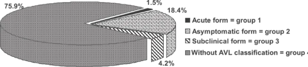
The study published by Badaró et al. (1986) has been adopted as a reference to characterize the subclinical form of AVL as nonspecific mild clinical manifestations lasting for more than 3 weeks, including fever, cough, diarrhea, malaise, mild hepatomegaly, and eventually splenomegaly with slight laboratory alterations, presenting a fluctuat-ing course that evolves over a prolonged period of time (mean, 35 months). The authors showed that individuals with antibodies to Leishmania can be divided into four groups according to their clinical evolution: those who remain asymptomatic (23.2%), those who develop the dis-ease but without progression from the subclinical form (17.4%), those with the subclinical form who progress to
Financial support: Fapesp, CNPq, Laboratório de Patologia de Moléstias Infecciosas/Universidade de São Paulo (LIM50) +Corresponding author. Fax number: +55- 98-227.0791. E-mail:
mgama@elo.com.br Received 8 June 2004 Accepted 12 November 2004
disease (15.1%), and those with the subclinical form that is resolved after a prolonged period of time (44.2%). In most cases, the children were initially indistinguishable. Professionals working in endemic AVL areas often face doubts about children with nonspecific symptoms and about the evolution and clinical behavior of the subclini-cal form of AVL and its diagnostic confirmation. Doubts also arise about whether suitable therapy should be insti-tuted since the determinant factors of the evolution to disease are not established and because of the confusion with others causes of mild symptoms. Thus, the objective of the present study was to define the clinical and labora-tory profile of the subclinical form of AVL.
PATIENTS AND METHODS
A cohort study was carried out in endemic areas of AVL, in the state of Maranhão, Brazil, between January/ 1998 and Dececember/2000.
Areas of investigation - The areas selected were Vila Nova and Vila Bom Viver (villages in the municipal district of Raposa, MA, Brazil - Fig. 1) known to be endemic for AVL, with an incidence of 3.3 cases/1000 inhabitants per year. The district is located 28 km from the state capital, São Luis. The Center for Tropical Pathology and Social Medicine, Federal University of Maranhão (UFMA) and the National Health Foundation (Funasa, MA) have been conducting studies in this area for 15 years with the ob-jective of controlling the more common diseases (AVL and malaria). Chagas disease, mansonic schistosomiasis or American tegumentary leishmaniasis have not been re-ported in this area (Caldas et al. 2001).
890 890 890 890
890 Subclinical Form of the AVL • M ônica Elionor Alves Gama et al.
(L.) chagasi antigen was carried out to discriminate be-tween individuals infected or not to Leishmania. Regard-less of the serological results, all children were clinically evaluated and submitted to regular monthly visits for the possible detection of nonspecific symptoms. The health agent of Funasa/MA, and the community health agents of the municipal district helped to locate the children. If the children presented any symptoms, the adults respon-sible for them and the agents were instructed to return immediately for clinical evaluation and laboratory analy-sis (blood count, protein count, blood sedimentation rate, and transaminases determination). The children with symp-toms were evaluated every 3 days until they could be classified and complementary exams were requested if necessary.
Children were assembled into four different groups according to the clinical and laboratory initial manifesta-tion and to their final clinical course. The symptoms con-sidered were fever, diarrhea, cough, pallor, hepatomegaly and/or splenomegaly. 1) AVL acute form/Group 1-12 chil-dren (classic symptoms of AVL); 2) asymptomatic form/
Group 2-144 children (positive ELISA), oligosymptomatic or subclinical; 3) form/Group 3-33 children (two or more mild symptoms, with positive ELISA); and 4) children
with-out AVL classification/Group 4-595 children (specific clinical situation, not classified as a clinical form of AVL). After the children were classified as oligosymptomatic or as having the acute form of the disease, specific tests for the diagnosis of AVL were done – ELISA with leishmanial antigen, again, and a bone marrow aspirate to search for
Leishmania amastigotes forms. A single pediatrician per-formed all clinical evaluations.
The clinical data were recorded and analyzed utilizing the SPSS-PC program (Statistical Package for Social Sci-ences, PC version 10.0). First, the incidence of the all vari-ables [fever, cough, diarrhea, pallor, hepatomegaly, sple-nomegaly, leukopenia, lymphocytosis, anemia, thromb-ocytopenia, hypoalbuminemia, hyperglobulinemia, in-creased blood sedimentation rate (BSR) and transami-nases] was calculated for each group and compared among groups using analysis of variance (ANOVA) or the Kruskal-Wallis test. A multiple discriminant analysis was performed to identify the combination of variables that would permit the discrimination between groups.
The study was approved by the Ethics Committees of the University of São Paulo and the Federal University of Maranhão and fulfilled the requirements of the Resolu-tion of the NaResolu-tional Council for Health 196/96 for research involving humans.
RESULTS
Fig. 2 shows the distribution of children into the four groups based on their final clinical course. After the chil-dren identification with positive or not ELISA (infected or not to Leishmania) it was proceeded its monthly follow-up. Along the clinical reevaluations, it was observed that 12 children who had developed to AVL acute form out of 4 presented positive ELISA previously and from 33 chil-dren classified with subclinical form only 8 of them pre-sented an infection in the serum conspicuosness observed previously to the classification.
The incidence of the variables was calculated for each group and a comparative analysis of the data by ANOVA or the Kruskal-Wallis test showed that all variables dif-fered significantly among the 4 groups, except cough (p = 0.630).
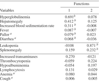
A multiple discriminant analysis (MDA) was performed to determine the combination of variables that character-ized the subclinical form. Two functions were derived to distinguish the oligosymptomatic group from the others (Table I). The importance of each variable within each function is expressed by its correlation coefficient and its importance for the diagnosis of the subclinical form was Fig. 1: municipal district of Raposa, in the São Luís Island, state of
Maranhão, Brazil.
891 891 891 891 891 Mem Inst Oswaldo Cruz, Rio de Janeiro, Vol. 99(8), D ecem ber 2004
compared with the other groups. To validate the multiple discriminant models, its functions were applied to the validation sample – these can represented 99.9% of the variations of the original measurement. Cross-validation showed that the model correctly classified 82% of the all cases.
The combination of fever, hepatomegaly, hyperglobu-linemia, and increased BSR (function 1) distinguished the subclinical form from the asymptomatic cases and from children without AVL classification. MDA also showed that the combination of splenomegaly and leukopenia (function 2) distinguished the acute form from the sub-clinical cases. Therefore, when function 1 values are high, the subclinical form is more likely to be present, and when
function 2 values are high, the acute form is more likely to be present. Fig. 3 shows the difficulty in dividing group 2 (asymptomatic) and group 4 (without AVL classification) into different clinical forms. The other variables contrib-uted little to the statistical model.
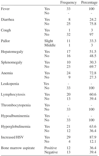
For the subclinical form, the clinical and laboratory findings were variable and fever was the major complaint (Table II). The duration of symptoms ranged from 10 to 40 days, with no intermittent course during follow-up. It is considered hepatomegaly and splenomegaly when the guts were felt below the flange joint in the back, and it was observed that the hepatomegaly varied from 3 to 5 cm, and the splenomegaly from 3 to 5 cm, however they were not present at all children. The associations of signs and symptoms were varied (Table III). By the way, in the group with the acute form, there was always an associa-tion of four signs/symptoms – fever, hepatomegaly, sple-nomegaly, and pallor – in each individual.
Table IV shows the clinical and laboratory differences between the oligosymptomatic cases with a negative or positive search of Leishmania in the bone marrow aspi-rate. The duration of symptoms and the dimensions of the visceromegalies did not differ between the two groups (p # 0.05).
DISCUSSION
No previous studies have been performed with monthly follow-up of children infected and not to Leishmania to discriminate between the subclinical or oligosymptomatic cases from the other frequent and possible diagnoses.
The present study confirmed that the oligosympt-omatic form was more frequent than the acute form of AVL. However, in the literature, the subclinical form oc-curs in almost 60% of the infected subjects (Badaró et al. 1986) while in the present study it occurred in only 17.4%, probably because the other common causes of the mild symptomatology were evaluated and ruled out before the AVL classification. This means that, in an endemic area, the professionals must consider other usual diagnoses even in the presence of a reactive serological test. TABLE I
Rotated structure matrix correlation between the discriminant variables and the discriminant functions (variables ordered by
size of correlation within the function)
Functions
Variables 1 2
Hyperglobulinemia 0.691b 0.078
Hepatomegaly 0.412b 0.125
Increased blood sedimentation rate 0.311b -0.008
Fever 0.087b -0.007
Pallor a 0.079b 0.023
Diarrhea a 0.068b -0.036
Leukopenia -0108 0.871b
Splenomegaly 0.159 0.232b
Increased transaminases 0.270 -0121
Thrombocytopenia -0.059 0.224
Hypoalbuminemia -0.054 0.197
Lymphocytosis 0.131 0.029
Anemia a 0.080 0.044
Cough a 0.006 0.005
a: variable not used in the analysis (with no significant contribution to the statistical model); b: absolute correlation between each variable and discriminant function.
892 892 892 892
892 Subclinical Form of the AVL • M ônica Elionor Alves Gama et al.
TABLE II
Distribution of the clinical and laboratory findings for the 33 children classified with the oligosymptomatic form
Frequency Percentage
Fever Yes 33 100
No -
-Diarrhea Yes 8 24.2
No 25 75.8
Cough Yes 1 3
No 32 97
Pallor Slight 11 33.3
Middle 1 3
Hepatomegaly Yes 17 51.5
No 16 48.5
Splenomegaly Yes 10 30.3
No 23 69.7
Anemia Yes 24 72.8
No 9 27.3
Leukopenia Yes -
-No 33 100
Lymphocytosis Yes 20 60.6
No 13 39.4
Thrombocytopenia Yes -
-No 33 100
Hypoalbuminemia Yes -
-No 33 100
Hyperglobulinemia Yes 21 63.6
No 12 36.4
Increased HSV Yes 29 87.9
No 4 12.1
Bone marrow aspirate Positive 12 36.4
Negative 13 39.4
TABLE IV
Clinical-laboratory characteristics of the 33 oligosymptomatics cases according to bone marrow positivity
(12 positive cases and 13 negative cases)
Search for Leishmania
Positive Negative NP
Clinical-laboratory findings Nr Nr Nr
Fever associated with:
Hepatomegaly 2 3 4
Pallor 2 2 1
Splenomegaly - 2 2
Diarrhea + pallor - 3
-Hepatomegaly + splenomegaly 1 1
-Diarrhea 1 - 1
Hepatosplenomegaly + pallor 2 -
-Hepatomegaly + pallor 1 1
-Hepatomegaly + diarrhea 1 1
-Splenomegaly + cough 1 -
-Splenomegaly + diarrhea 1 -
-Total 12 13 8
NP: not performed
of the subclinical group with spontaneous resolution was 8 years.
In this cohort, prolonged or intermittent mild illness was not observed, in agreement with Evans et al. (1992), who followed-up the infected subjects, although there are literature reports showing that the duration of symp-toms is up to 35 months in the subclinical form (Badaró et al. 1986). Different epidemiological situations can deter-mine different clinical behavior.
The spontaneous resolution of the subclinical form without progression to the acute form has been discussed since 1906, although many authors refer to the asymp-tomatic form as being synonymous of the subclinical form, a fact that impairs comparative analysis (Leishman 1906 apud Pampiglione 1974, Guerra et al. 1985, Holaday et al. 1993, Shiddo et al. 1995, D’Oliveira Jr et al. 1997)
Using the multiple discriminant analysis, the interac-tion of the all variables was determined to define the sub-clinical form profile. It showed that two associations (func-tions 1 and 2) represented almost 100% of the cases. The combination of fever, hepatomegaly, hyperglobulinemia, and increased BSR (function 1) can predict the oli-gosymptomatic form, and therefore this function can be interpreted as “nonspecific clinical and laboratory mani-festations related to AVL”. The occurrence of splenom-egaly and leukopenia (function 2) distinguished the acute form from the oligosymptomatic form and can therefore be interpreted as “specific manifestation of AVL disease” because it characterize the illness, occurring in almost 100% of the cases of acute form of AVL (Silva 1957, Pastorino 1993, WHO 1997).
On the basis of these findings, in the present analysis the subclinical form was characterized as a peculiar and specific clinical form which differs from the other diag-noses with mild symptomatology and which is not an early stage of AVL disease (acute or classic forms).
TABLE III
Associations of signs/symptoms observed in the 33 oligosymptomatics cases
Oligosymptomatic cases
Clinical associations Nr %
Fever associated with:
Hepatomegaly 9 27.3
Pallor 5 15.1
Splenomegaly 4 12.1
Diarrhea + pallor 3 9.1
Hepatomegaly + splenomegaly 2 6.1
Diarrhea 2 6.1
Hepatomegaly + splenomegaly + pallor 2 6.1
Hepatomegaly + pallor 2 6.1
Hepatomegaly + diarrhea 2 6.1
Splenomegaly + cough 1 3
Splenomegaly + diarrhea 1 3
893 893 893 893 893 Mem Inst Oswaldo Cruz, Rio de Janeiro, Vol. 99(8), D ecem ber 2004
It is important to observe the overlap of the groups of asymptomatic subjects and of subjects “without AVL” which emphasizes that, without a suggestive symptoma-tology, a positive search of anti-Leishmania antibodies does not indicate a parasitological investigation or spe-cific treatment. In spite of the peculiar profile, in the oligosymptomatic form the bone marrow aspirate did not contribute to the characterization of the disease and thus would not be essential in daily practice.
In the present study, the symptomatology was vari-able but the literature also reports many variations. Pampiglione et al. (1974) reported infected children with discrete splenomegaly and hyperglobulinemia; Ho et al. (1982) reported 34 cases with splenomegaly (12 with as-sociated fever and malaise). In a transverse study, Shiddo et al. (1995) reported 7 children infected to Leishmania
with splenomegaly (two with diarrhea and cough). In the study performed in Jacobina/Bahia, the subclinical group with spontaneous resolution presented diarrhea (18.4%), cough (13.1%), fever (10.5%), hepatomegaly (97.4%), and splenomegaly (28.9%), with 27.3% of positivity in the bone marrow aspirate – some cases with leukopenia too (Badaró et al. 1986). These data are divergent, probably due to the different study models.
This study demonstrated that there was a wide diver-sity of the symptoms associated with the oligosympt-omatic form, but the model of analysis proposed showed that, although mild and nonspecific, the clinical manifes-tations were of significant value to define the oli-gosymptomatic or subclinical form of AVL.
During the clinical analysis, the immunological mark-ers for the oligosymptomatic or subclinical form of VL were identifying. This can be seen in Gama et al. (2004).
ACKNOWLEDGMENT
To the health agent of Funasa/MA and the community health agents of the municipal district of Raposa/MA for their work with the children and their families.
REFERENCES
Badaró R, Jones T, Carvalho E, Sampaio D, Reed S, Barral A, Teixeira R, Johnson Jr W 1986. New perspectives on a subclinical form of visceral leishmaniasis. J Infect Dis 154: 1003-1011.
Caldas A, Silva D, Pereira C, Costa J 2001. Infecção por
Leishmania (Leishmania) chagasi em crianças de uma área endêmica de leishmaniose visceral americana, na Ilha de São Luís, Maranhão, Brasil. Rev Soc Bras Med Trop 34: 445-451.
D’Oliveira Jr A, Costa S, Barbosa A, Orge M, Carvalho E 1997. Asymptomatic Leishmania chagasi infection in relatives and neighbors of patients with visceral leishmaniasis. Mem Inst Oswaldo Cruz 92: 15-20.
Evans T, Teixeira M, MCauliffe I, Vasconcelos A, Sousa A, Lima J, Pearson R 1992. Epidemiology of visceral leishma-niasis in northeast Brazil. J Infect Dis 166: 1124-1132. Gama MEA, Costa, JML, Pereira, JCR, Gomes, CMC, Corbett,
CEP 2004. Serum cytokine profile in the subclinical form of visceral leishmaniasis. Braz J Med Biol Res 37: 129-36 Guerra M, Furtado T, Barros G, Sessa P, Daher V 1985. Infecção
subclínica na leishmaniose tegumentar. An Bras Dermatol 60: 365-369.
Ho M, Siongok K, Lyerly W, Smith D 1982. Prevalence and disease spectrum in a new focus of visceral leishmaniasis in Kenya. Trans R Soc Trop Med Hyg 76: 741-746.
Holaday B, Pompeu M, Evans T, Braga D, Teixeira M, Sousa A, Sadick M, Vasconcelos A, Abrams J, Pearson R, Locksley R 1993. Correlates of Leishmania-specific immunity in the clinical spectrum of infection with Leishmania chagasi. J Infect Dis 167: 411-417.
Leishman WB 1906. Handbuch der Tropenkrankheiten, 2nd ed., Leipzig apud Pampiglione S, Manson-Bahr P, Giungi F, Giunti G, Parenti A, Troti G 1974. Studies on Mediterra-nean leishmaniasis II. Asymptomatic cases of visceral leish-maniasis. Trans R Soc Trop Med Hyg 68: 349-358. Pampiglione S, Manson-Bahr P, Giungi F, Giunti G, Parenti A,
Troti G 1974. Studies on Mediterranean leishmaniasis II. Asymptomatic cases of visceral leishmaniasis. Trans R Soc Trop Med Hyg 68: 349-358.
Pastorino A 1993. Contribuição para o Estudo da Leishmaniose Visceral na Infância: Aspectos Clínico-laboratoriais de 78 casos, MSc Thesis, Universidade de São Paulo, São Paulo, 100 pp.
Shiddo S, Mohamed A, Akuffo H, Mohamud K, Herzi A, Mohamed H, Huldt G, Nilsson L, Ouchterlony A, Thorstensson R 1995. Visceral leishmaniasis in Somalia: prevalence of markers of infection and disease manifesta-tions in a village in an endemic area. Trans R Soc Trop Med Hyg 89: 361-365.
Silva J 1957. Leishmaniose visceral (calazar), Thesis, Serviço Nacional de Educação Sanitária, Rio de Janeiro, 498 pp. WHO-World Health Organization 1997. Manual de Controle