AGREEMENT BETWEEN MAGNETIC RESONANCE IMAGING
AND ULTRASONOGRAPHY IN THE CLASSIFICATION
OF SCHISTOSOMAL PERIPORTAL FIBROSIS, ACCORDING
TO NIAMEY’S CRITERIA*
Eduardo Scortegagna Junior1
, Alberto Ribeiro de Souza Leão2
, José Eduardo Mourão Santos3
, Danilo Moulin Sales1
, David Carlos Shigueoka4
, Luciane Aparecida Kopke de Aguiar5
, Paulo Eugênio Brant5
, Ramiro Colleoni Neto6
, Durval Rosa Borges7
, Giuseppe D’Ippolito8
OBJECTIVE: To evaluate the reproducibility of magnetic resonance imaging and the agreement between ultrasound and magnetic resonance imaging in the classification of periportal fibrosis in patients with schis-tosomiasis based on Niamey’s qualitative criteria. MATERIALS AND METHODS: A prospective, double-blinded study was conducted between February 2005 and June 2006 with 20 patients (10 men and 10 women, with ages ranging between 24 and 60 years, mean age 42.7 years) diagnosed with schistosomiasis mansoni. Both ultrasound and magnetic resonance images were independently evaluated by two experienced observ-ers. Interobserver agreement was evaluated for findings of periportal fibrosis on magnetic resonance images and in a comparison between magnetic resonance and ultrasound images. RESULTS: The analysis of mag-netic resonance images showed total interobserver agreement in 14 patients (70%). The comparison be-tween ultrasound and magnetic resonance imaging showed agreement bebe-tween images in only six cases (30%) by observer 1, and in eight cases (40%) by observer 2. CONCLUSION: Magnetic resonance imaging presents a good reproducibility in the evaluation of periportal fibrosis in later stages of schistosomiasis, however, the correlation between magnetic resonance imaging and ultrasound is poor.
Keywords: Schistosomiasis; Periportal fibrosis; Ultrasound; Magnetic resonance imaging.
Avaliação da concordância entre ressonância magnética e ultra-sonografia na classificação da fibrose peri-portal em esquistossomóticos, segundo a classificação de Niamey.
OBJETIVO: Avaliar a reprodutibilidade da ressonância magnética e a concordância entre a ultra-sonografia e a ressonância magnética na classificação da fibrose periportal em pacientes esquistossomóticos, segundo os critérios qualitativos de Niamey. MATERIAIS E MÉTODOS: Foi realizado estudo prospectivo e duplo-cego, entre fevereiro de 2005 e junho de 2006, em 20 pacientes (10 homens e 10 mulheres, idades entre 24 e 60 anos, média de 42,75 anos) com diagnóstico de esquistossomose mansônica. As imagens de ultra-sonogra-fia e de ressonância magnética foram avaliadas por dois examinadores experientes de forma independente. Foi medida a concordância interobservador para a ressonância magnética e entre a ressonância magnética e a ultra-sonografia. RESULTADOS: A ressonância magnética apresentou resultados concordantes entre os observadores em 14 pacientes (70%). Quando comparamos a ressonância magnética com a ultra-sonogra-fia, obtivemos concordância em apenas seis pacientes pelo observador 1 (30%) e em oito pacientes pelo observador 2 (40%). CONCLUSÃO: A ressonância magnética tem boa reprodutibilidade na avaliação de fi-brose periportal em pacientes com esquistossomose avançada, porém sua concordância com a ultra-sono-grafia é fraca.
Unitermos: Esquistossomose; Fibrose periportal; Ultra-sonografia; Ressonância magnética. Abstract
Resumo
* Study developed in the Department of Imaging Diagnosis and Disciplines of Clinical and Surgical Gastroenterology at Uni-versidade Federal de São Paulo/Escola Paulista de Medicina (Unifesp/EPM), São Paulo, SP, Brazil.
1. MD, Trainee in Radiology, Department of Imaging Diag-nosis at Universidade Federal de São Paulo/Escola Paulista de Medicina (Unifesp/EPM), São Paulo, SP, Brazil.
2. MD, Fellow PhD degree, Department of Imaging Diag-nosis at Universidade Federal de São Paulo/Escola Paulista de Medicina (Unifesp/EPM), São Paulo, SP, Brazil.
3. Master in Clinical Radiology, Universidade Federal de São Paulo/Escola Paulista de Medicina (Unifesp/EPM), São Paulo, SP, Brazil.
4. PhD, Clinical Radiology, MD, Department of Imaging Di-agnosis at Universidade Federal de São Paulo/Escola Paulista de Medicina (Unifesp/EPM), São Paulo, SP, Brazil.
5. MD, Fellow PhD degree, Department of
Gastroenterol-INTRODUCTION
Schistosomiasis is a millenarian disease affecting more than 200 million people in about 76 countries in Africa, Asia and Americas, and represents a significant problem of public health in Brazil(1,2).
Periportal fibrosis is the main cause of complications resulting from schistosomia-sis and occurs in 4% to 8% of the patients who develop chronic infection. Hepatome-galy reflects the presence of granulomatous ogy at Universidade Federal de São Paulo/Escola Paulista de
Medicina (Unifesp/EPM), São Paulo, SP, Brazil.
6. Affiliate Professor, Discipline of Surgical Gastroenterol-ogy at Universidade Federal de São Paulo/Escola Paulista de Medicina (Unifesp/EPM), São Paulo, SP, Brazil.
7. Titular Professor, Discipline of Gastroenterology, Depart-ment of Medicine at Universidade Federal de São Paulo/Escola Paulista de Medicina (Unifesp/EPM), São Paulo, SP, Brazil.
8. Associate Professor Doctor, Department of Imaging Di-agnosis at Universidade Federal de São Paulo/Escola Paulista de Medicina (Unifesp/EPM), São Paulo, SP, Brazil.
inflammation and occurs early in the dis-ease progression. Periportal collagen depo-sition leads to a progressive portal vein occlusion, portal hypertension, and a pos-sible progression to varices, high digestive hemorrhage, splenomegaly and hyper-splenism. Periportal fibrosis is characteris-tic of the disease and may be identified by means of several diagnostic methods(2).
Ultrasonography (US) has been the method most frequently utilized in this group of patients, demonstrating a typical pattern of abnormalities(3), especially in-cluding thickening along the walls of the portal vein and its branches characterized by periportal hyperechogenic bands(4).
Aiming at establishing standardized protocol for diagnosing and quantifying periportal fibrosis in schistosomal patients, ultrasonographic criteria were initially de-fined in a World Health Organization con-sensus meeting held in Cairo in 1991(5), and
later reviewed in the meeting on “Ultra-sound Schistosomiasis” held in 1996 in Niamey, resulting in the evolution of the standard ultrasound scoring protocol to include qualitative criteria, considering the hepatic texture as a whole, besides the pre-vious quantitative criteria utilizing the ob-jective measurement of the portal branch wall thickening(4).
US, although widely adopted in the as-sessment of patients with schistosomiasis, has demonstrated a moderate reproducibil-ity in the classification of portal fibrosis(6).
On the other hand, some studies have dem-onstrated the usefulness of magnetic reso-nance imaging (MRI) in the evaluation of hepatosplenic and vascular alterations in schistosomal patients, with high reproduc-ibility(7). Additionally, the MRI capacity in
the diagnosis of hepatic fibrosis has been well demonstrated(7).
However, the role of MRI in the char-acterization and quantification of peripor-tal fibrosis in schistosomal patients is still to be established. Considering its high re-producibility, anatomical detailing and high spatial resolution, some authors be-lieve that MRI may be a more sensitive method than US for indicating the disease progression, grading and therapeutic re-sponse, as a result of a more comprehen-sive evaluation of the abdominal cavity(8).
Therefore, it is important to establish the
MRI value in the evaluation of periportal fibrosis.
The present study was aimed at evalu-ating the MRI reproducibility and the agreement between US and MRI in the grading of periportal fibrosis in schistoso-mal patients, according to qualitative cri-teria defined in the ultrasound consensus meeting of Niamey for assessing peripor-tal fibrosis.
MATERIALS AND METHODS
In the period between February/2005 and June/2006, a prospective, transversal, observational, double-blinded study was developed with 20 patients (10 men, and 10 women in the age range between 24 and 60 years, mean 42.75 years) referred by the clinic of schistosomiasis, Discipline of Clinical Gastroenterology, Universidade Federal de São Paulo/Escola Paulista de Medicina (Unifesp/EPM), with diagnosis of schistosomiasis mansoni. Such diagno-sis had been obtained either by means of rectal biopsy or consistent clinical labora-tory (signs of portal hypertension and/or positive fecal parasitologic examination) or epidemiological evidence (contact with contamined water of lakes, ponds, rivers in endemic areas), and subclassified into the hepatosplenic form of the disease because of the finding of portal hypertension and splenomegaly.
Exclusion criteria were: patients with contraindication for MRI (cardiac pace-maker, cochlear implants, claustrophobia, cerebral aneurysm clips, allergy to para-magnetic contrast agents), patients with a previous history of alcoholism (ingestion of more than 160 g of ethanol/week),
posi-tive serology for hepatitis B or C viruses, with a previous history of proven autoim-mune disease, and use of hepatotoxic drugs.
The patients underwent US and MRI examinations with a minimum seven-day interval between studies. The results inter-pretation was performed by two indepen-dent observers, both resiindepen-dents in imaging diagnosis (A.R.S.L. and D.M.S.) and with at least two-year experience in the diagnos-tic methods utilized, with training in ultra-sonography specifically for assessment of schistosomal patients. In a later moment, the observers met to reach a consensus as regards the sonographic classification of periportal fibrosis which later on this study was utilized as a gold standard in the com-parison between readings and MRI studies.
Ultrasound scan
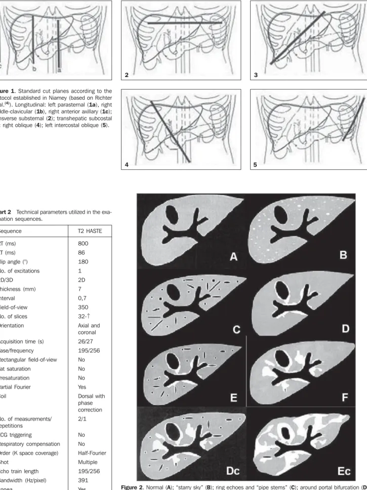
US scans were performed with a Philips EnVisor® model with a convex, multi-fre-quency, 2.5–4.5 MHz transducers, in pa-tients after eight-hour fasting, according to the Niamey standard protocol(4), with
lon-gitudinal (left paraesternal, right hemi-clavicular, right anterior axillary), subster-nal, subcostal, Intercostal left and right oblique scans (Figure 1). The patterns ob-served were classified according the spe-cific protocol (Chart 1 and Figure 2).
Magnetic resonance imaging
MRI examinations were performed in a Siemens Sonata model system operating in a high field (1.5 T), with synergy coil and MRI sequences configured for periportal fibrosis assessment(9) (Chart 2).
The periportal fibrosis classification was performed similarly to the one
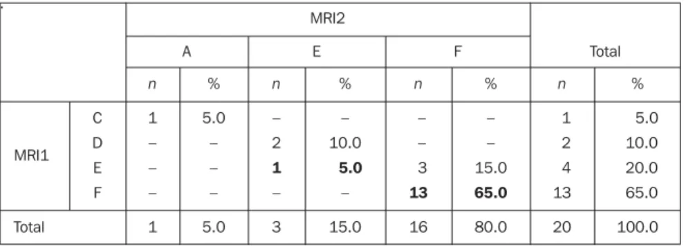
per-Chart 1 Patterns of hepatic parenchyma and periportal fibrosis scores.
Reference patterns
A B C D E
F
Periportal fibrosis score
0 1 2 4 6
8 Image description
Normal.
Starry sky (diffuse echogenic foci). Ring echoes and pipe stems.
Hyperechogenicity around portal bifurcation.
Hyperechoic patches extending from the portal vessels to the parenchyma.
Hyperechoic bands extending to the hepatic parenchyma, where they retract the organ surface.
Figure 1. Standard cut planes according to the protocol established in Niamey (based on Richter et al.(4)). Longitudinal: left parasternal (1a), right
middle-clavicular (1b), right anterior axillary (1c); transverse substernal (2); transhepatic subcostal (3); right oblique (4); left intercostal oblique (5).
1 2 3
4 5
Figure 2. Normal (A); “starry sky” (B); ring echoes and “pipe stems” (C); around portal bifurcation (D); focal (E); “bird’s claw” (F): D + C combination (Dc); E + C combination (Ec). (Based on Richter et al.(4)). Chart 2 Technical parameters utilized in the
exa-mination sequences.
Sequence
RT (ms) ET (ms) Flip angle (°) No. of excitations 2D/3D
Thickness (mm) Interval Field-of-view No. of slices Orientation
Acquisition time (s) Fase/frequency Rectangular field-of-view Fat saturation
Presaturation Partial Fourier Coil
No. of measurements/ repetitions
ECG triggering
Respiratory compensation Order (K space coverage) Shot
Echo train length Bandwidth (Hz/pixel) Apnea
T2 HASTE
800 86 180 1 2D 7 0,7 350 32-↑ Axial and coronal 26/27 195/256 No No No Yes Dorsal with phase correction 2/1
formed by US, according to the previously mentioned criteria of the Niamey protocol adapted for MRI.
The observers had no previous informa-tion on the sonographic classificainforma-tion.
The statistical analysis was based on the calculation of the interobserver agreement between MRI and the consensual gold stan-dard obtained by sonographic evaluation.
RESULTS
The analysis of the MRI results by ob-servers 1 and 2 demonstrated a global interobserver agreement of 70% (14/20 patients) (Table 1).
A poor agreement was found in a com-parison between MRI results analyzed by the observer 1 and US results considered as gold standard. Agreement was found in only six cases (30%) (Figure 3), with dis-crepancies in the classification of the 14 remaining patients (70%) (Table 2).
The results of the observer 2 were not very different. Agreement was found only in eight patients (40%) (Table 3).
The highest variation in the classifica-tion level occurred with a patient classified as level E at US and Levels A and C at MRI (Figure 4).
The other differences occurred from a classification level to its subsequent level, i.e., D to E in two cases, and from E to F in three cases (Figure 5).
DISCUSSION
Schistosomiasis is extremely important both at national and international levels, because of its high prevalence and morbid-ity in patients at more advanced stages of disease(2,10).
Generally, its treatment leads to a de-crease in the levels of infection and im-provement in some clinical findings such as hepatosplenomegaly and periportal fi-brosis. The reduction of periportal fibrosis possibly prevents the emergence of portal hypertension and may result event in de-crease of the portal pressure(11), a fact that
highlights the significance of methods with high reliability and reproducibility for the disease follow-up.
Ultrasonography has been widely uti-lized for evaluation of this disease,
consid-Table 1 Interobserver agreement in the evaluation of magnetic resonance imaging. .
MRI2
A E F Total
n
1 – – –
1 %
5.0 – – –
5.0 C
D E F
n
– 2
1
–
3
%
– 10.0
5.0
–
15.0
n
– – 3
13
16 %
– – 15.0
65.0
80.0
n
1 2 4 13
20
%
5.0 10.0 20.0 65.0
100.0 MRI1
Total
MRI, magnetic resonance imaging; n, number of patients; A–F, fibrosis degrees according the Niamey qualitative classification(5). Note: Agreeing cases in bold letters.
Table 2 Observer 1 results for magnetic resonance imaging reading.
US
E F Total
n
1 1
1
8
11
%
5.0 5.0
5.0
40.0
55.0 C
D E F
n
0 1 3
5
9
%
0.0 5.0 15.0
25.0
45.0
n
1 2 4 13
20
%
5.0 10.0 20.0 65.0
100.0 MRI
Total
US, ultrasound; MRI, magnetic resonance imaging; n, number of patients; A–F, fibrosis degrees according the Niamey qualitative classification(5). Note: Agreeing cases in bold letters.
Figure 3. Ultrasound (A) and magnetic resonance imaging (B) of a patient with F scoring at both meth-ods. Periportal fibrosis aspect (arrows).
Table 3 Observer 2 results for magnetic resonance imaging reading.
US
E F Total
n
1
1
9
11
%
5.0
5.0
45.0
55.0 A
E F
n
0 2
7
9
%
0.0 10.0
35.0
45.0
n
1 3 16
20
%
5.0 15.0 80.0
100.0 RM
Total
US, ultrasound; MRI, magnetic resonance imaging; n, number of patients; A, E, F, fibrosis degrees according the Niamey qualitative classification(5). Note: Agreeing cases in bold letters.
ering its non-invasiveness, low cost and wide availability, constituting the ideal method for assessment of the disease in large population groups.
This method can be utilized for detect-ing portal fibrosis and hypertension (dila-tation of portal and splenic veins and portosystemic collaterals). Evidently, US is more reliable than clinical methods for the diagnosis of hepatosplenic diseases. Peri-portal fibrosis, the essential lesion observed in schistosomiasis, is generally identified years after the infection onset, but it has already been report at earlier stages of in-festation(4). On the other hand,
ultrasonog-raphy has demonstrated just a moderate reproducibility in the assessment of schis-tosomal patients(6).
MRI, on its turn, has progressively emerged as an extremely useful method in the evaluation of focal or diffuse hepato-pathies(12), and with the high
reproducibil-ity(13) necessary for consistent results,
stimulating the method adoption. Similarly, MRI has demonstrated to be capable of accurately identifying hepatic fibrosis(7,12).
Notwithstanding, until the present moment there is no study in the literature compar-ing the performances of MRI and US in the evaluation of periportal fibrosis, so, as far as we are concerned, the present study is the first one to do it.
The portal fibrosis aspects have already been described. On T1-weighted sequences, fibrosis presents as a hypointense band in relation to the liver, while on T2-weighted sequences it presents a high signal(11), the
T2-weighted images being the most com-prehensively utilized with this purpose ac-cording to the literature(8,9,13,14), a fact ob-served in the present study, although with a poor agreement with sonographic find-ings.
The present study presents some limi-tations that must be pointed out. All of the patients receive Niamey sonographic ref-erence patterns E or F, characterizing an advanced stage of periportal fibrosis and limiting the evaluation of the method for this group of patients. The method behav-ior in the assessment of patients with milder degrees of periportal fibrosis is still to be known.
This also has prevented the kappa cal-culation of interobserver agreement for Figure 4. Ultrasound (A) and magnetic resonance imaging (B) of a patient with sonographic scoring E
and interobserver disagreement on magnetic resonance imaging readings (respectively scored as C and A).
MRI results because there was no patient with all the levels of fibrosis according to the Niamey criteria. On the other hand, the patients who most benefited from a com-prehensive assessment like that provided by MRI, would be those with more ad-vanced stages of the disease with higher risks for developing complications. This was precisely the group included in the present study.
The adaptation of the Niamey ultra-sonographic criteria to MRI demonstrated a good reproducibility of the method (70%), however, a satisfactory relation between US and MRI classifications. Agreement between both methods was found in less than 50% of studied cases. A possible explanation for this results may be found in the subtle morphological variation between E and F reference patterns, whose the only difference is the parenchymal re-traction in F, a finding that, like the others, presents a certain subjectivity. It is impor-tant to note that, even utilizing ultrasonog-raphy, the interobserver variability is con-siderable, which has been demonstrated in another study developed by the authors(6). Another factor to be considered is that the present study is based on the assumptions that US is the gold standard in the evalua-tion of periportal fibrosis, despite the ab-sence of a consensus on this matter. Cor-relation between US, MRI and
anatomo-pathological analysis of periportal fibrosis is still to be established to define which method presents the higher efficacy.
Additional studies will be necessary to establish a proposal of a simplified classi-fication model for periportal fibrosis to meet the needs of the professionals in-volved in the follow-up of this group of patients. Also studies involving a higher number of patients in early stages of the disease will be necessary to validate an eventual MRI usefulness.
Finally, in the evaluation of periportal fibrosis according to the criteria defined in Niamey and adapted for MRI, the method has presented a good reproducibility, but low rate of agreement with US results, so its applicability is still controversial, par-ticularly in schistosomal patients.
REFERENCES
1. Alves Jr A, Fontes DA, Melo VA, Machado MCC, Cruz JF, Santos EAS. Hipertensão portal esquis-tossomótica: influência do fluxo sangüíneo por-tal nos níveis séricos das enzimas hepáticas. Arq Gastroenterol 2003;40:203–208.
2. Ross AG, Bartley PB, Sleigh AC, et al. Schisto-somiasis. N Engl J Med 2002;346:1212–1220. 3. Homeida M, Ahmed S, Dafalla A, et al.
Morbid-ity associated with Schistosoma mansoni infec-tion as determined by ultrasound: a study in Ge-zira, Sudan. Am J Trop Med Hyg 1988;39: 196– 201.
4. Richter J, Hatz C, Campagne G, Berquist NR, Jenkins JM. Ultrasound in schistosomiasis. A practical guide to the standardized use of ultra-sonography for the assessment of
schistosomia-sis-related morbidity. Second International Work-shop, Niamey, Niger, October 22–26, 1996. 5. International conference on schistosomiasis.
Cairo, Egito, 1995.
6. Santos GT, Sales DM, Shigueoka DC, et al. Re-produtibilidade da classificação ultra-sonográ-fica de Niamey na avaliação da fibrose peripor-tal na esquistossomose mansônica. Radiol Bras 2006;39 (Supl 2):57.
7. Bezerra ASA, D’Ippolito G, Caldana RP, Cecin AO, Szejnfeld J. Avaliação hepática e esplênica por ressonância magnética em pacientes portado-res de esquistossomose mansônica crônica. Ra-diol Bras 2004;37:313–321.
8. Patel SA, Castillo DF, Hibbeln JF, Watkins JL. Magnetic resonance imaging appearance of he-patic schistosomiasis, with ultrasound and com-puted tomography correlation. Am J Gastroente-rol 1993;88:113–116.
9. Kashitani N, Kimoto S, Tsunoda M, et al. Portal blood flow in the presence or absence of diffuse liver disease: measurement by phase contrast MR imaging. Abdom Imaging 1995;20:197–200. 10. Hatz CF. The use of ultrasound in
schistosomia-sis. Adv Parasitol 2001;48:225–284.
11. Homeida MMA, Eltoum IA, Ali MM, et al. The effectiveness of annual versus biennial mass che-motherapy in reducing morbidity due to schisto-somiasis: a prospective study in Gezira-Managil, Sudan. Am J Trop Med Hyg 1996;54:140–145. 12. Vitellas KM, Tzalonikou MT, Bennett WF, Vas-wani KK, Bova JG. Cirrhosis: spectrum of find-ings on unenhanced and dynamic gadolinium-en-hanced MR imaging. Abdom Imaging 2001;26: 601–615.
13. Lambertucci JR, Silva LC, de Queiroz LC, Pinto-Silva RA. Magnetic resonance imaging and ultra-sound in hepatosplenic schistosomiasis mansoni. Rev Soc Bras Med Trop 2004;37:333–337. 14. Herborn CU, Akkoyunlu B, Ruehm G.