IX
Radiol Bras. 2010 Jul/Ago;43(4):IX–XI
Marcelo Souto Nacif1
, Teresa Cristina de Castro Ramos Sarmet dos Santos2
, Justin Huang3 , Eliane Lucas4
, Christopher T. Sibley5
, Alair Augusto Sarmet Moreira Damas dos Santos6
Study developed at Radiology and Imaging Center of Hospital de Clínicas de Niterói (HCN), Niterói, RJ, Brazil, and at the Department of Radiology and Imaging Sciences, National Institutes of Health (NIH) Clinical Center, Bethesda, MD, USA. 1. MD, PhD, Professor at Radiology Department, Faculdade de Medicina da Universidade Federal Fluminense (UFF), Niterói, RJ, Brazil, Fellow of Radiology and Imaging Sciences, National Institutes of Health (NIH) Clinical Center, Bethesda, MD, USA. 2. MD, Radiologist at Radiology and Imaging Center of Hospital de Clínicas de Niterói (HCN), Niterói, RJ, Brazil. 3. Summer Student at the Depart-ment of Radiology and Imaging Sciences, National Institutes of Health (NIH) Clinical Center, Bethesda, MD, USA. 4. MD, Head of the Unit of Pediatric Cardiol-ogy, Hospital Federal de Bonsucesso, Rio de Janeiro, RJ, Brazil. 5. MD, Staff Clinician, Department of Radiology and Imaging Sciences, National Institutes of Health (NIH) Clinical Center, Bethesda, MD, USA. 6. MD, PhD, Titular Professor, Course of Post-Graduation in Radiology and Imaging Diagnosis at Instituto de Pós-Graduação Médica Carlos Chagas (IPGMCC), Coordinator for Imaging Center of Hospital de Clínicas de Niterói (HCN), Niterói, RJ, Brazil. Mailing address: Dr. Marcelo Souto Nacif. Cordell Avenue 4583, 20814 Bethesda, MD, USA. E-mail: [email protected]. Web site: www.msnacif.med.br
0100-3984 © Colégio Brasileiro de Radiologia e Diagnóstico por Imagem
Which is your diagnosis?
• Qual o seu diagnóstico?
Nacif MS, Santos TCCRS, Huang J, Lucas E, Sibley CT, Santos AASMD. Which is your diagnosis? Radiol Bras. 2010;43(4):IX–XI.
A two-year-old male child was brought with complaints of fatigue and recurrent pneumonia. Chest radiography demon-strated pulmonary congestion and enlarged left heart chambers. Echocardiography confirmed situs solitus and
ventriculoar-terial and atrioventricular concordance. The left heart chambers were dilated with a double-orifice mitral valve without func-tional stenosis. An anomalous intraven-tricular muscle band was seen dividing the ventricle into two cavities. The patient was
referred to the Radiology and Imaging Center of Hospital de Clínicas de Niterói for evaluation by cardiac magnetic reso-nance imaging (MRI).
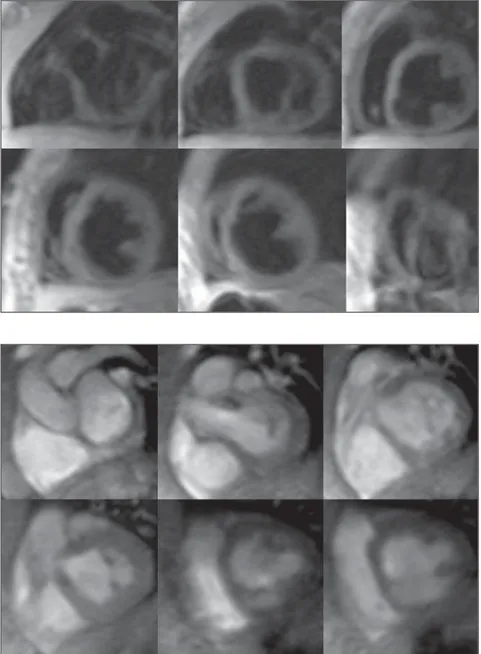
Figure 2. ECG-gated cine-MRI acquisition in short axis covering the whole extent of the left ventricle from the base to the left ventricular apex during diastole.
X Radiol Bras. 2010 Jul/Ago;43(4):IX–XI Images description
Figure 1. ECG-gated MRI morphology, black-blood, T1-weighted sequence with fat suppression in short axis view, cover-ing the whole extent of the left ventricle from the base to the left ventricular apex at mid-diastole. An abnormal muscle band divides the ventricle into two cavities.
Figure 2. ECG-gated cine-MRI acqui-sition in short axis covering the whole ex-tent of the left ventricle from the base to the left ventricular apex during diastole.
Diagnosis: Double-chambered left ven-tricle.
COMMENTS
Outpouching of the left ventricle is a rare condition with heterogeneous causes ranging from congenital abnormalities, such as diverticula or muscle bands, to complications secondary to myocardial infarction, such as aneurysm and pseudo-aneurysm. Distinguishing amongst these etiologies is challenging but of great clini-cal importance given the wide range of risks and implications involved(1–3).
Double-chambered left ventricle (DCLV) is characterized by the division of the ventricular chamber into two chambers by abnormal muscular tissue. It is best dif-ferentiated from left ventricular aneurysms and pseudoaneurysms by the fact that the double-chambered ventricle exhibits con-tractile motion during systole. Ventricular aneurysm lacks complete layering of the ventricular wall, and thus expands slightly due to the increased pressure during sys-tole(4,5).
The differentiation between double-chambered left and right ventricles is clear as they have different pathophysiology. Double-chambered right ventricle (DCRV) is more common and is often presented with murmur and exertional dyspnea. Stud-ies have found that DCRV is associated with septal defects, tetralogy of Fallot, and transposition of great arteries. Conversely, DCLV is commonly asymptomatic. DCRV is often caused by a progressive thickening of the right ventricular septum due to the presence of anomalous muscle bundles. This causes a pressure gradient, and two chambers in series develop. In contrast, the
chambers of a DCLV are in parallel and present less of a pressure gradient, as both contract synchronously. The DCLV etiol-ogy is less known, but the anomaly is thought to be congenital and non-progres-sive(6,7).
Usually, DCLV is incidentally found in the course of an evaluation for other car-diovascular abnormalities. As this is an extremely rare finding, no definite data regarding the prognosis, outcomes and potential complications, such as risk of embolism, of DCLV are available. It is generally believed that DCLV poses little risk to the patient. Treatment, if any, is usu-ally guided by the presence of other asso-ciated abnormalities(8,9).
Cardiac magnetic resonance imaging
Cardiac magnetic resonance imaging is useful in the assessment of associated con-ditions and to better understand the disease. Magnetic resonance imaging can dem-onstrate changes in the ventricular contrac-tility, presence or absence of fibrosis and is useful in the follow-up of patients with DCLV (Figure 3).
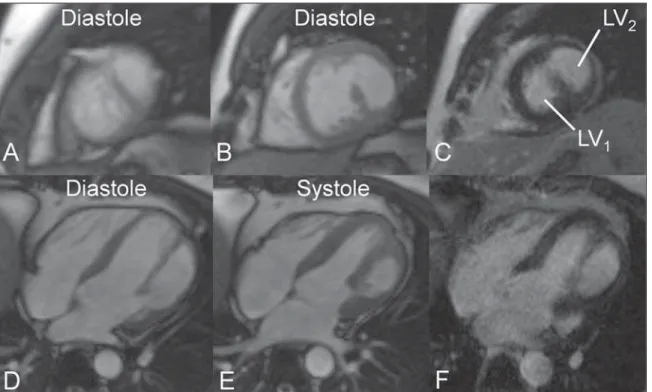
Figure 3. A 29-year-old male patient presented with myocardial infarction signs. Cardiac MRI with the cine (A,B,D,E) and late enhancement (C,F) techniques.
A: Cine-MRI, short axis view at the apical portion of left ventricle showing both cavities. B: Cine-MRI, short axis view at the middle portion of left ventricle showing both cavities. C: Delayed enhancement, short axis view at the middle portion of left ventricle without scar/fibrosis. D: Cine-MRI four-chamber view at diastole showing both cavities. E: Cine-MRI, four-chamber view at systole showing the thickening of the lateral wall of the LV2. F: Delayed enhancement,
XI
Radiol Bras. 2010 Jul/Ago;43(4):IX–XI Final considerations
The authors emphasize the rarity of this congenital anomaly in association with double mitral valve lesion and the diagnos-tic approach utilizing echocardiography and cardiac MRI.
REFERENCES
1. Gerlis LM, Partridge JB, Fiddler GI, et al. Two chambered left ventricle. Three new varieties. Br Heart J. 1981;46:278–84.
2. Vaidiyanathan D, Prabhakar D, Selvam K, et al. Isolated congenital left ventricular diverticulum in adults. Indian Heart J. 2001;53:211–3. 3. Krasemann T, Gehrmann J, Fenge H, et al.
Ven-tricular aneurysm or diverticulum? Clinical dif-ferential diagnosis. Pediatr Cardiol. 2001;22: 409–11.
4. Caron KH, Hernandez RJ, Goble M, et al. MR imaging of double chambered left ventricle. J Comput Assist Tomogr. 1991;15:140–2. 5. Harikrishnan S, Sivasankaran S, Tharakan J.
Double chambered left ventricle. Int J Cardiol. 2002;82:59–61.
6. Cil E, Saraçlar M, Ozkutlu S, et al.
Double-cham-bered right ventricle: experience with 52 cases. Int J Cardiol. 1995;50:19–29.
7. Hoffman P, Wójcik AW, Róza½ski J, et al. The role of echocardiography in diagnosing double cham-bered right ventricle in adults. Heart. 2004;90: 789–93.
8. Kay PH, Rigby M, Mulholland HC. Congenital double chambered left ventricle treated by exclu-sion of accessory chamber. Br Heart J. 1983;49: 195–8.