MONIQUE RENON ELLER
ISOLATION AND CHARACTERIZATION OF A PSEUDOMONAS-SPECIFIC PHAGE AND ITS USE TO CONTROL MILK
PROTEOLYSIS
VIÇOSA
MINAS GERAIS - BRASIL 2012
Ficha catalográfica preparada pela Seção de Catalogação e Classificação da Biblioteca Central da UFV
T
Eller, Monique Renon, 1986-
E45i Isolation and characterization of a Pseudomonas-specific 2012 phage and its use to control milk proteolysis / Monique Renon
Eller. – Viçosa, MG, 2012.
xiv, 115f. : il. ; (algumas color.) ; 29cm.
Texto em português e inglês
Orientador: Sérgio Oliveira de Paula
Tese (doutorado) - Universidade Federal de Viçosa. Inclui bibliografia.
1. Bacteriófagos. 2. Pseudomonas. 3. Genoma. 4. Leite - Qualidade. 5. Leite - Contaminação. 6. Leite - Microbiologia. I. Universidade Federal de Viçosa. Departamento de
Microbiologia. Programa de Pós-Graduação em Microbiologia Agrícola. II. Título.
_____________________________ Prof.ª Daniele da Glória de Souza MONIQUE RENON ELLER
ISOLATION AND CHARACTERIZATION OF A PSEUDOMONAS-SPECIFIC PHAGE AND ITS USE TO CONTROL MILK
PROTEOLYSIS
APROVED: December, 06th, 2012
______________________
Dissertation thesis presented to the Universidade Federal de Viçosa as part of the requirements of the PostGraduate Program in Agricultural Microbiology, to obtain the title of Doctor Scientiae.
_____________________________ Prof. Antônio Fernandes de Carvalho
(Co-advisor)
_____________________________ Prof. Luciano Gomes Fietto
_____________________________ Prof. Sérgio Oliveira de Paula
(Advisor) _____________________________
iv Acknowledgments
To God, for my life, health, friends, family, and for the providence. To Universidade Federal de Viçosa and to my masters and colleagues of the Department of Microbiology, for the opportunity, structure, and material and technical support. Special thanks to Nilcéia, for the patience and help.
To CAPES, for the finantial support to this research.
To my advisor, professor Sérgio Oliveira de Paula, for advice and encouragement, the good ideas and availability. To my co-advisors for their dedication, advice and availability.
To the colleagues of the Laboratory of Molecular Immunovirology, the the technicians, João and Jorge, for the friendship, teachings and availability. Special thanks to Roberto and Priscilla, for the daily work and learning. To Pedro, for the encouragement and technical aid.
To my friends:
Zamira, for the eternal friendship.
Raphael, Chris and Pituca, for the laughtes, tranquility and cheer on harder days.
v To Rafael, my dedicated husband, for the fellowship, patience, laughters, professional assistance and for make unforgettable moments from my routine. To his mother, Maria, for her dedication, teachings, faith and love of each day.
To all my family, for the good advices, talkings and concepts of life. In particular, to my parents, Romeu and Valéria, for the faith and unconditional strength; to my sisters, Pâmela and Allana, for the friendship and complicit; and to my brother Luan, for the lessons of life and love.
vi
List of Figures
Figure 3-1. Transmission Electron Microscopy of the φUFV-P2. ... 42
Figure 3-2. The φUFV-P2 genomic organization. ... 53
Figure 3-3. Dot plot alignment between the φUFV-P2 genome and the PaP3, MR299-2, LUZ24 and tf genomes. ... 55
Figure 3-4. Evolutionary relationships between φUFV-P2 and other phages based on generally conserved proteins. ... 56
Figure 3-5. Terminase small subunit aminoacid sequences alignment of UFV-P2, MR299-2, PAP3, LUZ24, and tf phages. ... 66
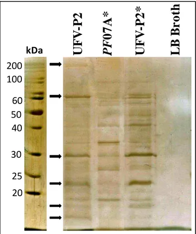
Figure 4-1. Electrophoretic pattern of the UFV-P2 proteins. ... 73
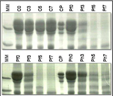
Figure 4-2. SDS-PAGE of the proteolysis assay.. ... 74
Figure 4-3. Densitometric analysis of the casein bands from the proteolysis assay.. ... 75
Figure 4-4.. ... 76
Figura 5-1. Campos de Aplicação - Bacteriófagos ... 89
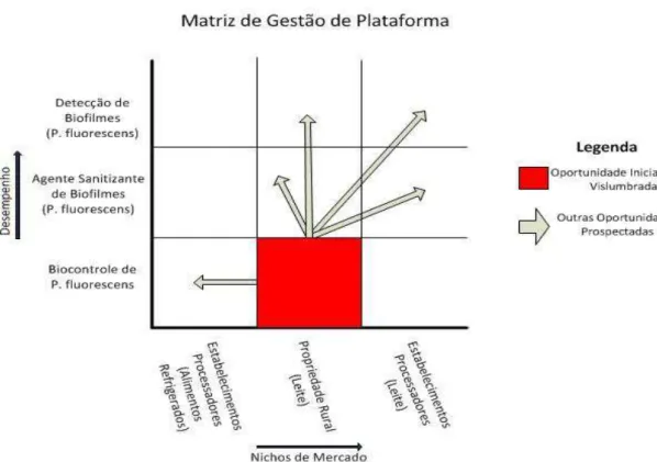
Figura 5-2. Matriz de Gestão de Plataforma ... 93
Figura 5-3. Cadeia de Valor do Leite ... 95
Figura 5-4. Produção de Leite no Brasil ... 98
Figura 5-5. Forças de Mercado ... 102
Figura 5-6. Estágio de Desenvolvimento do Projeto ... 106
vii
List of Tables
viii
Table of Contents
LIST OF FIGURES ... VI LIST OF TABLES ... VII RESUMO ... XI ABSTRACT ...XIII
1. GENERAL INTRODUCTION ... 1
2. LITERATURE REVIEW ... 4
USE OF PHAGES TO CONTROL MILK PROTEOLYSIS BY TERMOSTABLE ENZYMES OF PSYCHROTROPHIC BACTERIA... 5
Abstract ... 5
Use of phages to control food contaminants ... 5
Proteolysis in the dairy industry ... 6
Bacterial biofilms in food industry ... 10
Control points ... 12
Limitations in the use of phages in the food industry ... 14
Importance in the study of phage genomics and proteomics ... 17
Current Status ... 18
References ... 20
ix
COMPLETE GENOME SEQUENCE OF THE PSEUDOMONAS
FLUORESCENS BACTERIOPHAGE UFV-P2 ... 31
Abstract ... 31
Article ... 31
References ... 33
GENOME ANNOTATION OF THE PSEUDOMONAS PHAGE UFV-P2: A NEW MEMBER OF ―LUZ24-LIKE VIRUSES‖ ... 35
Abstract ... 35
Introduction ... 35
Materials and methods ... 36
Results and discussion ... 41
Conclusions ... 57
References ... 58
Supplementary material ... 63
4. CONTROL OF MILK PROTEOLYSIS BY THE PHAGE UFV-P2 . 67 CONTROL OF MILK PROTEOLYSIS BY THE TEMPERATE PSEUDOMONAS PHAGE UFV-P2 ... 68
Abstract ... 68
Introduction ... 69
Materials and methods ... 70
Results ... 72
x
Conclusions ... 79
References ... 80
5. ANÁLISE TÉCNICA, COMERCIAL E DE IMPACTO AMBIENTAL E SOCIAL (EVTECIAS) ... 87
UTILIZAÇÃO DE BACTERIÓFAGO DE PSEUDOMONAS FLUORESCENS NO CONTROLE DA GELIFICAÇÃO DO LEITE. ... 88
Tecnologia, produtos e oportunidades de negócio ... 88
Aspectos regulatórios ... 96
Mercado ... 97
Equipe do projeto... 104
Estágio de desenvolvimento do projeto ... 106
Plano de desenvolvimento tecnológico ... 108
Barreiras e riscos relacionados ao projeto ... 110
Impacto ambiental e relevância social ... 111
Conclusão sobre o projeto ... 111
xi
Resumo
ELLER, Monique Renon. D.Sc. Universidade Federal de Viçosa, dezembro de 2012. Isolamento e caracterização de um fago de Pseudomonas e seu uso no controle da proteólise do leite. Orientador: Sérgio Oliveira de Paula; Co-orientadores: Antônio Fernandes de Carvalho, Cynthia Canedo da Silva, Leandro Licursi de Oliveira e Maria Cristina Dantas Vanetti.
xiii
Abstract
ELLER, Monique Renon. D.Sc. Universidade Federal de Viçosa, December, 2012. Isolation and characterization of a Pseudomonas-specific phage and its use to control milk proteolysis. Advisor: Sérgio Oliveira de Paula; Co-advisors: Antônio Fernandes de Carvalho, Cynthia Canedo da Silva, Leandro Licursi de Oliveira and Maria Cristina Dantas Vanetti.
1
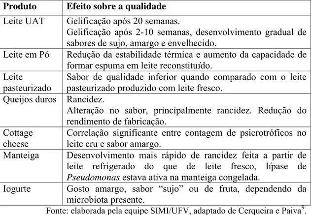
2 Milk proteolysis, when uncontrolled or unwanted, cause serious flavor and taste defects in the product, which cannot be passed to the consumer. The problem is even greater when it is caused by enzymes of psychrotrophic bacteria that maintain their activity even after heat treatments, including UHT. These enzymes slowly hydrolyze the milk caseins, increasing the milk viscosity and causing the effect called gelation of UHT milk, very difficult to be discovered by the manufacturers before its transfer to the final consumer.
Thus, preventive measures consist of the only way to eliminate this problem in the dairy industry, and the use of phages could be an economical, practical and safe alternative. Bacteriophages are viruses capable of killing the host bacterium or partially inhibit its metabolism. The addition of these particles in the food or even as cleaning agents doesn’t cause changes in the characteristics of the food nor risks to human health.
The main genus of psychrotrophic bacteria found in milk is Pseudomonas. The studies about specific phages against these bacteria are limited mainly to the use of phages to control P. aeruginosa in hospitals, and there are few isolated and characterized phages for the control of P. fluorescens, the main species found in the dairy environment. Thus, we propose the isolation and characterization of a phage that infects the bacteria P. fluorescens and the analysis of its potential use as a food preservative or as a component of active sanitizers in food industries.
4
5
Use of phages to control milk proteolysis by termostable enzymes of
psychrotrophic bacteria
Abstract
Studies involving the control of food spoilage using biological agents have emerged in the last decade because biocontrol generally has no consequences on human health and food characteristics. The new interest in the use of phages to control pathogenic bacteria expanded to industrial microbiology and is shown to be a safe, economic, and efficient technology. This study reviews the main aspects of the use of phages to control food contaminants, with an emphasis on the control of milk proteolysis by proteases from psychrotrophic bacteria, the main cause of UHT milk gelation.
Key words: Bacteriophage; Milk proteolysis; Biocontrol; Food microbiology; Food spoilage; Pseudomonas.
Use of phages to control food contaminants
6 and Mann, 2009). Moreover, interest has increased in the use of these viruses for the biocontrol of bacterial contaminants in industrial environments. In particular, this increased interest is reflected in studies on the use of these agents in the food industry to control potentially pathogenic bacteria such as Listeria monocytogenes and Salmonella sp. (Jones et al., 1991; Loessner and Carlton, 2005). More recently, phages were found to inhibit growth of spoilage bacteria, which can cause significant economic losses every year (Arcuri et al., 2008; Azeredo and Sutherland, 2008; Sillankorva et al., 2008). Despite this information, studies on bacteriophages in the food industry are often related to the presence of phages that infect starter cultures, causing fermentation processes to fail (Eller et al., 2012).Research on the isolation of Pseudomonas phages still concentrates on the control of hospital contamination by bacteria P. aeruginosa, but there are few studies on the isolation of phages to control P. fluorescens (Sillankorva et al., 2008), an important agent of milk proteolysis. To date, only 78 Pseudomonas phage genome sequences exist in the EMBL-EBI database (http://www.ebi.ac.uk/genomes), a low number considering the importance of this contaminant and its estimated viral diversity. Of these, only three sequences correspond to P fluorescens phages, the UFV-P2 (Eller et al., 2013), phi-2 (Paterson et al., 2010) and phiIBB-PF7A (Sillankorva et al., 2011) phages.
Proteolysis in the dairy industry
7 during the maturation process are used as substrates for a variety of catabolic reactions that generate compounds important to the development of the characteristic flavor of each kind of cheese and that depend on the dairy native microbiota (for example, non-starter lactic acid bacteria) or microorganisms added to the curd before ripening (Yvon and Rijnen, 2001). However, although proteolysis has beneficial effects and is essential for the development of desirable qualities of food products, uncontrolled proteolysis can negatively affect food quality.
The development of psychrotrophic bacteria infecting raw milk and the proteolysis caused by their proteolytic heat-resistant enzymes can cause serious effects on the quality of dairy products, such as off-flavors in pasteurized milk, hard cheeses, cottage cheeses, butter, and yogurt; reduced shelf life in pasteurized and Ultra-High-Temperature (UHT) milk; foaming formation in pasteurized milk and reconstituted milk powder; reduced thermal stability in milk powder and UHT milk; and reduced yields in cheese production (Sorhaug and Stepaniak, 1997).
8 Independent studies performed by Deeth et al. (2002) and López-Fandiño et al. (1993) showed significant differences in the behavior of UHT skimmed and whole milk contaminated with psychrotrophic microorganisms. Although skimmed milk exhibited a predominantly bitter flavor, whole milk presented a sour and acid flavor. The studies indicated that the different flavors occurred from increased proteolysis in skim milk caused by larger production of protease and susceptibility of proteins to attack by this enzyme. Furthermore, whole milk also involves the process of lipolysis caused by substantial amounts of lipase produced by contaminants (Deeth et al., 2002). This different behavior should be considered when establishing processing conditions.
9 The undesirable changes occurring in the UHT milk produced from contaminated raw milk are also caused, or at least accelerated, by the casein hydrolysis and subsequent release of the β-lactoglobulin-κ-casein complex (βκ complex) formed during heat treatment. The complex forms a three-dimensional network of interconnected proteins, which causes gel formation (MacMahon, 1996).Thus, any processing (e.g. UHT treatment) or storage (e.g. refrigeration) condition that accelerates (or delay) the release of βκ complex from the casein micelle will accelerate (or decelerate) the time for gelation of UHT milk (Datta and Deeth, 2003).
Several studies reported the biochemical properties of proteases produced by some strains of P. fluorescens associated with dairy product spoilage (Dufour et al., 2008; Marchand et al., 2009). In milk, proteases of psychrotrophic bacteria preferentially hydrolyze κ-casein, β-casein, and then αS1-casein. Whey proteins are only weakly hydrolyzed (Chen et al., 2003; Dufour et al., 2008; Koka and Weimer, 2000; Rajmohan et al., 2002).
Baruzzi and colleagues (2012) analyzed the proteolytic activity of proteases of Pseudomonas in mozzarella cheese. They observed a higher proteolytic activity on the outside, which was in direct contact with a liquid contaminated with the proteolytic samples, originally used in the conservation of cheese. According to them, this phenomenon is consistent with the wrinkling and exfoliation that occur on the surface of mozzarella cheese, the progressive release of cheese cuts, and an increase in the turbidity of the liquid.
10 attributable to protease activity of psychrotrophics after heat treatment. This phenomenon in coagulation is particularly important for UHT milk because it is intensified when heating at higher temperatures.
The genus Pseudomonas represents a number of bacterial groups found in a wide variety of environments. Some are plants, animals, and humans pathogens, whereas others are responsible for food contamination (Palleroni, 1992). Pseudomonas spp. are the major contaminating microorganisms that limit the shelf life of the processed fluid milk during refrigeration (Alatossava and Alatossava, 2006).
Among the bacteria belonging to this genus, the species P. fluorescens is the main food spoilage, especially in products of dairy origin (Dogan and Boor, 2003).This species comprises a large and heterogeneous group, which has been subdivided into five biotypes based on phenotypic characteristics (Palleroni, 1992). Beyond P. fluorescens, other species are associated with milk deterioration, such as P. putida, P. fragi,P. maltophila, P. putrefaciens, and, less frequently, P. aeruginosa (Alatossava and Alatossava, 2006; Wiedmann et al., 2000).
Bacterial biofilms in food industry
11 proteins, pili, DNA, RNA, lipids, and, especially, polysaccharides (Flemming and Wingender, 2010; Rendueles et al., 2012). In the dairy industry, these biofilms consist primarily of milk organic components, in particular proteins and calcium phosphate, constituting a rich source for biofilm formation in equipment (Marchand et al., 2012; Sillankorva et al., 2008; Sorhaug and Stepaniak, 1197).Commonly, bacteria in the biofilm are catalysts of chemical reactions that corrode steel equipment (Gibson et al., 1999; Gram et al., 2007; Oliver et al., 2005).The presence of biofilms may also clog pipes, reduce heat flux through the surfaces, and contaminate food, although several studies found a decrease in the adhesion of certain microorganisms in the presence of milk or milk proteins (Al-Makhlafi et al., 1995; Barnes et al., 1999; Kumar and Anand, 1998).
12 the adhesion of others. For example, microorganisms from rinse water, particularly Pseudomonas, Aeromonas, and Legionella spp., are able to form biofilms and act as a substrate for the anchoring of others with less potential adhesion microorganisms (Marchand et al., 2012). Sasahara and Zottola (1993) observed that Listeria monocytogenes formed a biofilm more efficiently when associated with a primary colonizing organism, the bacteria Pseudomonas fragi, which was attributed to polysaccharide production by this microorganism (Sasahara and Zottola, 1993). Currently, Pseudomonas fragi and Flavobacterium spp. are known to act as primary surface colonizers, facilitating the adhesion of L. monocytogenes cells, whereas the primary adhesion by Bacillus spp. and Staphylococcus spp. reduces adherence and biofilm formation by that bacterium (Moretro and Langsrud, 2004).
Control points
The use of phages in the control of contaminants in foodstuffs industries is based on three main points: limiting the growth of microorganisms in the raw material, control of biofilm in the points of the industrial process, and rapid and cheap detection of contaminated surfaces.
13 Using phage as sanitizers: The second point is the use of phages to decontaminate surfaces and eliminate biofilms on equipment and utensils. Given its high penetration ability, phages in suspension may be used in places that are hardly reached and that commonly have bacterial biofilms. Furthermore, phages are able to efficiently reduce the bacterial population in biofilms even without prolonged exposure times, as demonstrated by Sillankorva and colleagues using Pseudomonas-specific phages (Sillankorva et al., 2010).However, infection of cells in an EPS matrix depends on biofilm chemical composition and environmental factors such as temperature, stage of development, physico-chemical conditions of the medium, and phage concentration, which increase the need for exhaustive studies until the phages can be used in foodstuffs (Chaignon et al., 2007; Sillankorva et al., 2010). In this context, genetic engineering has been of a great assistance in optimizing biofilm inactivation techniques and even increasing viral efficiency for this purpose, as was done in the work of Lu and Collins (2007). They altered the viral genome by inserting a gene that encodes an enzyme capable of degrading the polysaccharide matrix of biofilms. The modified phage was able to reduce E. coli biofilms by approximately 99.997% (Lu and Collis, 2007).
14 Salmonella phage is commercially used to detect this pathogen in foods (VIDAS™UP Salmonella – BioMérieux Industry, França) as a modified and optimized protocol of the traditional method VIDAS, and results are achieved within 24 h. Likewise, a specific L. monocytogenes phage containing the lux gene was used to detect this contaminant in meat and showed great sensitivity and practicality, even in the presence of large numbers of natural competitors (Loessner et al., 1996). Thus, phages specific to the major agents of food spoilage, such as P. fluorescens, could be used to detect these microorganisms, helping to control contamination and enabling rapid implementation of corrective actions where necessary.
In industries, phages could act in combination with other components to improve the efficiency of treatment, such as antibiotics, enzymes, and chemical and/or physical agents which do not reduce viral activity. For example, some patents suggest using phages as active ingredients in disinfectants formulations (Breeuwer et al., 2005; Kang et al., 2012; Sulakvelidze et al., 2004; Yoon et al., 2011). In this case, the use of phages would decrease the need for high concentrations of sanitizers, thus reducing the risk of toxic residues in products brought to consumers.
Limitations in the use of phages in the food industry
15 The use of phages in the food industry, either as additives or as active agents of sanitizers, also has additional implications.
Specificity: Generally, phages have a narrow host range, which can easily be countered using mixtures (cocktails) of phages specific for different hosts and / or host receptors. Research on the use of viral consortia and the interaction between different phages, and phages with different molecules present in food and in biofilms, is also fundamental to finding the best way to use these particles for biocontrol of food contaminants. However, phage specificity is also a positive point for its use. In addition to ensuring consumer safety, its specific activity does not destroy indigenous bacteria in raw material—unlike other microbial control agents—which is often required in the production of fermented derivatives.
Resistance to acceptance: The addition of viral particles in foods may be a factor for rejection by consumers. However, marketing programs emphasizing the beneficial effects of the use of a biological agent over a chemical reagent to control food spoilage could turn this limitation in a strategic advantage.
16 Complex environments: One of the major problems that the application of phages in food must overcome is the diversity and complexity of the environments in which they will act. Viral activity is highly dependent on environmental conditions, and small changes could cause large loss in activity (Coffey et al., 2011). Therefore, research on viral activity over food contaminants must be carried out in the conditions in which the phages will be used, including composition, presence of food preservatives, pH, and temperature. The current demand for milder food preservation methods and the standardization of processes will assist in maintaining the activity of the viral particles.
Emergence of resistant strains: The constant possibility of the emergence of bacteria resistant to phages used for biocontrol is one of the main points regarding the viability of studies in this area. However, phages have a great ability to readapt and circumvent the bacterial defense mechanisms, one of the most significant advantages on their use to control contaminants. In most environments, a wide variety of phages and hosts is involved in continuous cycles of coevolution in which the emergence of phage-resistant hosts is important for the preservation of bacterial strains (Rohwer, 2003), whereas constant phage co-resistance threatens these new bacterial strains (Labrie et al., 2010).
17 reducing the effects of each treatment on the physico-chemical and sensorial characteristics of the food, and on consumer health.
Importance in the study of phage genomics and proteomics
18 The study of phages plays a central role in some of the most significant discoveries in the biological sciences, from the identification of DNA as the genetic material to the development of recombinant technology. Phage-derived proteins or recombinant proteins produced in viral particles are often used as diagnostic and therapeutic agents and for drug discovery (Liu et al., 2004; Loeffler et al., 2001; Schuch et al., 2002; Smith et al., 2001). The continuous introduction of new sequencing technologies in the last decade has caused a dramatic increase in the number of completely sequenced phage genomes: from 40 in January 1997 to 1089 in October 2012 (Ceyssens, 2009). However, the annotation of a new sequenced genome generates approximately 50% of ORFs encoding proteins with unknown functions. Eller and colleagues (2013) conducted a study on the sequencing of a genome of a Pseudomonas fluorescens phage and found 92 predicted ORFs, of which only 41 (44.6%) showed significant similarity to ORFs already described. Of these 41, 15 (36.6%) were related to ORFs that encoded proteins of unknown function.
Current Status
19 example, the patent of Breeuwer and colleagues (2005) deals with the use of Cronobacter sakasakii-specific phages in foods, sanitizers, and possible infections, and part of its specification refers to the use of these viruses to control this contaminant in dairy products. The patent registered by Sulakvelidze and colleagues (2004) is on a formulation containing phages for biocontrol of contaminants on surfaces and equipment, giving special attention to the methodology for the application of these agents. Despite these numerous patents, only one phage until today has been recognized as GRAS (Generally Recognized As Safe) by the U.S. Food and Drug Administration, and its application is limited to controlling L. monocytogenes in certain meat products (FDA, 2006).
Ceyssens and colleagues (2011) noted that small genomic variations ―intra -species‖ have phenotypic consequences on essential applications of different phages. Distinct host ranges were observed between isolates with identical tail fibers and/or genomic regions of early genes, implying that smaller genomic alterations can cause a significant change in the spectrum of viral infectivity. Thus, the importance of genomic studies on the largest number of phages and a comparison of different sequences that combine the biological characteristics of the virus with their respective gene sequences can be realized. Then, expanding the knowledge of these organisms will be possible to ensure greater reliability and agility on the registration processes for use as food additives or as sanitizer agents for biological control in the dairy industry.
Acknowledgments
20 Aperfeiçoamento de Pessoal de Nível Superior (CAPES), and Conselho Nacional de Desenvolvimento Científico e Tecnológico (CNPq). The funders had no role in the study design, data collection, analysis, decision to publish, or preparation of this manuscript.
References
Adams, D.M., Barach, J.T., Speck, M.L., 1975. Effect of psychrotrophic bacteria from raw milk on milk proteins and stability of milk proteins to ultrahigh temperature treatment. Journal Series of the North Carolina Agricultural Experiment Station; Paper Number 4746.
Alatossava, M.P., Alatossava, T., 2006. Phenotypic characterization of raw milk-associated psychrotrophic bacteria. Microbiol. Res. 161, 334-346. Al-Makhlafi, H., Nasir, A., McGuire, J., Daeschel, M., 1995. Adhesion of
Listeria monocytogenes to silica surfaces after sequential and competitive adsorption of bovine serum albumin and b-lactoglobulin. Appl. Environ. Microbiol. 61, 2013–2015.
Arcuri, E.F., da Silva, P.D.L., Brito, M.A.V.P., Brito, J.R.F., Lange, C.C., Magalhães, M.M.A., 2008. Counting, isolation and characterization of psychrotrophic bacteria from refrigerated raw milk. Ciência Rural 38, 2250-2255.
21 Barnes, L.M., Lo, M.F., Adams, M.R., Chamberlain, A.H.L., 199. Effect of Milk Proteins on Adhesion of Bacteria to Stainless Steel Surfaces. Appl. Environ. Microbiol. 65, 4543-4548.
Baruzzi, F., Lagonigro, R., Quintieri, L., Morea, M., Caputo, L., 2012. Occurrence of non-lactic acid bacteria populations involved in protein hydrolysis of cold-stored high moisture Mozzarella cheese. Food Microbiol. 30, 37-44.
Bore, E., Langsrud, S., 2005. Characterization of micro-organisms isolated from dairy industry after cleaning and fogging disinfection with alkyl amine and peracetic acid. J. Appl. Microbiol. 98, 96–105.
Breeuwer, P., Boissin-Delaponte, C., Joosten, H., Lardeau, A., 2005. Isolated phages and their use as disinfectants in food or for sanitation of factory environments. [EP 1533369 A1] Ref Type: Patent.
Bruynoghe, R., Maisin, J., 1921. Essais de thérapeutique au moyen du bacteriophage du Staphylocoque. J. Compt. Rend. Soc. Biol. 85, 1120-1121.
Ceyssens, P.J., Glonti, T., Kropinski, N.M., Lavigne, R., Chanishvili, N., Kulakov, L., et al., 2011. Phenotypic and genotypic variations within a single bacteriophage species. Virol. J. 8, 134-138.
Ceyssens, P.J., 2009. Isolation and characterization of lytic bacteriophages infecting Pseudomonas aeruginosa. PhD Thesis from Katholieke Universiteit Leuven, Leuven, Belgium.
22 enzymatic treatments depends on their chemical composition. Appl. Microbiol. Biotechnol. 75, 125–132.
Chen, J., Griffiths, M.W., 1996. Luminescent Salmonella strains as real time reporters of growth and recovery from sublethal injury in food. Int. J. Food Microbiol. 31, 27-43.
Chen, L., Daniel, R.M., Coolbear, T., 2003. Detection and impact of protease and lipase activities in milk and milk powders. Int. Dairy J. 13, 255-275. Coffey, B., Rivas, L., Duffy, G., Coffey, A., Ross, R.P., McAuliffe, O., 2011.
Assessment of Escherichia coli O157:H7-specific bacteriophages e11/2 and e4/1c in model broth and hide environments. Int. J. Food Microbiol. 147, 188-194.
Costerton, J.W., Cheng, K.J., Geesey, G.G., Ladd, T.I., Nickel, T.J., 1987. Bacterial biofilms in nature and disease. Annu. Rev. Microbiol. 41, 435– 464.
Datta, N., Deeth, H.C., 2001. Age gelation of UHT-milk - a review. Food Bioprod. Process 79, 197-210.
Datta, N., Deeth, H.C., 2003. Diagnosing the cause of proteolysis in UHT milk. Lebenson Wiss Technol. 36, 173-182.
Deeth, H.C., Khusniati, T., Datta, N., Wallace, R.B., 2002. Spoilage patterns of skim and whole milks. J. Dairy Res. 69, 227-241.
Dogan, B., Boor, K.J., 2003. Genetic diversity and spoilage potentials among Pseudomonas spp. isolated from fluid milk products and dairy processing plants. Appl. Environ. Microbiol. 69, 130–138.
23 Dufour, D., Nicodème, M., Perrin, C., Driou, A., Brusseaux, E., Humbert, G., et al., 2008. Molecular typing of industrial strains of Pseudomonas spp. isolated from milk and genetical and biochemical characterization of an extracellular protease produced by one of them. Int. J. Food Microbiol. 125, 188-196.
Eller, M.R., Dias, R.S., Moraes, C.A., Carvalho, A.F., Oliveira, L.L., Silva, E.A.M., et al., 2012. Molecular characterization of a new lytic bacteriophage isolated from cheese whey. Arch. Virol., In press. PMID: 22865166.
Eller, M.R., Salgado, R.L., Vidigal, P.M.P., Alves, M.P., Dias, R.S., de Oliveira, L.L., et al., 2013. Complete genome sequence of the Pseudomonas fluorescens bacteriophage UFV-P2. Genome Annoucement. In press.
FDA: Food and Drug Administration. 2006. Food additives permitted for direct addition to food for human consumption; Bacteriophage preparation. Federal Register: Rules and Regulations, 71[160]. Ref Type: Report.
Fischetti, V.A., 2010. Bacteriophage endolysins: a novel anti-infective to control Gram-positive pathogens. Int. J. Med. Microbiol. 300, 357-362. Flemming, H.C., Wingender, J., 2010. The biofilm matrix. Nature Rev.
Microbiol. 8, 623–633.
24 Gibson, H.J., Taylor, H., Hall, K.E., Holah, J.T., 1999. Effectiveness of cleaning techniques used in the food industry in terms of the removal of bacterial biofilms. J. Appl. Microbiol. 87, 41-48.
Goodridge, L., Chen, J., Griffiths, M., 1999. The use of a fluorescent bacteriophage assay for detection of Escherichia coli O157:H7 in inoculated ground beef and raw milk. Int. J. Food Microbiol. 47, 43-50. Górski, A., Miedzybrodzki, R., Borysowski, J., Weber-Dabrowska, B.,
Lobocka, M., et al., 2009. Bacteriophage therapy for the treatment of infections. Curr. Opin. Investig. Drugs. 10, 766-774.
Gram. L., Bagge-Ravn, D., Ng, Y.Y., Gymoese, P., Vogel, B.F., 2007. Influence of food soiling matrix on cleaning and disinfection efficiency on surface attached Listeria monocytogenes. Food Control 18, 1165– 1171.
Housby, J.N., Mann, N.H., 2009. Phage therapy. Drug Discov. Today 14, 536-540.
Jones, C. R., Rennie, G. K., & Moore, C. H. 1991. Use of viruses against undesirable microorganisms. [EP 0414304 A2] Ref Type: Patent.
Jonghe, V., Coorevits, A., Van Hoorde, K., Messens, W., Van Landschoot, A., De Vos, P., et al., 2001. Influence of storage conditions on the growth of Pseudomonas species in refrigerated raw milk. Appl. Environ. Microbiol. 77, 460-470.
25 Koka, R., Weimer, B.C., 2000. Isolation and characterization of a protease
from Pseudomonas fluorescens RO98. J. Appl. Microbiol. 89, 280-288. Koonin, E.V., Wolf, Y.I., 2012. Evolution of microbes and viruses: a
paradigm shift in evolutionary biology? Front. Cell. Infect. Microbiol. 119. Epub 2012 Sep 13. PMID: 22993722
Kumar, C.G., Anand, S.K., 1998. Significance of microbial biofilms in food industry: a review. Int. J. Food Microbiol. 42, 9–27.
Labrie, S.J., Samson, J.E., Moineau, S., 2010. Bacteriophage resistance mechanisms. Nature Rev. Microbiol. 8, 317-327.
Law, B.A., Andrews, A.T., Sharpe, M.E., 1977. Gelation of UHT sterilized milk by proteases from a strain of Pseudomonas fluorescens, isolated from raw milk. J. Dairy Res. 44, 145-148.
Liu, J., Dehbi, M., Moeck, G., Arhin, F., Bauda, P., Bergeron, D., et al., 2004. Antimicrobial drug discovery through bacteriophage genomics. Nature Biotechnol. 22, 185-191.
Loeffler, J.M., Nelson, D., Fischetti, V.A., 2001. Rapid killing of Streptococcus pneumoniae with a bacteriophage cell wall hydrolase. Science 294, 2170-2172.
Loessner, M., Carlton, R.M., 2005. Virulent phages to control Listeria monocytogenes in foodstuffs and in food processing plants. [US 2005/0175594 A1] Ref Type: Patent.
26 López-Fandiño, R., Olano, A., Corzo, N., Ramos, M., 1993. Proteolysis during storage of UHT milk: differences between whole and skim milk. J. Dairy Res. 60, 339-347.
Lu, T.K., Collins, J.J., 2007. Dispersing biofilms with engineered enzymatic bacteriophage. P. Natl. Acad. Sci. USA 104, 11197-202.
Marchand, S., Block, J.D., Jonghe, V.D., Coorevits, A., Heyndrickx, M., Herman, L., 2012. Biofilm formation in milk production and processing environments; influence on milk quality and safety. Compr. Rev. Food Sci. F. 11, 133-147.
Marchand, S., Vandriesche, G., Coorevits, A., Coudijzer, K., De Jonghe, V., Dewettinck, K., et al., 2009. Heterogeneity of heat-resistant proteases from milk Pseudomonas species. Int. J. Food Microbiol. 133, 68-77. McMahon, D.J., 1996. Age-gelation of UHT milk: Changes that occur during
storage, their effect on shelf life and the mechanism by which age-gelation occurs. Heat treatments and alternative methods. International Dairy Federation ref S.I. 9602. Brussels, Belgium, International Dairy Federation, 315-325.
Meer, R.R., Bakker, J., Bodyfelt, F.W., Griffiths, M.W., 1991. Psychotrophic Bacillus spp. in fluid milk products: a review. J. Food Protect. 54, 969-979.
Møretrø, T., Langsrud, S., 2004. Listeria monocytogenes: biofilm formation and persistence in food processing environments. Biofilms 1, 107-121. Nielsen, S.S., 2002. Plasmin System and Microbial Proteases in Milk:
27 Oliver, S.P., Jayarao, B.M., Almeida, R.A., 2005. Foodborne pathogens in milk and the dairy farm environment: food safety and public health implications. Foodborne Pathog. Dis. 2, 115-129.
Palleroni, N.J., 1992. Human- and animal-pathogenic pseudomonads. In: Balows, A., Truper, H.G., Dworkin, M., Harder, W., Schleifer, K.-H. (Eds.), The Prokaryotes. A Handbook on the Biology of Bacteria: Ecophysiology, Isolation, Identification, Applications. Springer-Verlag, New-York, pp. 3086-3103.
Pasternack, G.R., Sulakvelidze, A., 2009. Listeria monocytogenes bacteriophage and uses thereof. [US 7,507,571] Ref Type: Patent.
Paterson, S., Vogwill, T., Buckling, A., Benmayor, R., Spiers, A.J., Thomson, N.R., et al., 2010. Antagonistic coevolution accelerates molecular evolution. Nature 464, 275-278.
Rajmohan, S., Dodd, C.E., Waites, W.M., 2002. Enzymes from isolates of Pseudomonas fluorescens involved in food spoilage. J. Appl. Microbiol. 93, 205-213.
Rendueles, O., Kaplan, J.B., Ghigo, J.M., 2012. Antibiofilm polysaccharides. Environ. Microbiol., In press. doi:10.1111/j.1462-2920.2012.02810. Rohwer, F., 2003. Global phage diversity. Cell 113, 41.
Sasahara, K., Zottola, E.A., 1993. Biofilm formation by Listeria monocytogenes utilizes a primary colonizing microorganism in flowing systems. J. Food Protect. 56, 1022-1028.
28 Sillankorva, S., Kluskens, L.D., Lingohr, E.J., Kropinski, A.M., Neubauer, P., Azeredo, J., 2011. Complete genome sequence of the lytic Pseudomonas fluorescens phage jIBB-PF7A. Virol. J. 8, 142-147.
Sillankorva, S., Neubauer, P., Azeredo, J., 2008. Isolation and characterization of a T7-like lytic phage for Pseudomonas fluorescens. BMC Biotechnol. 8, 80-91.
Sillankorva, S., Neubauer, P., Azeredo, J., 2010. Phage control of dual species biofilms of Pseudomonas fluorescens and Staphylococcus lentus. Biofouling 26, 567-575.
Smith, D.E., Tans, S.J., Smith, S.B., Grimes, S., Anderson, D.L., Bustamante, C., 2001. The bacteriophage ⱷ29 portal motor can package DNA against a large internal force. Nature 413, 748-752.
Sorhaug, T., Stepaniak, L., 1997. Psychrotrophs and their enzymes in milk and dairy products: Quality aspects. Trends Food Sci. Tech. 8, 35-41.
Sulakvelidze, A., Morris, J.G.Jr., Alavidze, Z., Pasternack, G.R., Brown, T.C., 2004. Method and device for sanitation using bacteriophages. [US 2004/0029250 A1] Ref Type: Patent.
Sulakvelidze, A., Sozhamamnnan, S., Pasternack, G.R., 2010. Salmonella bacteriophage and uses thereof. [US 7,674,467] Ref Type: Patent.
Suttle, C.A., 2005. Viruses in the sea. Nature 437, 356-361.
29 Ulitzur, S., Kuhn, J., 2000. Construction of lux bacteriophages and the determination of specific bacteria and their antibiotic sensitivities. Method Enzymol. 305, 543-557.
Wiedmann, M., Weilmeier, D., Dineen, S.S., Ralyea, R., Boor, J.K., 2000. Molecular and phenotypic characterization of Pseudomonas spp. isolated from milk. Appl. Environ. Microbiol. 66, 2085-2095.
Yoon, S., Kang, S., Kyoung, S., Choi, Y., Son, J., 2011. Bacteriophage having killing activity specific to Staphylococcus aureus. [US 8,071,352] Ref Type: Patent.
30
3.
STRUCTURAL AND GENOMIC
31
Complete genome sequence of the Pseudomonas fluorescens
bacteriophage UFV-P2
Abstract
Milk proteolysis caused by Pseudomonas fluorescens is a serious problem in the dairy industries due to their ability to grow under refrigeration. The use of phages to control contaminants in food has been considered an alternative to traditional methods; therefore, thorough understanding of such organisms is vital for their use. In the present study, we show the complete genome sequence and analysis of a P. fluorescens phage isolated from waste water of a dairy industry in Brazil.
Article
32 examination to ensure its safety and effectiveness. Therefore, it was essential to determine the complete genome sequence of phage UFV-P2, a P. fluorescens phage with a high ability to reduce casein proteolysis in milk.
Phage UFV-P2 was isolated and purified from the waste water of a dairy industry in Brazil, and then its genomic DNA was extracted and sequenced in Illumina Genome Analyzer II by CD Genomics (New York, USA). The viral genome was assembled and analyzed using CLC Genomics Workbench version 5.1 (CLCBio). The reads were assembled in contigs that considered more stringent parameters, in which 90% of each read had to cover the other read with 90% identity. This assembly produced the UFV-P2 genome sequence with coverage of 30,655-fold. Around 92 open reading frames (ORFs) were predicted using the Bacterial Genetic Code (NCBI translation table 11) and alternatives start codons (AUG,CUG, and UUG). All predicted ORFs were functionally annotated using Blastx searches against GenBank (http://www.ncbi.nlm.nih.gov/genbank) and UniProt (http://www.uniprot.org) databases. Only 41 ORFs (44.57%) presented significant similarities to known proteins and were considered in genome annotation. Additionally, the presence of tRNA genes was predicted using tRNAscan-SE program version 1.21 (8).
33 Finally, 15 ORFs (36.6%) hits with genes encoding enzymes, including one lysozyme, the terminase small and large subunits, a exonuclease, a endonuclease, a primase/helicase and two parts of the DNA polymerase. The bioinformatics analyses showed 53.61% of identity with the genome of the temperate Pseudomonas phage PaP3 (9). Knowledge about this group of phages is still limited, and further analyses are needed to confirm UFV-P2’s safety and its potential as an agent for biocontrol of milk contaminants.
Nucleotide Sequence Accession Number. The complete genome sequence of P. fluorescens phage UFV-P2 is available in GenBank under accession number JX863101.
Acknowledgments
We would like to thank to Cristina Dantas Vanetti, of the Federal University of Viçosa, for providing materials and technical support. This study was supported by grants from the Fundação de Amparo à Pesquisa do Estado de Minas Gerais (FAPEMIG), Coordenação de Aperfeiçoamento de Pessoal de Nível Superior (CAPES) and Conselho Nacional de Desenvolvimento Científico e Tecnológico (CNPq). The funders had no role in the study design, data collection, analysis, decision to publish, or preparation of this manuscript.
References
34 2. Mu Z, Du M, Bai Y. 2009. Purification and properties of a heat-stable enzyme of Pseudomonas fluorescens Rm12 from raw milk. Eur. Food Res. Technol. 228:725-734.
3. Nörnberg MFBL, Friedrich RSC, Weiss RDN, Tondo EC, Brandelli A. 2009. Proteolytic activity among psychrotrophic bacteria isolated from refrigerated raw milk. Int. J. Dairy Technol. 63:41-46.
4. Dufour, D, Nicodeme M, Perrin C, Driou A, Brusseaux E, Humbert G, Gaillard JL, Dary A. 2008. Molecular typing of industrial strains of Pseudomonas spp. isolated from milk and genetical and biochemical characterization of an extracellular protease produced by one of them. Int. J. Food Microbiol. 125:188-196.
5. Kives J, Guadarrama D, Orgaz B, Rivera-Sem A, Vazquez J, SanJose C. 2005. Interactions in biofilms of Lactococcus lactis ssp. cremoris and Pseudomonas fluorescens cultured in cold UHT milk. J. Dairy Sci. 88:4165-4171.
6. Karl T. 2004. Old dogma, new tricks—21st century phage therapy. Nat. Biotechnol. 22:31-36.
7. Dixon B. 2004. New dawn for phage therapy. Lancet Infect. Dis. 4:186. 8. Lowe TM and Eddy SR. 1997. tRNAscan-SE: A program for improved
detection of transfer RNA genes in genomic sequence. Nucleic Acids Res. 25:955-964.
35
Genome annotation of the Pseudomonas phage UFV-P2: a new
member of “LUZ24
-like virus
es”
Abstract
Phages infecting spoilage microorganisms have been considered as alternative biocontrol agents, and the study of their genomes is essential to their safe use in foods. UFV-P2 is a new Pseudomonas fluorescens-specific phage that has been tested for its ability to inhibit milk proteolysis. It belongs to the Podoviridae family and has a dsDNA genome of 45,517 bp, which contains at least 41 ORFs and a genome organization similar to the MR299-2, PaP3 and LUZ24 phage genomes, recently grouped as LUZ24-like viruses. In the present study, the structural genome analysis and the comparison of phylogenetic hypotheses lead us to propose the classification of φUFV-P2 in the LUZ24-like genus. Additionally, we propose the inclusion of the previously unclassified φtf in this genus.
Introduction
According to ICTV classification
36 LUZ24 (8) and MR299-2 (1). Two unassigned species, the phages 119X (17) and F166 (6), are also classified in Podoviridae. This classification is based on biological characteristics and genome organization. Additionally, a search in the current databases showed that Pseudomonas-infecting Podoviridae still comprehends the genus of N4-like viruses with the phages LIT1 and LUZ7 (7) and others unclassified phages, including Bf7 (21), tf (16), and PaP2 (NC_005884).
Recently, we reported the genome announcement of Pseudomonas fluorescens bacteriophage UFV-P2 (12), a phage with a high ability to reduce casein proteolysis in milk. Milk proteolysis caused by thermoresistant enzymes produced by psychrotrophics is responsible for serious losses in the dairy industry due to negative effects on the quality and reduced shelf life of dairy products. In this environment, Pseudomonas spp. are prevalent contaminants (3, 18, 20), mainly P. fluorescens (2, 9). The use of phages in biocontrol has been suggested as an alternative to the use of chemicals (4, 23, 24), but they must be used with caution. In addition of proteolysis and biofilm inhibition studies and their host range definitions, it is necessary to understand phages’ genome and proteome to make possible their use as biocontrol agents.
To expand our understanding about the P. fluorescens-specific phage UFV-P2 (12), we present in detail the analysis of its structural genome and comparisons to other phage genomes available in GenBank database.
Materials and methods
37 The phage UFV-P2 was isolated from wastewater of a dairy industry in Minas Gerais, Brazil, and propagated in a strain of P. fluorescens 07A, courtesy of Laboratory of Food Microbiology, located at Federal University of Viçosa, Brazil, at 30 °C in LB medium.
Morphological analysis of UFV-P2
An aliquot of the viral extract (50 mL) was purified with 10 % PEG 8000 and used for electron microscopy studies. Ten microliters of a 10X diluted viral suspension was added to a 200-mesh grid that was covered with Formvar® for 5 min. The excess liquid was removed with filter paper, and the reaction was covered with 10 µl of 2 % uranyl acetate for 20 sec. The samples were visualized with a transmission electron microscope (Zeiss EM 109 TEM) operating at 80 kV at the Nucleus of Microscopy and Microanalysis (NMM) at UFV.
Genome extraction and composition
38 Genomic DNA sequencing and assembly
UFV-P2 genome was extracted and sequenced in Illumina Genome Analyzer II by CD Genomics (New York, USA) and was assembled and analyzed using CLC Genomics Workbench version 5.1 (CLCBio). The sequenced reads were assembled in contigs considering more stringent parameters, in which 90% of each read had to cover the other read with 90% identity. The data from initial analyses, including genome assembly and ORFs prediction, were submitted by Eller et al. (12) and are available in GenBank database under accession number JX863101.
Bioinformatics analysis
To detect homologous proteins and functionally annotate each predicted ORF, BLASTX searches were carried out against GenBank (http://blast.ncbi.nlm.nih.gov) and UniProt (http://www.uniprot.org/blast) databases. Promoter sequences were predicted by BPROM Prediction of bacterial promoters (http://linux1.softberry.com/berry.phtml) and BDGP
Neural Network Promoter Prediction
(http://www.fruitfly.org/seq_tools/promoter.html). Transcriptional terminators were predicted by FindTerm (http://linux1.softberry.com/berry.phtml) and RibEx Riboswitch Explorer (http://132.248.32.45/cgi-bin/ribex.cgi). Putative
tRNA-genes were analyzed by tRNA tRNAscan-SE
(http://selab.janelia.org/tRNAscan-SE/). Additionally, a search for direct terminal repeats (DTRs) was carried out using Pygram (10).
39 matrix was calculated using CLC Genomics Workbench version 5.1 (CLCBio). Dot plot analysis was carried out using Nucleic Acid Dot Plots (http://www.vivo.colostate.edu/molkit/dnadot/index.html), considering a window size of 13 and a mismatch limit of 0.
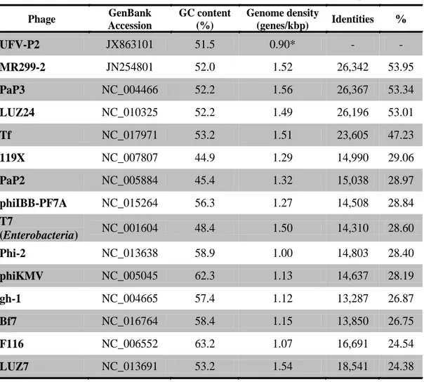
Table 3-1. Comparison of Pseudomonas φUFV-P2 and others phage genomes.
Phage UFV-P2
Phage GenBank Accession
GC content (%)
Genome density
(genes/kbp) Identities %
UFV-P2 JX863101 51.5 0.90* - -
MR299-2 JN254801 52.0 1.52 26,342 53.95
PaP3 NC_004466 52.2 1.56 26,367 53.34
LUZ24 NC_010325 52.2 1.49 26,196 53.01
Tf NC_017971 53.2 1.51 23,605 47.23
119X NC_007807 44.9 1.29 14,990 29.06
PaP2 NC_005884 45.4 1.32 15,038 28.97
phiIBB-PF7A NC_015264 56.3 1.27 14,508 28.84
T7
(Enterobacteria) NC_001604 48.4 1.50 14,310 28.60
Phi-2 NC_013638 58.9 1.00 14,803 28.40
phiKMV NC_005045 62.3 1.13 14,637 28.19
gh-1 NC_004665 57.4 1.12 13,287 26.87
Bf7 NC_016764 58.4 1.15 13,850 26.75
F116 NC_006552 63.2 1.07 16,691 24.54
LUZ7 NC_013691 53.2 1.54 18,541 24.38
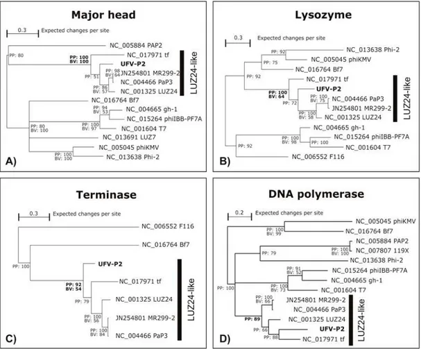
40 Phylogenetic trees
For clustering UFV-P2 phage in an evolutionary way, phylogenetic hypotheses were inferred by Bayesian inference (BI) and maximum likelihood (ML) using MrBayes v3.1.2 (15) and GARLI 2.0 (28), respectively.
Coding sequences (CDS) of major head protein (MH), lysozyme (LYS), terminase large subunit (TERM), and DNA polymerase part I (DNAP) were selected in the UFV-phage genome. Homologous CDS of these proteins were also selected in genomic reference sequences (RefSeq) of related phages (see Table 1). The codons of these four sequence sets (MH, LYS, TERM, and DNAP) were aligned using MUSCLE v.3.8.31 9 (11), a local alignment algorithm. Alignments were manually inspected, and the sites with gaps were excluded. To expedite the construction of phylogenetic trees, a model of nucleotide substitution was estimated using the jModeltest program (19). The GTR + I + G substitution model was selected as the best DNA evolution model for MH sequence set, the TIM2+I+G for LYS and TERM, and the TPM2uf+G for DNAP, according to the Akaike Information Criterion (AIC).
41 The respective substitution models of each sequence set were selected in the GARLI settings, and the statistical support of the ML phylogenetic trees was calculated by 1,000 bootstrap replicates. The 50% majority rule consensus trees of all bootstrap replicates was summarized using the SumTrees of DendroPy 3.8.0 (25).
Results and discussion
Isolation and morphology
42 Figure 3-1. Transmission Electron Microscopy of the φUFV-P2. Virions have isometric capsids of 40-50 nm and very short tails (arrows). Scale bars = 100 nm.
Functional genomic organization
The viral genome was extracted, and different aliquots were digested with DNase I or RNase A. While DNAse I digestion degraded the viral genome, RNase A digestion had no effect, indicating that it was composed of DNA (data not shown). The phage UFV-P2 has a linear 45,517 bp DNA genome with a GC content of 51.5%, no DTRs, and was sequenced with coverage of 30,655 fold (Table 3-1).
44 Table 3-2. Functional genomic annotation based of phage UFV-P2.
ORF Start End Length Sense Start
codon Product Similarity (GenBank access) E-value*
1 673 1146 474 positive ATG terminase small subunit Pseudomonas phage tf
(YP_006382530) 7,00E-63
2 1040 1573 534 positive CTG lysozyme Pseudomonas phage tf
(YP_006382529) 2,00E-53
3 1577 3022 1446 positive ATG terminase large subunit Pseudomonas phage LUZ24
(YP_001671939) 0,00E+00
4 3001 5139 2139 positive TTG portal protein Pseudomonas phage MR299-2
(AFD10682) 0,00E+00
5 5380 6366 987 positive ATG Scaffolding protein Pseudomonas phage LUZ24
(YP_001671936) 8,00E-100
6 6385 7338 954 positive ATG major head protein Pseudomonas phage PaP3
(NP_775251) 0,00E+00
7 7384 7710 327 positive TTG hypothetical protein Pseudomonas phage MR299-2
45 8 7714 8343 630 positive ATG phage particle protein Pseudomonas phage LUZ24
(YP_001671933) 1,00E-77
9 8192 8542 351 positive TTG hypothetical protein Pseudomonas phage LUZ24
(YP_001671932) 2,00E-26
10 8764 9399 636 positive ATG tail fiber protein Pseudomonas phage tf
(YP_006382516) 4,00E-39
11 9407 10948 1542 positive ATG phage particle protein Pseudomonas phage tf
(YP_006382515) 0,00E+00
12 10894 11658 765 positive CTG hypothetical protein Pseudomonas phage LUZ24
(YP_001671928) 5,00E-51
13 11592 12089 498 positive TTG hypothetical protein Pseudomonas phage LUZ24
(YP_001671927) 2,00E-55
14 12070 13014 945 positive ATG phage particle protein Pseudomonas phage tf
(YP_006382512) 1,00E-85
15 13011 13415 405 positive ATG phage particle protein Pseudomonas phage LUZ24
(YP_001671925) 9,00E-07
16 13417 15129 1713 positive ATG phage particle protein Pseudomonas phage LUZ24
46 17 15135 18299 3165 positive ATG phage particle protein Pseudomonas phage LUZ24
(YP_001671923) 0,00E+00
18 18310 19197 888 positive ATG phage particle protein Pseudomonas phage LUZ24
(YP_001671922) 5,00E-142
19 19199 19555 357 positive ATG hypothetical protein Pseudomonas phage MR299-2
(AFD10699) 2,00E-38
20 20456 21211 756 negative ATG hypothetical protein Pseudomonas phage LUZ24
(YP_001671917) 5,00E-128
21 21177 21737 561 negative TTG endonuclease Pseudomonas phage tf
(YP_006382505) 9,00E-40
22 21445 22440 996 negative ATG hypothetical protein Pseudomonas phage tf
(YP_006382504) 3,00E-38
23 22415 23260 846 negative CTG 5'-3' exonuclease Pseudomonas phage PaP3
(NP_775229) 2,00E-138
24 23507 23884 378 negative CTG hypothetical protein Pseudomonas phage tf
(YP_006382502) 3,00E-30
25 24023 24481 459 negative CTG hypothetical protein Pseudomonas phage LUZ24
47 26 24450 25046 597 negative ATG hypothetical protein: DNA-binding protein
Pseudomonas phage tf
(YP_006382500 YP_001526518)
1,00E-68
27 25113 26636 1524 negative CTG DNA polymerase part II Pseudomonas phage tf
(YP_006382498) 0,00E+00
28 28852 29406 555 negative TTG DNA polymerase part I Pseudomonas phage tf
(YP_006382490) 1,00E-73
29 29342 31198 1857 negative ATG primase; helicase Pseudomonas phage PaP3
(NP_775217) 0,00E+00
30 31349 31789 441 negative ATG AIG2 family protein Pseudomonas phage tf
(YP_006382487) 1,00E-25
31 31737 32612 876 negative ATG glutathione synthase; ribosomal protein S6 modification enzyme (glutaminyl transferase)
Pseudomonas phage PaP3
(NP_775214) 6,00E-77
32 32974 34434 1461 negative ATG glutamine amidotransferase Pseudomonas phage tf
(YP_006382482) 7,00E-140
33 34447 34965 519 negative CTG HNH endonuclease Enterobacteria phage BA14
(YP_002003475) 2,00E-18
34 34860 36017 1158 negative ATG amidoligase Pseudomonas phage tf
48 35 35993 36577 585 negative CTG hypothetical protein Pseudomonas phage PaP3
(NP_775210) 1,00E-38
36 36550 37347 798 negative ATG hypothetical protein: putative COOH.NH2 ligase-type 2
Pseudomonas phage tf
(YP_006382479) 2,00E-110
37 38229 39143 915 negative TTG hypothetical protein Pseudomonas phage tf
(YP_006382478) 5,00E-04
38 39804 40652 849 negative CTG Transposase fusion protein Burkholderiathailandensis Bt4
(ZP_02389877) 4,00E-46
39 41060 41458 399 negative ATG hypothetical protein Pseudomonas phage tf
(YP_006382473) 3,00E-13
40 43535 44071 537 negative CTG hypothetical protein Pseudomonas phage tf
(YP_006382463) 1,00E-35
41 44035 44448 414 negative CTG hypothetical protein Pseudomonas phage PaP3
(NP_955002) 0,002
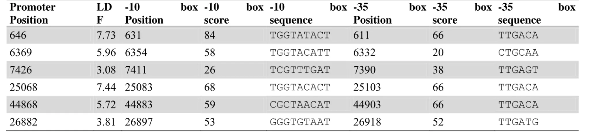
49 The searches for consensus sequences of bacterial promoters revealed the presence of 14 promoters (supplementary material). Promoter sequences were evaluated according to annotated ORFs, and those without biological sense were not considered. Among the predicted bacterial promoters, 10 were found in the negative strand initiating the transcription of ORFs that codify for early proteins, which is a common feature of viral genomes that need bacterial transcription factors to start their infection cycle. The 4 other promoters were located in late genes modules. These genes are usually transcribed by viral transcription factors
Two sequences of Rho-independent transcription terminators were predicted, both in the positive strand. The first is located after the gene encoding the major head protein and was predicted by the RibEx software. The second, predicted by the program FindTerm, is located in the end of the ORF19, which encodes a hypothetical protein. The low number of sequences of Rho-independent terminators compared to the number of predicted ORFs may be due to the existence of other types of terminators or the presence of transcriptional modules and the generation of polycistronic mRNAs.
The predicted UFV-P2 genes were functionally classified as its promoters, predicted order of transcription, and its possible functions.
Biosynthesis and DNA replication
50 between which there is a large non-coding intergenic region (about 2,000 bp); and ORF29 encodes a primase/helicase.
In addition, ORF38 encodes a transposase fusion protein related to the processes of integration of viral DNA in the bacterial chromosome. This protein presents the conserved HflC Band 7 domain (CDD accession cd03405,E-value 1.40e-03). According the Conserved Domains Database (CDD), this group includes proteins that are components of a complex that regulates the decision between the lysogenic and lytic cycles growth during lambda phage infection. In BLASTX searches, this protein presented significant hits with several bacterial proteins, including a hypothetical SPFH domain/Band 7 family protein of Pseudomonas aeruginosa ATCC 700888 (GenBank accession EKA49278; E-value 6e-58) and a transposase fusion protein of Burkholderia thailandensis Bt4 (GenBank accession ZP_02389877; E-value 4e-46). We also observed the presence of two endonucleases encoded by ORF21 and ORF33, a HNH endonuclease, a group I homing endonuclease. As described by Hertveldt et al. (14), these enzymes may be related to the presence of introns in the UFV-P2 genome; this remains to be confirmed in further studies.
51 the early genes module are hypothetical proteins without information about their functions.
Virion assembly and host lysis
Nineteen genes (ORFs 1-19) related to composition and assembly of the viral particle, DNA packaging, and host lysis were found in the UFV-P2 genome positive strand, named late genes (Figure 3-2). Two transcriptional clusters were found based on predicted bacterial promoters and terminators. The first cluster is located in the initial part of the genome (ORFs 1-6), and the second module, starting immediately after the first, corresponds to ORFs 7-19. The first transcriptional cluster terminator is partially overlapped to the sequence of the second transcriptional cluster promoter (Figure 3-2).
54 Structural genomic comparisons and evolutionary clustering
The alignment of genomic sequences and pairwise comparisons revealed that MR299-2, PaP3, LUZ24, and tf are the most closely related phages to UFV-P2. Genomic sequences of these phages presented an identity to the UFV-P2 genome ranging from 47% to 53%, while to other phages genome sequences, it ranged from 24% to 29% (see Table 3-1).
55 Figure 3-3. Dot plot alignment between the φUFV-P2 genome and the
PaP3, MR299-2, LUZ24 and tf genomes.
56 LUZ24-like genus. Based on the evolutionary relationships, we propose the classification of phages UFV-P2 and tf as LUZ24-like viruses.
Figure 3-4. Evolutionary relationships between φUFV-P2 and other phages based on generally conserved proteins.
57 value corresponds to bootstrap values (BV) (expressed as percentages) that define the clusters in the maximum likelihood tree.
Although not all phages have presented homologous sequences to analyzed coding sequences, we can observe that viruses of T7-like genus were also included in distinct monophyletic clades. On the other hand, it was not possible to define a phylogenetic clustering pattern to PhiKMV-like and the other unassigned viruses.
Conclusions
We have presented the functional annotation of UFV-P2, a new Pseudomonas fluorescens-specific phage. Based on structural genomic comparison and phylogenetic analyses, we suggest the classification of UFV-P2 in the LUZ24-like genus. Additionally, we propose the inclusion of φtf, a previously unclassified phage, in this genus.
Acknowledgments
58 References
1. Alemayehu D, Casey PG, McAuliffe O, Guinane CM, Martin JG, Shanahan F, Coffey A, Ross RP, Hill C. 2012. Bacteriophages φMR299-2 and φNH-4 can eliminate Pseudomonas aeruginosa in the murine lung and on cystic fibrosis lung airway cells. mBio 3:e00029– 12.
2. Arcuri EF, Aparecida M, Paiva V, Lange CC. 2008. Contagem , isolamento e caracterização de bactérias psicrotróficas contaminantes de leite cru refrigerado. Ciência Rural 38:2250–2255.
3. Baruzzi F, Lagonigro R, Quintieri L, Morea M, Caputo L. 2012. Occurrence of non-lactic acid bacteria populations involved in protein hydrolysis of cold-stored high moisture Mozzarella cheese. Food microbiology 30:37–44.
4. Baum MM, Kainović A, O’Keeffe T, Pandita R, McDonald K, Wu S, Webster P. 2009. Characterization of structures in biofilms formed by a Pseudomonas fluorescens isolated from soil. BMC microbiology 9:103.
5. Buckling A, Rainey PB. 2002. Antagonistic coevolution between a bacterium and a bacteriophage. Proceedings. Biological sciences / The Royal Society 269:931–6.
59 7. Ceyssens P-J, Brabban A, Rogge L, Lewis MS, Pickard D, Goulding
D, Dougan G, Noben J-P, Kropinski A, Kutter E, Lavigne R. 2010. Molecular and physiological analysis of three Pseudomonas aeruginosa phages belonging to the ―N4-like viruses‖. Virology 405:26–30.
8. Ceyssens P-J, Hertveldt K, Ackermann H-W, Noben J-P, Demeke M, Volckaert G, Lavigne R. 2008. The intron-containing genome of the lytic Pseudomonas phage LUZ24 resembles the temperate phage PaP3. Virology 377:233–8.
9. Dogan B, Boor KJ. 2003. Genetic diversity and spoilage potentials among Pseudomonas spp. isolated from fluid milk products and dairy processing plants. Applied and environmental microbiology 69:130–8. 10. Durand P, Mahé F, Valin A-S, Nicolas J. 2006. Browsing repeats in
genomes: Pygram and an application to non-coding region analysis. BMC bioinformatics 7:477.
11. Edgar RC. 2004. MUSCLE: multiple sequence alignment with high accuracy and high throughput. Nucleic acids research 32:1792–7. 12. Eller MR, Salgado RL, Vidigal PMP, Alves MP, Dias RS, de
Oliveira LL, da Silva CC, de Carvalho AF, de Paula SO. 2013. Complete Genome Sequence of the Pseudomonas fluorescens temperate bacteriophage UFV-P2. Genome Announcement.