UNIVERSIDADE FEDERAL DE MINAS GERAIS
INSTITUTO DE CIÊNCIAS BIOLÓGICAS
DEPARTAMENTO DE BIOLOGIA GERAL
PROGRAMA DE PÓS-GRADUAÇÃO EM GENÉTICA
ESTUDO DE MICDOPADTÍCULAS
NA PDÉ-ECLÂMPSIA GDAVE
ORIENTADO: Fabiana Kalina Marques
ORIENTADOR: Prof
a. Dr
a. Karina Braga Gomes Borges
Prof
a. Dr
a. Luci Maria Sant’Ana Dusse
FABIANA KALINA MARQUES
ESTUDO DE MICROPARTÍCULAS NA PRÉ-ECLÂMPSIA
GRAVE
Diss ertação apresentada ao programa de Pós-graduação em Genética do Instituto de Ciências Biológicas da Universidade Federal de Minas Gerais, como requisito parcial para obtenção do título de mestre em Genética. Orientadora: Profa. Dra. Karina Braga Gomes
Borges
Co-orientadora: Profa. Dra. Luci Maria
Sant’Ana Dusse
Instituto de Ciências Biológicas Belo Horizonte – MG
Marques, Fabiana Kalina.
Estudo de micropartículas na pré-eclâmpsia grave. [manuscrito] / Fabiana Kalina Marques. – 2012.
72 f. : il. ; 29,5 cm.
Orientadora: Karina Braga Gomes Borges. Co-orientadora: Luci Maria Sant’Ana Dusse.
Dissertação (mestrado) – Universidade Federal de Minas Gerais, Instituto de Ciências Biológicas.
1. Coagulação – Teses. 2. Inflamação – Teses. 3. Pré-eclâmpsia - Teses. 4. Genética – Teses. 5. Micropartículas derivadas de células. I. Borges, Karina Braga Gomes. II. Dusse, Luci Maria Sant’Ana. III. Universidade Federal de Minas Gerais. Instituto de Ciências Biológicas. IV. Título.
Mestranda: Fabiana Kalina Marques
Orientadora: Profa. Dra. Karina Braga Gomes Borges
Co-orientadora: Profa. Dra. Luci Maria Sant’Ana Dusse
Colaboradores: Dra. Andréa Teixeira de Carvalho
Dra. Fernanda Magalhães Freire Campos
Dr. Olindo Assis Martins Filho
Linha de Pesquisa
Biotecnologia
Área de Conhecimento
Genética
Instituições participantes
Instituto de Ciências Biológicas – UFMG Faculdade de Farmácia – UFMG
Centro de Pesquisas René Rachou / Fundação Oswaldo Cruz Maternidade Odete Valadares
Santa Casa de Misericórdia de Belo Horizonte Hospital Municipal Odilon Behrens
AGRADECIMENTOS
À Deus, por me dar força e sabedoria para seguir apesar das adversidades.
À professora Dra. Karina Braga Gomes Borges, pelos ensinamentos, amizade e
dedicação na orientação deste trabalho.
À professora Dra. Luci Maria Sant’Ana Dusse, por contribuir com sua experiência na
co-orientação deste trabalho.
À Lara, Patrícia, Melina e Letícia, por ajudarem com suas experiências e pela fundamental parceria na coletas.
À Dra. Andréa Teixeira de Carvalho e à Dra. Fernanda Magalhães Freire Campos, pela
dedicação, ensinamentos e importante contribuição nos experimentos e análise dos resultados. Ao Dr. Olindo Assis Martins Filho, por abrir as portas do seu laboratório e contribuir para a realização deste estudo.
A todos os amigos do setor de Citogenética do Hermes Pardini, pelo incentivo e torcida. Agradeço em especial às coordenadoras Cristiane Saraiva Ferreira e Keila Rivelly Pinheiro Dias, pelo apoio e compreensão.
A todos do Centro de Pesquisas René Rachou, em especial aos funcionários Laboratório de Biomarcadores de Diagnóstico e Monitoração e da Citometria de Fluxo, pela recepção e ajuda.
Aos funcionários Laboratório de Análises Clínicas da Maternidade Odete Valadares, pelo auxílio nas coletas.
À equipe de médicos e enfermeiros da Maternidade Odete Valadares, Santa Casa de Misericórdia de Belo Horizonte, Hospital Municipal Odilon Behrens e Centro de Saúde Guanabara – Betim, pela recepção e auxílio nas coletas.
Em especial as todas as mulheres participantes deste estudo, pois tornaram possível a realização do mesmo.
Ao CNPq e FAPEMIG, pelo apoio e financiamento.
SUMÁRIO
LISTA DE FIGURAS ... VIII LISTA DE TABELAS ... IX LISTA DE ABREVIATURAS ... X
INTRODUÇÃO ... 1
RESUMO ... 4
CAPÍTULO 1 – Artigo de revisão “Interaction of Microparticles and Preeclampsia”.. 5
OBJETIVOS ... 22
CAPÍTULO 2 – Artigo “Microparticles in Severe Preeclampsia” ... 24
DISCUSSÃO ... 44
CONCLUSÕES ... 53
REFERÊNCIAS BIBLIOGRÁFICAS ... 55
ANEXOS... 63
Anexo 1 – Aprovação do projeto pelo Comitê de Ética da UFMG... 64
Anexo 2 – Termo de Consentimento Livre e Esclarecido... 65
Anexo 3 – Ficha Clínica...66
Anexo 4 – Comprovante de submissão do artigo do capítulo 2 para publicação... 71
LISTA DE FIGURAS
Figura 1 (A) MPs isolated from the plasma were gated based on the basis of their forward (FSC) and side (SSC) scatter distribution. (B) Mouse IgG FITC and PE conjugated isotype control monoclonal antibodies were used to accurately place the gates... 31 Figura 2 Data points and medians for total numbers of MPs in women with severe preeclampsia, normotensive pregnant women, and non-pregnant women 35 Figura 3 Flow cytometry plots of MPs derived from erythrocytes and trophoblasts in non-pregnant woman, normotensive pregnant women, and women with severe preeclampsia……….. 36 Figura 4 Absolute number of MPs in women with severe PE, normotensive
LISTA DE TABELAS
LISTA DE ABREVIATURAS
APC – allophycocyanin, aloficocianina
BMI – body mass index, índice de massa corporal CD – cluster of differentiation, cluster de diferenciação CID – coagulação intravascular disseminada
COEP – Comitê de Ética e Pesquisa
COX-2 – cyclooxygenase-2, ciclooxigenase-2
CXCR4 –CXC chemokine receptor type 4, receptor de quimiocina CXC do tipo 4 Cy5 – cyanine 5, cianina 5
DBP – diastolic blood pressure, pressão sanguinea diastólica FITC – fluorescein isothiocyanate, isotiocianato de fluoresceína FSC – foward scatter, dispersão frontal
GA – gestational age, idade gestacional
GLA – gama-carboxyglutamic acid, ácido gama-carboxiglutâmico
HELLP – hemolysis, elevated liver enzymes and low platelet, hemólise, enzimas hepáticas elevadas e plaquetas baixas
I-CAM 1 – intercellular adhesion molecule 1, molécula de adesão intercelular 1 IgG1 – immunoglobulin G1, imunoglobulina G1
IgM – immunoglobulin M, imunoglobulina M INFγ – interferon γ
iNOS – inducible nitric oxide synthase, óxido nítrico sintase induzível IL – interleukin, interleucina
mmHg – milímetro de mercúrio MP – microparticle, micropartícula
mRNA – messenger ribonucleic acid, ácido ribonucléico mensageiro
NDOG2 – trophoblast monoclonalantibody (clone NDOG2), anticorpo monoclonal anti-trofoblasto (clone NDOG2)
NO – nitric oxide, óxido nítrico
PBS – phosphate buffered saline, tampão fosfato salino PE – preeclampsia, pré-eclâmpsia
PE – phycoerythrin, ficoetrina
PerCP –peridinin chlorophyll protein, proteína clorofila peridinina PMP – platelet microparticle, micropartícula de plaqueta
PS – phosphatidylserine, fosfatidilserina
ROS – reactive oxygen species, espécies oxigênio reativas SBP – systolic blood pressure, pressão sanguinea sistólica
STBM – syncytiotrophoblast microparticles, microparticula do sinciciotrofoblasto SSC – side scatter, dispersão lateral
TF/FT – tissue factor, fator tissular
TNF-α – tumor necrosis factor-alpha, fator de necrose tumoral alfa TTP – púrpura trombocitopênica trombótica
A Pré-eclâmpsia (PE) é uma doença multisistêmica específica da gestação, que caracteriza-se clinicamente pelo aparecimento de hipertensão e proteinúria após a 20ª semana de gestação.
Por ser uma doença cuja única resolução baseia-se na interrupção da gestação, a PE é responsável por 10% a 15% de mortes maternas em todo mundo e é ainda importante causa de morte fetal devido à restrição ao crescimento intrauterino e prematuridade.
É importante classificar e diferenciar os casos de PE leve e grave. Segundo o American College of Obstetricians and Gynecologists (2002): a PE leve é caracterizada por
hipertensão com pressão sistólica ≥140mmHg e diastólica ≥90mmHg em pelo menos duas medições separadas por intervalo de 4 horas; e proteinúria ≥300mg em urina de 24 horas ou ≥1+ pelo método de fita. A PE grave é caracterizada por hipertensão com pressão sistólica ≥160mmHg e diastólica ≥110mmHg em pelo menos duas medições separadas por um intervalo de 4 horas; e proteinúria ≥5g na urina de 24 horas ou ≥3+ pelo método de fita. A forma grave da PE pode evoluir para outras manifestações clínicas de risco, como a eclâmpsia, a Síndrome HELLP (Hemolysis, elevated liver enzymes and low platelet) e a coagulação intravascular disseminada (CID).
A gestação normal está associada a adaptações anatômicas e funcionais do sistema cardiovascular da gestante para acomodar as novas demandas fisiológicas, no entanto na PE esta adaptação é inadequada. Embora o conhecimento seja limitado, já foram identificados fatores de risco para o desenvolvimento da PE como: primiparidade, gestação múltipla, obesidade, PE prévia, fatores genéticos e comorbidades maternas. Várias hipóteses têm sido levantadas na tentativa de explicar a patogênese da PE, mas apesar da extensiva pesquisa, os mecanismos envolvidos nesta disfunção vascular ainda não são bem compreendidos. Recentemente, pesquisas têm reportado elevados níveis de micropartículas (MP) na PE e sugerido seu envolvimento nas manifestações clínicas associada a esta doença, em especial a hipertensão.
As MP são conhecidas como uma população heterogênea de pequenos fragmentos liberados da membrana das células durante ativação celular e apoptose. Muitos tipos celulares, como células endoteliais, plaquetas e leucócitos, liberam estas MP in vitro, mas vários estudos têm demonstrado a presença destes fragmentos in vivo. Sabe-se que as MP são liberadas durante o remodelamento da membrana plasmática. O súbito aumento dos níveis de cálcio citosólico muda o estado transmembrana, resultando em externalização de fosfatidilserina e ativação de enzimas citosólicas, levando à clivagem do citoesqueleto. Este fenômeno resulta em vesiculação da membrana e liberação das MP para o meio.
potentes vetores de informação biológica e protagonistas na rede de comunicação celular, tais como indução de modificações endoteliais, angiogênese e diferenciação. As MP in vivo parecem estar envolvidas na regulação da coagulação e função vascular, pois estas atuam como potentes indutores pró-inflamatórios e modificadores da expressão gênica nas células endoteliais.
Sabe-se que os processos de coagulação e inflamação co-existem na PE. Desta forma, a principal motivação para o desenvolvimento deste trabalho foi elucidar a relação entre as MP e a PE grave, uma vez que, pelo nosso conhecimento, há poucos trabalhos envolvendo esta associação, tendo como limitante a menor variedade nos tipos de MP avaliadas e o tamanho amostral. Como até o momento nenhum marcador laboratorial mostrou-se efetivo no diagnóstico da doença, sendo hoje feito essencialmente pelas características clínicas e proteinúria apresentadas pela gestante, torna-se oportuno conhecer possíveis analitos biológicos que permitam diagnosticar ou acompanhar a evolução da PE.
Apesar de inúmeras pesquisas sobre essa condição, a etiologia da PE permanece por ser elucidada e não há como prever a ocorrência da mesma antes do aparecimento dos sintomas. Sendo assim, o presente estudo tem como objetivo avaliar a origem e o número de MP e associá-los ao desenvolvimento da PE grave.
DESUMO
Objetivo: O presente estudo teve como objetivo avaliar micropartículas (MPs) a partir de
fontes diferentes em gestantes com pré-eclâmpsia grave (PE), em comparação com gestantes normotensas e mulheres não gestantes.
Estudo: Este estudo de caso-controle avaliou 28 gestantes com PE grave, 30 gestantes
normotensas e 29 mulheres não gestantes. MPs de neutrófilos, células endoteliais, monócitos, plaquetas, leucócitos, eritrócitos e sinciciotrofoblastos foram avaliados usando citometria de fluxo.
Desultados: Foi observado um aumento no total de MPs nas gestantes com PE grave, em
comparação com gestantes normotensas e mulheres não gestantes (P = 0,004 e P = 0,001, respectivamente). MPs derivadas de eritrócitos estavam aumentadas nas gestantes com PE grave, comparativamente com gestantes normotensas (P = 0,002). Uma correlação positiva foi observada entre a contagem de plaquetas e do número de MPs derivados de plaquetas (P = 0,05). Uma correlação positiva também foi encontrada entre o número de MPs derivadas de células endoteliais e o número de MPs derivadas de plaquetas, leucócitos, neutrófilos e linfócitos (P <0,05).
Conclusão: a contagem de MP pode ser útil para o diagnóstico de PE grave, e as MPs
derivadas de eritrócitos parece ser um bom marcador para PE grave. Além disso, MPs derivadas de células endoteliais estão associados com a inflamação e coagulação em PE grave.
CAPÍTULO 1
Artigo de revisão intitulado: INTERACTION OF
Abstract
Preeclampsia (PE) is a multi-system disorder, characterized by hypertension and proteinuria, occurring after the twentieth week of pregnancy. Despite intensive research, PE is still one of the leading causes of maternal mortality, and reliable screening tests or effective treatments of this disease have yet to be discovered. The most common procedure is to deliver the baby and the placenta, often prematurely, in the interest of providing the most appropriate conditions for the baby or the mother. Therefore, improving the overall understanding of the role of microparticles in PE may well be useful for new clinical diagnoses and therapeutic approaches.
Microparticles (MPs) are small vesicles released after cell activation or apoptosis, which contain membrane proteins that are characteristic of the original parent cell. MPs have been proven to play key roles in thrombosis, inflammation, and angiogenesis, as well as to mediate cell-cell communication by transferring mRNAs and microRNA from the cell of origin to target cells. It has been suggest that MPs, mainly placenta-derived syncytiotrophoblast microparticles (STBMs), may well play an important role in the pathogenesis of PE.
PDEECLAMPSIA
Preeclampsia (PE) is a multi-system obstetric disorder, whose natural occurrence can only be found in primates and humans [1]. Two percent of women with PE will progress to eclampsia leading to convulsions and potential maternal and fetal death. PE is characterized either by a systolic blood pressure of ≥140mmHg or by a diastolic blood pressure of ≥90mmHg on two or more consecutive occasions, 4 hours apart; together with proteinuria (either ≥300mg protein/day or protinuria by dipstick urine >1+) occurring after the twentieth week of pregnancy in women who had presented no prior symptoms [2]. PE, as compared a normal pregnancy, is associated with increased intravascular coagulation [3, 4], fibrin deposition [5], and inflammatory response [6, 7].
Several hypotheses have been postulated in an attempt to explain the pathogenesis of PE, as described in Table 1 [1, 8, 9]. Although the PE etiology is still unknown, the theory most widely discussed emphasizes the abnormal placenta and describes the PE as a disorder that occurs in two stages. The first stage begins with the abnormal placentation and production of placental factors, such as proteins and cytoplasmic debris falling into the maternal circulation. The second, called the “mother stage”, is the multisystemic maternal syndrome of PE and depends not only on the action of these circulating factors, but also on the health of the pregnant woman, including diseases that affect the vascular system, including preexisting heart or renal diseases, metabolic diseases, genetic factors, and obesity [8, 9].
Table 1. Theories that explain the PE pathogenesis
• Placentation abnormalities (defects in the trophoblast and spiral arteries)
• Angiogenic factors
• Maladaptive cardiovascular and vasoconstriction
• Genetic predisposition
• Immunologic intolerance between maternal and fetal tissue
• Platelet activation
• Vascular endothelial damage or dysfunction
hypoxia. The inadequate placentation results in reduced blood flow in the fetal-placental unit, which can lead to poor fetal growth [1, 10, 11].
Currently, PE has been considered as a syndrome, and not a disease, caused by isolated or combined alterations, whose vascular endothelial changes are recognized as a central process [12].
Despite intensive research, PE is still one of the leading causes of maternal mortality, and reliable screening tests or effective treatments of this disease have yet to be discovered. [12]. The most common procedure is to deliver the baby and the placenta, often prematurely, in the interest of providing the most appropriate conditions for the baby or the mother. [13].
MICDOPADTICLES
Microparticles (MPs) were first described by Wolf in 1967 as a “dust” procoagulant formation around an activated platelet [14]. Today, MPs are known as a heterogeneous population of small fragments (0.05-1µm) released from the cell membrane during cell activation and apoptosis. Moreover, it is well established in the literature that all eukaryotic cells have the capacity to release MPs [15, 16].
The cell membrane is characterized by its distribution of phospholipids, with phosphatidylcholine and sphingomyelin on the outside, and phosphatidylethanolamine and phosphatidylserine (PS) on the inside. The initial step in the formation of MP is the remodeling of the membrane, with the formation of blebs within it. This step requires an increase in intracellular calcium levels, consequently resulting in the rearrangement and loss of the phospholipidic membrane’s asymmetry, coupled with the externalization of PS to the outer surface. Concomitant to the loss of membrane asymmetry, calcium-sensitive enzymes are activated and promote the cleavage of the filaments of the cytoskeleton leading to the formation of blebs on the membrane and the release of MPs [16, 17, 18].
membrane, MPs also expose a variable spectrum of bioactive substances, receptors, and adhesion molecules [15]. MP membranes also carry chemokines, cytokines, enzymes, growth factors, and signaling proteins [19, 20].
MPs have been proven to play key roles in thrombosis, inflammation, and angiogenesis, as well as to mediate cell-cell communication by transferring mRNAs and microRNA from the cell of origin to target cells [15]. MPs are considered potent tools in the cellular communication network, such as the induction of endothelial changes, angiogenesis, and differentiation [15, 16]. The nature and physical characteristics of the MPs need to be better studied, since most studies assess only their amount, origin, and biological activity. Current knowledge about the formation of MPs derived from experiments with isolated or cultured cells shows that activation and apoptosis promote the release of MPs in vitro; however, the mechanism in vivo remains unknown [18].
MPs are rich in phospholipids and can be derived from endothelial cells, erythrocytes, platelets and leucocytes [17, 21]. Although they are present in the peripheral blood of healthy individuals, with platelet-derived MPs representing approximately 70% to 90% of all circulating MPs, a significant increase in certain pathological conditions could be observed. These conditions include autoimmune diseases, diabetes, cancer, and infectious diseases [14, 16].
Sheremata et al. [22] observed an increase of platelet-derived MPs in patients with multiple sclerosis, when compared to a normal control group. Tramonato et al. [23] found a significant increase in MPs derived from endothelial cells in the plasma of diabetic patients, as compared to non-diabetic individuals. Kalinkovich et al. [24] showed an increase of MPs expressing C-X-C chemokine receptor type 4 (CXCR4) in the blood and bone marrow of patients with acute myeloid leukemia. Goswami et al. [25] observed increased levels of MPs derived from the syncytiotrophoblast in pregnant women with PE, as compared to a group of normotensive pregnant women with fetal growth restriction. Campos et al. [26] found a significant increase in circulating MPs derived from platelets, erythrocytes and leukocytes in patients infected with Plasmodium vivax.
apoptotic stimuli from those resulting from cell activation. For example, the comparison of the protein expression in MPs derived from microvascular endothelial cells revealed that the endothelial markers CD31 and CD62E are strongly expressed by MP when released from apoptotic cells; however, CD51 and CD54 are preferentially expressed in MP when released by cell activation [14]. Flow cytometry is the most widely used method to analyze MPs by employing antibodies to cell markers and specific binding of annexin V to phosphatidylserine [17, 18, 27].
MPs from different cell types have different in vitro effects on vascular and blood cells, and are commonly involved in regulating coagulation and vascular functions [20]. MPs act as potent pro-inflammatory mediators, beginning an array of signal transduction pathways and gene expression profiles in endothelial cells, thereby affecting their function. MPs derived from platelets stimulate the expression of cyclooxygenase-2 (COX-2) and prostacyclin [28], the production of cytokines in endothelial cells, and an increase in adhesion molecules on the endothelial surface, resulting in monocyte adhesion and platelet activation [29]. These can also directly activate and stimulate monocytes to produce cytokines and reactive oxygen species (ROS), resulting in an inflammatory response [30]. MPs derived from leukocytes also induce the increase of adhesion molecules on endothelial cells and initiate the production of interleukin 6 and 8 [31]. Those derived from endothelial cells can also activate neutrophils, resulting in endothelial adhesion [32].
Regarding the haemostatic function, The MPs present a high level of procoagulant activity, given that they contain anionic PS and express the tissue factor (TF). The PS facilitates the gathering of the components of the clotting cascade that contain gama-carboxyglutamic acid (GLA), such as factors VII (FVII), IX, X, and protrombin. PS-MPs are derived mostly from megakaryocytes and seem to express receptors for both collagen and the von Willebrand factor, as can be seen in the activated platelet [33].
TF is the main regulator of blood coagulation, since this is a receptor for FVII/VIIa. Circulating TF-MP may provide an alternative source of TF that would be recruited to the growing thrombus and reinitiate clotting [33]. The presence of PS may induce a conformational change in TF that increases its specific activity [34]. Some studies suggest that monocytes are likely to be the major source of TF-MPs in health and disease, while endothelial cells may release TF-MP in certain diseases [35, 36].
transcription factor NF-κB, leading to the enhanced expression of cyclooxygenase-2 (COX-2) with a subsequent increase in prostacyclin productions, respectively, resulting in vasodilatation [38].
MICDOPADTICLES AND PDEECLAMPSIA
Normal pregnancy is associated with extensive changes in hemostasis and generalized maternal inflammatory response. The hypercoagulability and inflammation states are increased in PE. Detailed understanding of the links between the blood coagulation and inflammation are imperative to the elucidation of the etiology of PE [39, 40]. Since MPs are involved in both processes, to understand the role of MPs in PE can contribute to a better understanding of the etiopathogenesis of this disease.
Studies have shown that MPs are commonly increased in pregnancy, since this is a medical condition associated with the anatomical and functional adaptation of the vascular system of a mother to accommodate the new physiological demands. However, this increase is especially important in pregnant women with PE, which shows an extensive activation of endothelial cells, leukocytes, and the coagulation system [30, 41, 42].
Recently, several groups reported high levels of circulating MPs in plasma of pregnant women with PE and suggest their involvement in hypertension associated with the disease [42, 43]. Some studies have shown not only MPs derived from platelets, endothelium, and leukocytes, but also from MPs derived from syncytiotrophoblast [44].
Elevated concentrations of erythrocyte-derived MPs have appeared in PE, which are most likely due to hemolysis and haemoconcentrations, since these process are often associated with this syndrome [12]. Increased MPs from T cells, monocytes, and granulocytes were reported in PE, and the number of granulocyte-derived MPs correlates with elastase, a marker of granulocyte activation and secretion [12, 45, 46].
E-selectin, and fibronectin in the plasma of pregnant women with PE [48, 49]. These adhesion molecules are expressed constitutively and regulate the trafficking of circulating inflammatory cells to sites of cellular damage [47].
Meziani et al. [41] found evidence that women with PE have increased levels of MPs derived from monocyte/lymphocyte and platelet (PMPs) when compared to normal pregnant women. Microparticles from preeclamptic, but not healthy pregnant women, induced an ex vivo vascular hyporeactivity toward serotonin in human omental arteries and mouse aortas. Hyporeactivity was associated with increased NO production and was reversed by an NO synthase inhibitor. In the presence of a COX-2 inhibitor, serotonin-mediated contraction was partially reduced in arteries treated with healthy microparticles but was abolished after treatment with MPs from preeclamptic women. MPs were associated with an up-regulation of inducible nitric oxide (iNOS) and COX-2 inflammatory proteins through the activation of the NF-kB transcription factor in the vascular wall. When PMPs and MPs from other sources were separated and tested for vascular reactivity, it was observed that only PMPs stimulated NO release, suggesting that its inflammatory properties would be associated with nitrosative and oxidative stress in the vascular wall and that the positive role of this type of MP would result in the sudden increase of blood pressure in the PE. However, it could be observed that MPs from other sources, most probably derived from leukocytes, induce the release of vasoconstrictor products and COX-2, especially the 8-isoprostane, whose increase has been observed in the placenta of preeclamptic women [42, 50].
Lok et al. [51] observed a positive correlation between the number of PMPs and the platelet count when compared preeclamptic and normotensive pregnant women during pregnancy and postpartum. In both groups, the PMPs represented the highest percentage of all MPs, but these were reduced in preeclamptic women as compared to normotensive pregnant. However, in postpartum, no difference could be observed among the groups as regards PMP. The reduction in platelet count often observed in PE should explain the lower levels of circulating PMPs in this group. However, the increase in the number of PMPs after birth is most likely due to elevation and normalization in the platelet count.
The syncytiotrophoblast membrane cell is an additional source of MPs during PE and their levels increase significantly during pregnancy, which is expected, since there is a gradual increase in the placental volume. Placental oxidative stress destabilizes the syncytiotrophoblast cells, resulting in an increased release of MPs containing oxidized lipids. These are surface membrane fragments shed from the outer layer of the placenta directly into the maternal blood. Other STBMs enter the maternal circulation via decidual veins, which should lead to maternal systemic effects [51, 53].
This type of MP has also been shown to cause endothelial dysfunction [16]. Due to oxidative stress in the intravillous space, the STBMs carrying TF accumulates in this area, starting the local effect on the placental hemostasis. Alternatively, the increased blood pressure, inflammation, and other pathological conditions should result in increased levels of maternal MPs that reach the intravillous placental space through maternal spiral arteries, affecting placental hemostasis [54, 55].
STBMs reach their highest level in the third trimester [12, 56]. Preeclamptic women in this period, as compared to normotensive pregnant, have increased STBMs, which is thought to directly reflect placental hypoxia and apoptosis [12, 56-60]. Indeed, hypoxia leads to excessive ROS generation in the placenta. In normal pregnancies ROS generation is low, and antioxidative pathways are able to inactivate endogenous ROS, thereby limiting placental damage. However, in PE these adaptive mechanisms are overwhelmed by an enhanced production of ROS, in turn leading to an apoptotic/necrotic cascade and STBM formation [61].
The presence of STBMs specifically promoted cell death and/or reduced the proliferation of endothelial cells, as well as activated superoxide production in neutrophils isolated from preeclamptic women [12, 56, 61]. Furthermore, in normotensive pregnant women, the walls of the uteroplacental arteries are invaded by trophoblasts. In PE, reduced trophoblast invasion is combined with an accumulation of apoptotic trophoblasts in the arteries walls, increasing the STMBs levels [62]. In PE, the networks of interstitial and endovascular trophoblast invasion are affected by maternal factors [63, 64]. The interstitial invasion is affected by the premature increase of oxygen in the placenta and a reduced proliferation, while the endovascular network is affected by macrophage-induced apoptosis of perivascular and intramural trophoblasts. Both events limit the number and extent of adaptation of spiral arteries, required for growth fetal [65].
higher levels of MPs derived from T lymphocytes, B lymphocytes, and granulocytes could be observed in pregnant women with PE, as compared to normotensive pregnant women [20, 66]. The increase in this particular subgroup of MPs in PE may well represent a possible mechanism for the development of vascular dysfunction and seems to reflect a modified status of immune system activation and higher inflammatory response [20]. Increased levels of MPs derived from lymphocytes may be due the release of activated lymphocytes in the maternal circulation, as these also tend to increase in the placental tissue during PE [67]. These MPs can cause direct or indirect endothelial injury by inducing new MPs by activating other cells, creating a vicious circle [20]. Studies have shown that, in PE, neutrophils are activated when they pass through the placenta, which would explain the increased levels of MPs derived from this cellular type [68]. The increased number of MPs derived from leukocytes observed in PE should reflect the activation of leukocytes, mainly monocytes and neutrophils, as it one of the core characteristics of this disease [69]. It could also observed that MPs derived from monocytes produce high levels of inflammatory cytokines [70].
CONCLUSION
The data reported in other studies in recent years has discussed the involvement of MPs in physiological and pathological conditions, mainly in inflammatory diseases.
Taking in account that the cellular origin, contents, and forms of MPs are variable, some may well represent target treatments of pathological states, in turn reducing their harmful effects linked to procoagulant and proinflammatory properties in the vessel wall and target organs, especially since the MPs are able to regulate the gene expression involved in inflammation and the regulation of oxidative stress caused by the vascular function.
Other studies are warranted to answer this question, given that the elucidation of the mechanisms involved in the effects of MPs may well represent a highly effective contribution to additional intervention strategies concerning PE.
Acknowledgments - CNPq (Conselho Nacional de Desenvolvimento Científico e
Tecnológico) for the financial support.
DEFEDENCES
1)
Trogstad L, Magnus P, Stoltenberg C: Pre-eclampsia: Risk factors and causal models. Best Pract Res Clin Obstet and Gynaecol 2011:25:329-342.2) American College of Obstetricians and Gynecologists (ACOG): Practice bulletin: Diagnosis and management of preeclampsia and eclampsia. Obstet Gynecol 2002:99:159-167.
3) Schjetlein R, Abdelnoor M, Haugen G, Husby H, Sandset PM, Wosloff M: Hemostatic variables asindependent predictors for fetal growth retardation in preclampsia. Acta Obstet Gynecol Scand 1999: 78:191-197.
4) Higgins JR, Walshe JJ, Darling MR, Norris L, Bonnar J: Hemostasis in the uteroplacental and peripheral circulations in normotensive and preeclamptic pregnancies. Am J Obstet Gynecol 1998:179:520-526.
5) Weiner CP, Brandt J: Plasma antithrombin III activity: an aid in the diagnosis of preeclampsia-eclampsia. Lancet 1977: 2:1249-1252.
6) Rákóczi I, Tallián F, Bagdány S, Gáti I: Platelet life-span in normal pregnancy and pre-eclampsia as determined by a non-radioisotope technique. Thromb Res 1979:15:553-556. 7) Redman CW, Bonnar J, Beilen L: Early platelet consumption in preeclampsia. Br Med J 1978:1:467-469.
9) Young BC, Levine RJ, Karumanchi SA: Pathogenesis of Preeclampsia. Annu Rev Pathol Mech Dis 2010:5:173-192.
10) Hawfield A, Freedman BI: Pre-eclampsia: the pivotal role of the placenta in its pathophysiology and markers for early detection. Ther Adv Cardiovasc Dis 2009:3:65-73. 11) Wang A, Rana S, Karumanchi SA: Preeclampsia: The Role of Angiogenic Factors in Its Pathogenesis. Physiology 2009:24:147-158.
12) Rodie VA, Freeman DJ, Sattar N, Greer IA: Pre-eclampsia and cardiovascular disease: metabolic syndrome of pregnancy? Atherosclerosis 2004:175:189- 202.
13) Coomarasamy A, Honest H, Papaionnou S, Gee H, Kahn KS: Aspirin for prevention of preeclampsia: a systemic review. Obstet Gynecol 2003:101:1319-1332.
14) Wolf P: The nature and significance of platelet products in human plasma. Br J Haematol 1967:13:269-288.
15) Mause SF, Weber C: Microparticles: Protagonists of a Novel Communication Network for Intercellular Information Exchange. Circ Res 2010:107:1047-1057.
16) Meziani F, Tesse A, Andriantsitohaina R: Microparticles are vectors of paradoxical information in vascular cells including the endothelium: role in health and diseases. Pharmacol Rep 2008:60:75-84.
17) Chironi GN, Boulanger CM, Simon A, George FD, Freyssinet JM, Tegui A: Endothelial microparticles in diseases. Cell Tissue Res 2009:335:143-151.
18) Boulanger CM, Amabile N, Tedgui A: Circulating Microparticles: A Potential Prognostic Marker for Atherosclerotic Vascular Disease. Hypertension 2006:48:180-186. 19) Tan KT, Lip GY: The potential role of platelet microparticles in atherosclerosis. Thromb Haemost 2005:94:488-492.
20) VanWijk M J, Nieuwland R, Boer K, Van der Post JAM, VanBavel E, Sturk A: Microparticle subpopulations are increased in preeclampsia: Possible involvement in vascular dysfunction? Am J Obstet Gynecol 2002:187:450-456.
22) Sheremata WA, Jy W, Horstman LL, Ahn YS, Alexander JS, Minagar A: Evidence of platelet activation in multiple sclerosis. J Neuroinflammation 2008:5:1-6.
23) Tramontano AF, Lyubarova R, Tsiakos J, Palaia T, DeLeon JR, Ragolia L: Circulating Endothelial Microparticles in Diabetes Mellitus. Mediators of Inflamm 2010:1-8.
24) Kalinkovich A, Tavor S, Avigdor A, Kahn J, Brill J, Petit I, Gonchberg P, Tesio M, Netzer N, Naparstek E, Hardan I, Nagler A, Resnick I, Tsimanis A, Lapidot T: Functional CXCR4-Expressing Microparticles and SDF-1 Correlate with Circulating Acute Myelogenous Leukemia Cells. Cancer Res 2006:66:11013-11020.
25) Goswami D, Tannetta DS, Magee L, Fuchisawa A, Redman CWG, Sargent IL, Von Dadelszen P: Excess Syncytiotrophoblast Microparticle Shedding is a Feature of Early-onset Pre-eclampsia, but not Normotensive Intrauterine Growth Restriction. Placenta 2006:27:56-61.
26) Campos FMF, Franklin BS, Teixeira-Carvalho A, Filho AL, de Paula SC, Fontes CJ, Brito CF, Carvalho LH: Augmented plasma microparticles during acute Plasmodium vivax infection. Malar J 2010:9:1-8.
27) Dey-Hazra E, Hertel B, Kirsch T, Woywodt A, Lovric S, Haller H, Haubitz M, Erdbruegger: Detection of circulating microparticles by cytometry: influence of centrifugation, filtration of buffer, and freezing. Vasc Health Risk Manag 2010:6:1125-1133.
28) Barry OP, Pratico D, Lawson JA, Fitzgerald GA: Transcellular activation of platelets and endothelial cells by bioactive lipids in platelets microparticles. J Clin Invest 1997:99:2118-2127.
29) Nomura S, Tandon NN, Nakamura T, Cone J, Fukuhara S, Kambayashi J: High-shear-stress-induced activation of platelets and microparticles enhances expression of cell adhesion molecules in THP-1 and endothelial cells. Atherosclerosis 2001:158:277-287.
31) Mesri M, Altieri DC: Endothelial cell activation by leukocyte microparticles. J Immunol. 1998:161:4382-4387.
32) Bizios R, Lai LC, Cooper JA, Del Vecchio PJ, Malik AB: Thrombin-induced adherence of neutrophils to cultured endothelial monolayers: increased endothelial adhesiveness. J Cell Physiol 1988:134:275-280.
33) Owens A, Mackman N: Microparticles in hemostasis and thrombosis. Circ Res 2011:108:1284-1297
34) Bach R: Tissue factor encryption. Arterioscler Thromb Vasc Biol 2006:26:456-461.
35) Bajaj MS, Ghosh M, Bajaj SP: Fibronectin-adherent monocytes express tissue factor
and tissue factor pathway inhibitor whereas endotoxin-stimulated monocytes primarily express tissue factor: physiologic and pathologic implications. J Thromb Haemost
2007:5:1493-1499.
36) Solovey A, Kollander R, Shet A: Endothelial cell expression of tissue factor in sickle mice is augmented by hypoxia/reoxygenation and inhibited by lovastatin. Blood
2004:104:840-846.
37) Sibai B, Dekker G, Kupferminc M: Pre-eclampsia. Lancet 2005:365:785-795.
38) Szaba FM, Smiley ST: Roles for thrombin and fibrinogen in cytokine/chemokine production and macrophage adhesion in vivo. Blood 2002:99:1053-1059.
39) Walter JJ: Pre-eclampsia. Lancet 2000:356:1260-1265.
40) Roberts JM, LAIN KY: Recent insights into the pathogenesis of pre-eclampsia. Placenta 2002:23:359-372.
41) Meziani F, Tesse A, David E, Martinez CM, Wangsteen R, Schneider F, Andriantsitohaina R: Shed membrane particles from preeclamptic women generate vascular wall inflammation and blunt vascular contractility. Am J Pathol 2006:169:1473-1483.
44) Redman CW, Sargent IL: Circulating microparticles in normal pregnancy and pre-eclampsia. Placenta 2008:29:73-77.
45) Kupferminc MJ, Fait G, Many A, Lessing JB, Yair D, Bar-Am A, Eldor A: Low molecular weight heparin for the prevention of obstetric complications in women with thrombophilia. Hypertens Pregnancy 2001:20:35-44.
46) Paternoster DM, Fantinato S, Manganelli F, Nicolini U, Milani M, Girolami A: Recent progress in the therapeutic management of pre-eclampsia. Expert Opin Pharmacother 2004:5:2233-2239.
47) González-Quintero VH, Smarkusky LP, Jiménez JJ: Elevated plasma endothelial microparticles: Preeclampsia versus gestational hypertension. Am J Obstet Gynecol 2004:191:1418-1424.
48) Austgulen R, Lien E, Vince G, Redman C: Increased maternal plasma levels of soluble adhesion molecules (ICAM-1, V-CAM-1, E-selectin) in preeclampsia. Eur J Obstet Gynecol Reprod Biol 1997:71:53-58.
49) Clausen T, Djurovic S, Brosstad F, Berg K, Henriksen T: Altered circulating levels of adhesion molecules at 18 week’s gestation among women with eventual preeclampsia: Indicators of disturbed placentation in absence of evidence of endothelial dysfunction? Am J Obstet Gynecol 2000:182:321-325.
50) Walsh SW, Vaughan JE, Wang Y, Roberts LJ: Placental isoprostane is significantly increased in preeclampsia. FASEB J 2000:14:1289-1296.
51) Lok CAR, Van Der Post JAM, Sargent IL: Changes in Microparticle Numbers and Cellular Origin During Pregnancy and Preeclampsia. Hypert Pregn 2008:27:344-360. 52) Cockell AP, Learmont JG, Smárason AK, Redman CW, Sargent IL, Poston L: Human placental syncytiotrophoblast microvillous membranes impair maternal vascular endothelial function. Br J Obstet Gynaecol 1997:104:235-240.
53) Redman CW, Sargent IL: Placental debris, oxidative stress and preeclampsia. Placenta 2000:597-602.
54) Aharon A, Brenner B: Microparticles and pregnancy complications. Thromb Res 2011:127:67-71.
56) Makrides M, Duley L, Oslen SF: Fish oil, and other prostaglandin precursor supllementation during pregnancy for reducing pre-eclampsia, preterm birth, low birth weight and intrauterine growth restriction. Cochrane Database Syst Rev 2001:4:CD003402.
57) McKay DG: Hematologic evidence of disseminated intravascular coagulation in eclampsia. Obstet Gynecol Surv 1972:27:399-417.
58) Raijmakers MTM, Dechend R, Poston L: Oxidative stress and preeclampsia. Rationale for antioxidant clinical trials. Hypertension 2004:44:374-380.
59) Chappell LC, Seed PT, Briley AL, Kelly FJ, Lee R, Hunt BJ, Parmar K, Bewley SJ, Shennan AH, Steer PJ, Poston L: Effect of antioxidants on the occurrence of pre-eclampsia in women at increased risk: a randomised trial. Lancet 1999:354:810-816. 60) Sibai BM: Prevention of preeclampsia: a big disappointment. Am J Obstet Gynecol 1998:179:1275-1278.
61) Aagaard-Tillery KM, Belford MA: Eclampsia: Morbidity, Mortality, and Management. Clin Obst Gynecol 2005:48: 12-23.
62) Messerli M, May K, Hansson SR, Schnneider H, Holzgreve W, Hahn S, Rusterholz C: Feto-maternal interactions in pregnancies: Placental microparticles activate peripheral blood monocytes. Placenta 2010:31:106-112.
63) Reister F, Frank HG, Heyl W, Kosanke G, Huppertz B, Schroder W: The distribution of macrophages in spiral arteries of the placental bed in pre-eclampsia differs from that in healthy patients. Placenta 1999:20:229-233.
64) Huppertz B, Kadyrov M, Kingdom JCP: Apoptosis and its role in the trophoblast. Am J Obstet & Gynecol 2006:195: 29-39.
65) Redman CWG, Sargent IL: Microparticles and immunomodulation in pregnancy and pre-eclampsia. J Reprod Immunol 2007:76:61-67.
66) Stallmach T, Hebisch G, Orban P, Lu X: Aberrant positioning of trophoblast and lymphocytes in the feto-maternal interface with pre-eclampsia. Virchows Arch 1999:434:207-211.
68) Mellembakken JR, Aukrust P, Olafsen MK, Ueland T, Hestdal K, Videm V: Activation of leukocytes during the uteroplacental passage in preeclampsia. Hypertension 2002:39:155-160.
69) Luppi P, Deloia JA: Monocytes of preeclamptic women spontaneously synthesize pro-inflammatory cytokines. Clin Immunol 2006:118:268-275.
1 Objetivo geral
Avaliar a origem e o número de micropartículas e associá-los ao desenvolvimento da pré-eclâmpsia grave e suas complicações clínicas e laboratoriais.
2 Objetivos específicos
Padronizar e validar, por citometria de fluxo, a análise das MPs originadas de: plaquetas, endotélio, leucócitos, eritrócitos, neutrófilos, células trofoblásticas, monócitos e linfócitos.
Identificar a origem celular das MPs e quantificá-las em mulheres com PE grave, comparado a um grupo composto por gestantes normotensas e um grupo formado por não-gestantes.
CAPÍTULO 2
Artigo original intitulado: MICROPARTICLES IN
MICDOPADTICLES IN SEVEDE PDEECLAMPSIA
Fabiana K. MARQUES1, MsC.; Fernanda M. F.CAMPOS2, Ph.D.; Olindo A. M. FILHO2,
Ph.D.; Andréa T. CARVALHO2, PhD., Luci. M. S. DUSSE3, Ph.D.; Karina B. GOMES1,3,
Ph.D.
Belo Horizonte, Minas Gerais, Brazil
1 - Departamento de Biologia Geral, Instituto de Ciências Biológicas, Universidade Federal de Minas Gerais, Belo Horizonte – MG, Brazil.
2 - Centro de Pesquisas René Rachou, Belo Horizonte – MG, Brazil.
3 - Departamento de Análises Clínicas e Toxicológicas, Faculdade de Farmácia, Universidade Federal de Minas Gerais, Belo Horizonte – MG, Brazil.
Disclosure: None of the authors have a conflict of interest
Financial support: Conselho Nacional de Desenvolvimento Científico e Tecnológico
(CNPq), Fundação de Amparo à Pesquisa de Minas Gerais (FAPEMIG) and Pró-Reitoria de Pesquisa - Universidade Federal de Minas Gerais (PRPq/UFMG).
Corresponding author:
Karina Braga Gomes
Departamento de Análises Clínicas e Toxicológicas, Faculdade de Farmácia, Universidade Federal de Minas Gerais.
Avenida Antônio Carlos, 6627, Zip Code: 31270-901. Belo Horizonte, Minas Gerais, Brazil.
CONDENSATION
Microparticles are associated with severe preeclampsia, and those derived from erythrocytes and endothelial cells seem to be good markers for the diagnosis of preeclampsia.
SHODT VEDSION OF THE ADTICLE TITLE
ABSTDACT
Objective: The present study aimed to evaluate microparticles (MPs) from different sources
in women with severe preeclampsia (PE) compared with normotensive pregnant women and non-pregnant women.
Study Design: This case-control study evaluated 28 pregnant women with severe PE, 30 normotensive pregnant women, and 29 non-pregnant women. MPs from neutrophils, endothelial cells, monocytes, platelets, leukocytes, erythrocytes, and syncytiotrophoblasts were evaluated using flow cytometry.
Desults: A higher total number of MPs was observed in women with severe PE compared
with normotensive pregnant women and non-pregnant women (P = 0.004 and P = 0.001, respectively). MPs derived from erythrocytes were increased in women with severe PE compared with normotensive pregnant women (P = 0.002). A positive correlation was observed between platelet count and the number of MPs derived from platelets (P = 0.05). A positive correlation was also found between the number of endothelial cell-derived MPs and the number of platelet-derived MPs, leukocyte-derived MPs, neutrophil-derived MPs, and lymphocyte-derived MPs (P < 0.05).
Conclusion: MP counts can be helpful for the diagnosis of severe PE, and
erythrocyte-derived MPs seem to be a good marker for severe PE. Moreover, endothelial cell-erythrocyte-derived MPs are associated with inflammation and coagulation in severe PE.
INTDODUCTION
Preeclampsia (PE) is a pregnancy-specific syndrome characterized clinically by hypertension and proteinuria after 20 weeks’ gestation.1,2 The etiology of PE remains
unknown, but it is a multifactorial disorder. The clinical spectrum ranges from mild to severe.3,4 In its severe form, PE is an important cause of maternal and fetal morbidity and
mortality worldwide.3,5 The origin of PE remains enigmatic despite considerable research, but
the placenta undoubtedly plays a role in its pathogenesis because delivery inevitably leads to recovery.6,7
Pregnancy is a controlled inflammatory state. It is believed that an excessive systemic inflammatory response is the basis of clinical manifestations of PE, but the causes of this inflammatory response in normal pregnancy and PE are not known.8,9 Some studies
have shown that all network components of intravascular inflammation (leukocytes, endothelial cells, and the coagulation cascade) contribute to exacerbation of the inflammatory response in PE.10 In addition to placental cytokines and angiogenic factors,
apoptotic fragments released into the maternal blood are candidates that trigger this systemic inflammatory process.9
Microparticles (MPs) are vesicles (0.05–1 µm) that are shed from the plasma membranes of several cell types in response to activation or apoptosis. The initial step in their formation is membrane remodeling with the formation of blebs. This step requires increased intracellular calcium levels resulting in the rearrangement and loss of phospholipidic membrane asymmetry with externalization of phosphatidylserine (PS). Concomitant to the loss of membrane asymmetry, calcium-sensitive enzymes are activated and promote cleavage of the cytoskeletal filaments, leading to bleb formation on the membrane and MP release.11,12
MPs are considered potent vectors of biological information and protagonists of cellular communication networks, such as the induction of endothelial modifications, inflammation, differentiation, and angiogenesis, because they mediate cell–cell communication by transferring through their surface receptor mRNAs and microRNA from the cell of origin to the target cells.11,12,13
MPs of various cellular origins are found in the plasma of healthy subjects, and their amounts increase under pathological conditions.12 Several groups have reported elevated
disease.14,15 Measurement of MP phospholipid content (mainly PS) has allowed their
quantification and characterization.12
Few studies have evaluated the MPs of different cells in severe PE. Because severe PE is associated with procoagulant and pro-inflammatory states, studies involving MP pathways should be conducted to clarify a possible role of MPs in PE.
The present study aimed to evaluate MPs from different sources in pregnant women with severe preeclampsia compared with normotensive pregnant women and non-pregnant women.
MATEDIAL AND METHODS
Study design
This study included 87 women: 28 pregnant women with severe PE, 30 normotensive pregnant women, and 29 non-pregnant women. Women with severe preeclampsia were selected from Maternidade Odete Valadares, Santa Casa de Misericórdia de Belo Horizonte, and Hospital Municipal Odilon Behrens - Belo Horizonte/Brazil. Normotensive pregnant women and non-pregnant women were selected from Centro de Saúde Guanabara, Betim/Brazil. Clinical data were obtained from the patients’ medical records.
Inclusion criteria
Severe PE was defined as systolic blood pressure ≥160 mmHg or diastolic blood pressure ≥110 mmHg on at least 2 consecutive occasions, 4 h apart; and proteinuria ≥2 g/L or at least 3+ protein by dipstick. The normotensive pregnant women had systolic/diastolic blood pressure ≤120/80 mmHg and no history of hypertension or proteinuria. The non-pregnant women had neither clinical alterations nor a history of PE or hypertension.
Exclusion criteria
Exclusion criteria common to the 3 groups were chronic hypertension, haemostatic abnormalities, cancer, diabetes, obesity, and cardiovascular, autoimmune, renal, and hepatic diseases.
This study was approved by the Ethics Committee of Universidade Federal de Minas Gerais (COEP), No. ETIC 0343.0.203.000-10, and informed consent was obtained from all participants.
Blood samples
Blood samples were drawn in sodium citrate (0.129 mol/L) in a 9:1 volume ratio. The
samples were centrifuged at 2,500 × g for 15 min to obtain plasma. Samples were aliquoted and stored at -70°C until analysis.
Flow cytometry assay
MPs were prepared as described elsewhere.16 Briefly, samples were centrifuged at
13,000 × g for 3 min to obtain platelet-free plasma, which was then diluted 1:3 in citrated phosphate buffered saline (PBS) containing heparin and centrifuged at 14,000 × g for 90 min at 15°C. The subsequent MP pellet was resuspended in 1× annexin V binding buffer (Sigma-Aldrich, MO, USA).
MPs isolated from plasma were gated on the basis of their forward (FSC) and side (SSC) scatter distribution of synthetic 0.7–0.9 µm SPHEROTM Amino Fluorescent Particles (Spherotech Inc., Libertyville, IL, USA) (Figure 1). Taking into account the presence of phosphatidylserine residues on the MP surfaces, events present in the gate were assessed for their positive staining for annexin V (Sigma-Aldrich) - a classical marker for microparticles - using fluorescein isothiocyanate (FITC) - conjugated monoclonal antibodies. Labeling with cell-specific monoclonal antibodies was corrected for isotype-matched control antibodies.
FITC-labeled immunoglobulin G1 (IgG1) and PE-labeled IgG1 isotype controls, monoclonal antibodies directed against neutrophils (CD66-PE), endothelial cells (CD51-PE), monocytes (CD14-PERCP), platelets (CD41-PERCP), leukocytes (CD45-APC), and erythrocytes (CD235a-PECy5), were purchased from BD Biosciences® (CA, USA). Monoclonal antibody directed against lymphocytes (CD3-PE) was purchased from Beckman Coulter Immunotech (Marseille, France).
incubated with unlabeled NDOG2, washed with PBS, and incubated with secondary antibody PE.
The samples were analyzed for 60 s in a Flow Cytometry FACSCalibur (Becton-Dickinson®, CA, USA). The following final dilutions of antibodies were used: anti-CD235a-PECy5 (1:400), NDOG2 (1:20), and anti-mouse IgM secondary antibody (1:25). The other antibodies were used in concentrations according to each manufacturer’s instructions.
Determination of MP plasma levels
To investigate the absolute MP plasma levels and to determinate the numbers of plasma MPs per microliter (MPs/µL), the cytometer was set to operate at a high flow rate setting for 60s for each sample. The MPs/µL of plasma was calculated as described elsewhere17: MPs/µL = (N × 400)/(60 × 100), in which N = number of events, 400 = total
volume of sample in the tube before analysis, 60 = sample volume analyzed, and 100 = original volume of MP suspension.
Statistical analysis
Statistical analyses were performed using SPSS software version 13.0 (SPSS Inc.,
Chicago, IL, USA). Shapiro-Wilk tests were used to verify if the variables were normally
Figure 1. (A) MPs isolated from the plasma were gated based on the basis of their forward (FSC)
distributed. Data not normally distributed were compared using the Kruskal-Wallis test. Comparison between 2 groups was done using the Mann-Whitney U test with Bonferroni’s correction (non-normal data) or t-test (normal data). Normal data are presented as mean and standard deviation, while non-normal variables are presented as median and interquartile range (25th–75th percentiles). Correlations were analyzed using the Pearson or Spearman two-sided test. Differences were considered significant when P < 0.05.
DESULTS
The characteristics of the women enrolled in this study are summarized in Table 1. All women with severe PE had significantly increased systolic (P < 0.001) and diastolic blood pressure (P < 0.001) compared with the 2 other groups. The mean proteinuria value (g/L/24 h) for pregnant women with PE was 4.16 ± 2.1, confirming the presence of severe PE.
No differences in gestational age were noted between the women with severe PE and the normotensive pregnant women. Body mass index (BMI) before pregnancy did not differ among the 3 groups. Differences were found in age among the 3 groups (P = 0.008).
Table 1: Characteristics of the women studied
Characteristic Severe PE Normotensive Nonpregnant p Value (n=28) pregnant women
(n=30) (n=29) Ag e (years) 29.0(26.0-34.5) 24.7(20.7-28.0) 22.0(18.5-30.0) 0.008* GA (weeks) 33.5+3.7 33.9+3.9 - 0.493** Platelets (/mm3) 193,741±75,586
-Parity
Nulliparous 11 (39%) 10 (33%) 12 (41%) Multiparous 17 (61%) 20 (67%) 17 (59%)
Ag
e – Values are presented as median (25th-75th percentiles). GA: gestacional age, platelets – Values are
presented as mean ± standard deviation. * Kruskal-Wallis test, ** T test, (-): does not apply.
Normotensive pregnant and non-pregnant women showed higher levels of circulating
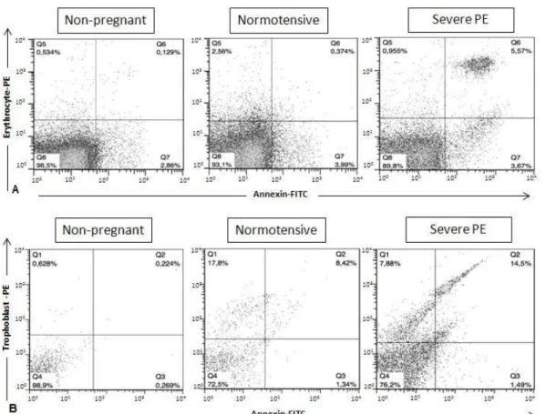
platelet-derived MPs. Unlike these 2 groups, most circulating MPs in women with severe PE originated from the endothelial cells. Numbers of erythrocyte-derived MPs were increased in women with severe PE compared with normotensive pregnant women (P = 0.002) and were higher in normotensive pregnant women compared with non-pregnant women (P = 0.005) (Figure 3A).
Trophoblast-derived MPs (NDOG2-positive) were detected in the circulation of women with severe PE and in normotensive pregnant women. Curiously, some trophoblast-derived MPs were detected in non-pregnant women. However, those levels were lower than what was seen in the women with severe PE or normotensive pregnant women (P = 0.002 in both cases) (Figure 3B).
Table 2. Cellular origin and numbers of circulating microparticles
MPs Severe PE Normotensive Non-pregnant P Value* pregnant women
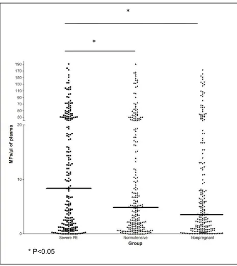
Total 8.43 (1.60-30.48) 4.87 (1.23-19.20) 3.53 (0.80-14.73) 0.004a 0.001b
0.154c
Platelet 30.93 (11.08-86.92) 38.27 (11.43-132.93) 60.13 (11.87-129.80) 0.726a 0.798b
0.915c
Endothelial 36.77 (5.48-73.03) 28.67(3.55-95.48) 7.93 (2.77-38.90) 0.453a
cell 0.132b
0.468c
Leukocyte 19.76 (5.20-63.77) 16.57 (2.70-58.07) 16.67 (4.43-79.70) 0.474a
0.873b
0.565c
Erythrocyte 12.77 (1.87-37.40) 5.27 (1.22-10.08) 2.73 (1.23-14.20)* 0.002a 0.814b
0.005c Neutrophil 9.13 (1.37-17.78) 3.00 (1.42-9.25) 3.47 (0.63-8.60) 0.123a
0.133b
0.808c
Trophoblast 6.37 (1.62-12.45) 5.00 (1.00-13.08) 2.00 (0.17-3.03) 0.555a
0.002b 0.002c
Monocyte 1.93 (0.55-5.40) 1.53 (0.48-2.70) 1.00 (0.23-3.60) 0.259a
0.238b
0.879c
Lymphocyte 0.90 (0.15-3.37) 1.20 (0.22-4.20) 0.73 (0.13-2.53) 0.674a
0.527b
0.215c
Dat a are presented as median (25th-75th centiles), MPs/µL. *Differences between two groups (Mann-Whitney U test and Bonferroni correction).
a= group 1 x group 2 b= group 1 x group 3 c= group 2 x group 3
* Two outliers were excluded in this analysis
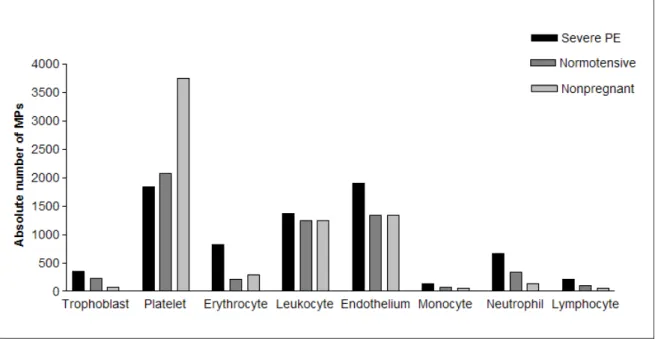
No significant differences were observed among the 3 groups regarding numbers of platelet-, endothelium-, leukocyte-, neutrophil-, monocyte-, and lymphocyte-derived MPs. Nevertheless, there was a clear reduction in platelet-derived MP levels in women with severe PE and normotensive pregnant women but an increase in neutrophil- and endothelial cell-derived MPs in women with severe PE (Figure 4).
Correlation analysis showed no correlation between MP levels and gestational age
considering all types of MPs in women with PE (P > 0.05). No correlation was observed between trophoblast-derived MPs and systolic or diastolic blood pressure (P > 0.05). Similarly, no correlation was found among trophoblast-, endothelial cell-, and platelet-derived MPs (P > 0.05). No correlation was found between platelet-, erythrocyte-, and leukocyte-derived MPs and their respective cell numbers in the circulation of women with* P<0.05
*
Figure 2. Data points and medians for total numbers of microparticles in
women with severe preeclampsia, normotensive pregnant women, and
non-pregnant women.
PE. However, we did find a positive correlation between platelet count (categorized according to cutoff = 150,000/mm3) and the number of MPs derived from platelets
(categorized considering the median of the control group) (r = 0.380; P = 0.05).
Positive correlations were found between numbers of endothelial cell-derived MPs and platelet-derived MPs (r = 0.483; P = 0.009), leukocyte-derived MPs (r = 0.519; P = 0.005), neutrophil-derived MPs (r = 0.394; P = 0.038), and lymphocyte-derived MPs (r = 0.616; P < 0.001).
Figure 3. Flow cytometry plots of microparticles derived from erythrocytes (A) and trophoblasts (B)
DISCUSSION
This study showed that MPs were significantly increased in women with severe PE compared with normotensive pregnant women and non-pregnant women. Similarly, Lok et al.18 and Orozco et al.19 demonstrated higher numbers of MPs in women with PE
compared with normotensive pregnant women.
The majority of circulating MPs detected in severe PE were derived from endothelial cells, while most MPs in normotensive pregnant women and non-pregnant women were derived from platelets. Although we observed reduced numbers of platelet MPs inwomen with severe PE compared with non-pregnant women, this difference was not significant, probably due to the high dispersion of the data in this variable. Similarly, Alijotas-Reig et al.20 found no difference in platelet-derived MPs in women with severe PE
vs. non-pregnant women. However, there was a positive correlation between platelet-derived MPsand platelet cell count when these variables were categorized. Lok et al.21 also
noted a reduction in platelet-derived MPs in women with severe PE and a correlation with platelet count.
The number of platelet-derived MPs may reflect the turnover of platelets in the plasma. Although platelet activation has been observed in PE, we were not able to identify increased numbers of platelet-derived MPs. A possible explanation for this finding could be
Figure 4. Absolute number of MPs in women with severe PE, normotensive pregnant women, and
that platelet MPs would remain trapped in the fibrin clots that are frequently evidenced in the placental microvasculature of women with severe PE.20 Therefore, a lower platelet
count in severe PE is associated with exacerbated platelet activation and high consumption.20,22 Thus, the decreased platelet counts in PE may explain the decreased
number of this MP type.21
PE is believedto be a disorder of the maternal endothelium.6 Although there was
a tendency for highernumbers of endothelial cells in women with severe PE compared with normotensive pregnant women and non-pregnant women, the difference was not significant. Contrarily, González-Quintero et al.23 documented higher numbers of
endothelial cell-derived MPs in women with PE compared with women with gestational hypertension and non-pregnant women. Endothelial cell activation may contribute to both inflammatory response and vasoconstriction. In the kidney, the endothelial defect can cause proteinuria and endothelium-dependent dilatation failure, which can contribute to hypertension and intense vasoconstriction in different organs.6 Therefore, endothelium
activation should be detectable by an increased number of endothelial cell-derived MPs in the circulation.24 Although we were not able to show a significant increase in numbers of
endothelial cell-derived MPs in women with severe PE compared with the other groups, the number of endothelial cell-derived MPs was associated with higher levels of lymphocyte-, leukocyte-, and platelet-derived MPs, which suggests a correlation between endothelium activation and these cell types.12
Our data showed increased numbers of erythrocyte-derived MPs in women with severe PE. This finding could be explained by hemolysis, which is commonly observed in this disease.25 Because fibrin clots have been observed in the microvasculature of women
with PE, one hypothesis is that erythrocytes are lysed by colliding with such clots and result in MP release.26 However, no correlation between erythrocyte-derived MPs and erythrocyte
numbers in the circulation was found.
Our data do not reveal significant differences in leukocyte-, monocyte-, lymphocyte-, and neutrophil-derived MPs, although there was a tendency toward increased numbers of neutrophil-derived MPs in women with severe PE. In contrast, monocyte-, lymphocyte-, and neutrophil-derived MPs were previously determined to be associated with
PE.21,27,28 Elevated numbers of leukocyte-derived MPs may reflect activation of these cells
because this disease is associated with the local inflammatory response that results in an enhanced leukocyte–endothelial interaction.29
Stallmach et al.30 observed higher numbers of activated lymphocytes in the