Implication of Tumor Necrosis Factor - Alpha in Preeclampsia
Dan MIHU
*, Nicolae COSTIN, Ligia Daniela BLAGA, Septimiu CIUCHIN
Ă
,
Raluca Bogdana POP
”Dominic Stanca” Clinic of Obstetrics - Gynecology. ”Iuliu Haţieganu” University of Medicine and Pharmacy Cluj-Napoca, 13 Emil Isac, 400023 Cluj-Napoca, Cluj, Romania.
E-mail*: dan.mihu@yahoo.com
* Author to whom correspondence should be addressed; Tel.: 0722837213; Fax: 0264596446.
Abstract:Introduction: Preeclampsia is an exacerbation of a generalized inflammatory response, physiologically present in the third trimester of pregnancy. Aim: The aim of the study consists in the evaluation of proinflammatory cytokine TNF-α in the context of preeclampsia. Material and Method: A transversal study was performed in three groups of patients: non-pregnant patients, patients with normal pregnancies in the third trimester, patients with preeclampsia. Serum TNF-α levels were determined using the immunometric sandwich EIA method. Results: The results obtained confirm a significant increase (p<0.01) in circulating TNF-α levels in the last trimester of pregnancy, compared to the non-pregnant status. Significantly increased serum TNF-α concentrations (p<0.001) were also found in pregnant patients with preeclampsia, compared to normotensive pregnant women. Conclusion: This proinflammatory cytokine can be a potential marker of the severity of the preeclamptic syndrome, without being an indicator of the fetal status at birth.
Keywords: Preeclampsia; Normal pregnancy; Tumor necrosis factor-α; Proinflammatory state.
Introduction
The absence of the rejection of the semiallogenic fetus by the activated maternal immune system is a true immunological paradox which is not yet completely understood.
Preeclampsia as well as recurrent abortion are considered by the majority of obstetricians the result of the alteration of immune physiological pregnancy maintaining processes.
During the course of human pregnancy, there are two immunologically distinct maternal-fetal interfaces, which are spatially and even temporally separate. The first interface is situated at decidual level and includes the interactions between the immune maternal decidual cell population and the invasive extravillous cytotrophoblast. This interface is dominant in early pregnancy and almost disappears in the third trimester, with the regression of invasive trophoblast. The second interface includes the interactions between the circulating maternal immune cells and the syncytiotrophoblast, which forms the villous surface of the human hemochorial placenta. These interactions involve the systemic maternal immune response [1].
This interface is activated with the onset of uteroplacental circulation and is enhanced with the growth of the placenta, to become dominant in the third trimester of pregnancy.
Material and Method
A transversal study was performed in the following three groups:
o Group 1 (preeclampsia - PE) included 40 pregnant women selected according to the following inclusion criteria: pregnant patients with preeclampsia (moderate or severe form), third trimester of pregnancy, single fetus pregnancy. For the diagnosis of preeclampsia, the classification system proposed by U.S. NHBPEP Working Group 2000 and adopted by ACOG was used:
- blood pressure values > 140/90 mmHg/ on two examinations repeated at an interval of minimum 6 hours and maximum 7 days
- proteinuria ≥ 30 mg/dl (in two urine samples taken at an interval of at least 4-6 hours)
- arterial hypertension and proteinuria occurred after 20 weeks of amenorrhea in a previously normotensive pregnant woman and were normalized within 12 weeks postpartum.
o Group 2 (normal pregnancy - NP) included 40 pregnant women with normal pregnancies, according to the following inclusion criteria: normotensive pregnant women during the entire period of pregnancy and postpartum, third trimester of pregnancy, single fetus pregnancy, pregnancy evolution and birth within physiological limits.
o Group3 (control group - C) was represented by 30 patients selected according to the following inclusion criteria: healthy non-pregnant women during the reproductive period.
After the obtaining of the informed consent, for each person included in the study, a form containing general and anthropometric data, family history and personal history of disease was completed. For the pregnant women included in the study, data relating on the current pregnancy were additionally recorded. The pregnant women were followed up during the pregnancy and birth period and for 12 weeks postpartum. At birth, the type of delivery (vaginal delivery or cesarean section), fetal weight and the Apgar score were recorded. Subsequently, the data resulting from the clinical and laboratory examination were added to the forms. The results obtained were introduced in a Microsoft Office Access data base.
From each subject included in the study, 10 ml blood were taken by venous puncture and collected in test tubes without anticoagulant. The serum obtained by centrifugation was divided into triplicates and stored in 600µl freezing tubes at a temperature of -200 C, until testing, in order to avoid repeated freezing-defreezing cycles.
Serum TNF-α levels were determined by the immunometric sandwich EIA technique, using the human TNF-α EIA kit 589201, Cayman Chemical Company, USA. The detection limit was 1.5 pg/ml.
The results obtained were expressed as representative indices and indicators, which were illustrated depending on relevance as figures, tables or diagrams. For numerical variables, dispersion and centrality indices were calculated: mean, median, standard deviation, standard error. For the comparison of quantitative variables and of the differences between their means, the t Student test and the ANOVA test were used.
Results
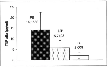
The mean serum TNF-α concentration in the three analyzed groups were:
o Group 1 (PE), the mean serum TNF- α concentration was 14.15 pg/ml; variation limits 0-32.67 (CI 95%; 11.48-16.38); SD ± 8.37.
o Group 2 (NP) the mean serum TNF-α concentration was 5.71 pg/ml; variation limits 0-14.8 (CI 95%; 4.63-6.79); SD ±3.38.
o Group 3 (C) the mean serum TNF-α concentration was 2.01 pg/ml; variation limits 0-4.01 (CI 95%; 1.52-2.49); SD ± 1.29 (Fig. 1).
Figure 1. Mean serum TNF-α concentration ± SD for the group preeclampsia (PE), normal pregnancy (NP) and control (C); p< 0.001
Statistical calculations: t test and ANOVA test
o Group NP versus group C: difference: -3.70 [CI 95%: ( -5) – ( -2.4)], p <0.001.
o Group PE versus group NP: difference: - 8.44 [CI 95%: ( - 11.28 ) – ( - 5.6 )], p < 0.01. ANOVA test: Highly statistical = 46.35, p < 0.001.
By following the relationship between the serum TNF-α concentration and the markers of the severity of preeclampsia, we obtained the following results:
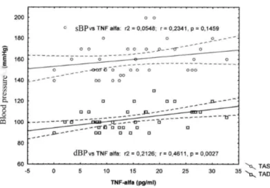
o Regarding the relationship between serum TNF-α concentration and systolic BP values in preeclampsia, no significant correlation was obtained. The correlation was positive and highly significant between serum TNF-α concentration and diastolic BP (Fig. 2).
o For group 1 (PE), a significant positive correlation was found between serum TNF-α and uric acid concentrations (Fig. 3).
Figure 2. Relationship between serum TNF-α concentration and systolic/diastolic blood pressure in preeclampsia
By analyzing the relationship between serum TNF-α concentration and some fetal parameters at birth, we obtained the following results:
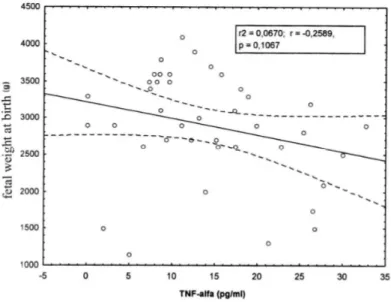
- For group 1 (PE) a negative but statistically insignificant correlation was found both between serum TNF-α concentration and fetal weight at birth and between serum TNF-α concentration and Apgar score at birth (Fig. 5, 6).
Figure 5. Relationship between serum TNF-α concentration and fetal weight at birth (PE group)
Teran et al. evaluated serum TNF-α concentrations in a group of 26 pregnant women with normal pregnancies compared to 21 healthy non-pregnant women and obtained similar results to our study (8.31 ± 1.55 pg/ ml in the third trimester of pregnancy versus 2.76 ± 0.41 pg/ml in non-pregnant women; p < 0.001) [4].
A number of longitudinal studies have assessed the influence of pregnancy on circulating TNF-α levels. Although an increase in circulating TNF-α levels is generally found during the course of pregnancy, this does not reach statistical significance in all the presented studies. The differences between the results might be at least partly explained by the different methods as well as by the reduced plasma half life of this cytokine [5,6].
Following our researches, we detected significantly increased serum TNF-α concentrations (p<0.001) in pregnant women with preeclampsia compared to normotensive pregnant women with the same gestational age (p=0.07).
Due to the pleiotropic effects of TNF-α, this cytokine has been intensely studied in preeclampsia in connection to various pathophysiological and immunological aspects found in this syndrome. Many studies in the literature have evaluated circulating TNF-α levels in preeclampsia as a bioactive or immunoreactive agent, with different results depending on the chosen study method. The results obtained are extremely controversial, increased, unchanged or even insignificantly decreased circulating TNF-α levels being reported in pregnant women with preeclampsia compared to normal pregnancy in the last trimester [3,4,7-10].
The biological effects of TNF-α are initiated after the binding of the cytokine to one of the two high affinity receptors situated at the surface of the target cells: TNF – RI (TNF p 55, CD 120a) and TNF – RII (TNF p 75, CD 120 b) [11].
It has been demonstrated that during gestation, soluble TNF-α receptors (sTNF-R) have a parallel evolution to that of TNF-α. Soluble receptors have a predictive value in the development of preeclampsia, increased concentrations of both receptors being detected in the third trimester of pregnancy in women subsequently developing preeclampsia [12].
Many authors have investigated sTNF-R levels during the last trimester of pregnancy in pregnant women with preeclampsia compared to normal pregnant women and reported high concentrations of the two receptors in preeclampsia [13,14].
There are only a few studies in the literature that have evaluated the predictive value of plasma TNF-α concentrations in preeclampsia [15,16].
The source of high circulating levels of inflammatory cytokines, including TNF-α, in pregnant women with preeclampsia has not yet been identified. TNF-α is located in the placenta in villous stromal cells, in the cytotrophoblast and the syncytiotrophoblast, as well as in the invasive trophoblast. One of the most studied hypotheses is the excessive production and release of TNF-α from the preeclamptic placenta as a consequence of hypoxia [17-19].
Another potential source of TNF-α in preeclampsia are activated maternal leukocytes.
As monocytes/ macrophages are generally the main reservoir of proinflammatory cytokines and are the first cells to be activated in non-specific immune responses, these can be good candidates for excessive TNF-α synthesis in preeclampsia [20,21].
Activated neutrophils are another TNF-α source in normal and pathological pregnancy [22]. Decidual macrophages and neutrophils can also be the source of this proinflammatory cytokine, these cells being activated and in higher numbers in preeclampsia [23].
In our study, maternal serum TNF-α concentration seems to be a marker of the severity of the preeclamptic syndrome, being positively and significantly correlated with both diastolic BP values and serum uric acid concentration.
In vitro studies suggest the fact that TNF-α may limit the migration of trophoblast cells in maternal tissues and may inhibit trophoblast DNA synthesis [25].
It has also been noted that excessive cytokine release in preeclampsia may restrict the invasion of endovascular cytotrophoblast at the level of spiral arteries [26].
Physiological TNF-α synthesis plays a protective role, but the overexpression of the cytokine as the result of genetic or environmental stimuli induces direct structural and functional changes in endothelial cells [27].
Conclusions
1. Serum immunoreactive TNF-α concentrations were significantly increased in the last trimester of gestation in preeclamptic women compared to normotensive pregnant women and the control group.
2. The positive and significant correlation of TNF-α with diastolic BP and uric acid levels makes this cytokine a potential marker of the severity of the preeclamptic syndrome.
3. TNF-α is not an indicator of fetal status at birth in preeclampsia.
4. Preeclampsia is an exacerbation of a generalized inflammatory response, physiologically present in the last trimester of pregnancy.
References
1. Sargent IL, Borzychowski AM, Redman CW. NK cells and human pregnancy-an inflammatory view. Trends Immunol 2006;27(9):399-404.
2. Redman CWG, Sargent IL. Pre-eclampsia, the placenta and the maternal systemic inflammatory response- a review. Placenta 2003;24:S21-S27.
3. Anim-Nyame N, Gamble J, Sooranna S.R, Johnson MR, Steer PJ. Microvascular permeability is related to circulating levels of tumour necrosis factor-alpha in pre-eclampsia. Cardiovascular Research 2003;58:162-169.
4. Teran E, Escudero C, Moyaw, Flores M, Vallance P, Lopez Jaramillo P. Elevated C-reactive protein and pro-inflammatory cytokines in Andean women with preeclampsia. Int J Gynecol Obstet 2001;75:243-249.
5. Melczer Z, Banhidy F, Csomor S, Kovacs M, Siklos P, Winkler G, Cseh K. Role of tumour necrosis factor-alpha in insulin resistance during normal pregnancy. Eur J Obstet Gynecol Reprod Biol 2002;105:7-10.
6. Coussons-Read M.E, Okun M, Nettles C.Psychosocial stress increases inXammatory markers and alters cytokine production across pregnancy. Brain, Behavior, and Immunity 2007;21:343-350.
7. Muzammil S, Singhal U, Gulati R, Bano I. Serum tumor necrosis factor-alpha in pre eclampsia.Indian J Physiol Pharmacol 2005;49:236-240.
8. Hayashi M, Ueda Y, Yamaguchi T, Sohma R, Shibazaki M, Ohkura T, Inaba N. Tumor necrosis factor-a in the placenta is not elevated in pre-eclamptic patients despite its elevation in peripheral blood. AJRI 2005; 53:113-119.
9. Jonsson Y, Ruber M, Matthiesen L, Berg G, Nieminen K, Sharma S, Emerudh J. Cytokine mapping of sera from women with preeclampsia and normal pregnancies. J Reprod Immunol 2006;70:83-91.
13. Nunez-Gonzalez JR, Sanabria-Vera CJ, Romero-Adrian T. Serum measurements of TNF-a and TNFa-soluble receptors in normal pregnancies and preeclampsia. Invest Clin 2001;42:171-182.
14. Madazli R, Aydin S, Uludag S, Vildan O, Tolun N. Maternal plasma levels of cytokines in normal and preeclamptic pregnancies and their relationship with diastolic blood pressure and fibronectin levels. Ada Obstet Gynecol Scand 2003;82:797-802.
15. Eneroth E, Remberger M, Vahlne A, Ringten O. Increased serum concentrations of interleukin-2 receptor in the first trimester in women who later developed severe preeclampsia. Acta Obstet Gynecol Scand 1998;77:591-593.
16. Serin YS, Ozcelik B, Bapbud M, Kylyc H, Okur D, Erez R. Predictive value of tumor necrosis factor alpha in preeclampsia. Euro J Obstet Gynecol Reprod Biol 2002;100: 143-145.
17. Chen HL, Yang YP, Hu XL, Yelavarthi KK, Fishback JL, Hunt JS. Tumor necrosis factor a mRNA and protein are present in human placental and uterine cells at early and late stages of gestation. Am J Pathol 1991;139:327-335.
18. Rinehart BK, Terrone DA, Lagoo-Deenadayalan, Barber WH, Hale EA, Martin JN, Bennett WA. Expression of the placental cytokines tumor necrosis factor a, interleukin 1b, and interleukin 10 is increased in preeclampsia. Am J Obstet Gynecol 1999;181:915-920.
19. Benyo DF, Miles TM, Conrad KP. Hypoxia stimulates cytokine production by villous explants from the human placenta. J Clin Endocrinol Metab 1997;82:1582-1588.
20. Luppi P, DeLoia JA. Monocytes of preeclamptic women spontaneously synthesize pro-inflammatory cytokines. Clinical Immunology 2006; 118:268-275.
21. Azizieh F, Raghupathy R, Makhseed M. Maternal cytokine production patterns in women with pre-eclampsia. AJRI 2005; 54:30-37.
22. Vaughan JE, Walsh SW. Neutrophils from pregnant women produce thromboxane and tumor necrosis factor-a in response to linoleic acid and oxidative stress. Am J Obstet Gynecol 2005;193:830-5.
23. Peracoli JC, Rudge MVC, Perac, Oli MTS. Tumor necrosis factor-alpha in gestation and puerperium of women with gestational hypertension and pre-eclampsia. Am J Reprod Immunol 2007;57:177-185.
24. Radaelli T, Uvena-Celebrezze J, Minium J, Huston-Presley L, Catalano P. Maternal lnterleukin-6: Marker of Fetal Growth and Adiposity. Soc Gynecol Investig 2006;13:53-57. 25. Hunt JS, Soares MJ, Lei MG, Smith RN, Wheaton D, Atherton RA, Morrison DC. Products
of lipopolysaccharide-activated macrophages (tumor necrosis factor-alpha, transforming growth factorbeta)but not lipopolysaccharide modify DNA synthesis by rat trophoblast cells exhibiting the 80- kDa lipopolysaccharide-binding protein. J Immunol 1989;143:1603-1613. 26. Redman CWG. Cytotrophoblasts: masters of disguise. Nat Med 1997;3:610-611.
27. Slowik MR, De Luca LG, Fiers W, Pober JS. Tumor necrosis factor activates human endothelial cells through the p55 tumor necrosis factor receptor but the p75 receptor contributes to activation at low tumor necrosis factor concentration. Am J Patfio 1993;143:1724-1730.