230 J Vasc Bras. 2013 Jul.-Set.; 12(3):230-233 http://dx.doi.org/10.1590/jvb.2013.045
T H E R A P E U T I C
C H A L L E N G E
INTRODUCTION
Aneurysms can form in several different visceral branches of the abdominal aorta1,2. Although uncommon, with an incidence that accounts for around 5% of intra-abdominal aneurysms, the risk of mortality is great if this kind of aneurysm ruptures. This is why it is important to raise awareness of this vascular disease3,4.
The most common site of splanchnic aneurysms is at the splenic artery4 (60% of cases), with a prevalence in the general population of 0.1 to 2%3. Pregnancy is one of the clinical conditions that favors the appearance these aneurysms4. The explanation is
that there is an increase of the splenic blood flow,
in addition to changes at the elastin of the vessels. Around 40% of women diagnosed with splenic artery aneurysms with no obvious cause are multiparous and have had at least six pregnancies. Other clinical conditions that may be responsible for the emergence of these aneurysms are portal hypertension and/or splenomegaly, accounting for 10% of cases. The incidence of rupture is 2%, with 10% of mortality3. Due to the relative rarity of these cases, their severity,
the diagnostic difficulties involved and the numerous
treatment options, this paper discusses the therapeutic challenge posed by a splenic artery aneurysm.
PART I: CLINICAL CASE
A 56-year-old female patient with hypertension, chronic obstructive pulmonary disease, dyslipidemia and heart disease presented at the hospital with a history of abdominal pain in the left flank. Abdominal ultrasound image was suggestive of splenic artery aneurysm, which had been
confirmed later by magnetic resonance angiography.
Patient history included bilateral saphenectomy,
left-side oophorectomy and two caesarean deliveries. General physical examination revealed nothing of significance. Her preoperative cardiac risk assessment was considered low risk.
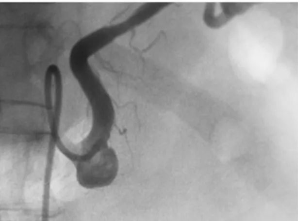
The angiography study identified a saccular aneurysm of the proximal third of the splenic artery, with a diameter of 11 mm by 16 mm and a wide neck, more than 6 mm in circumference (Figure 1). There were no signs of rupture, nor of anatomic variations
or significant changes to other visceral arteries. Even
though diameter was smaller than 2.0 cm, since the aneurysm was saccular and symptomatic, surgery was indicated because of the risk of rupture.
Possible treatment options in this case were as follows3:
• Conservative, nonsurgical treatment;
• Proximal and distal ligature with aneurysmectomy and preservation of the spleen;
• Aneurysmectomy with splenectomy;
• Videolaparoscopic ligature of the splenic artery; • Embolization of the aneurysm sac with coils and/or
cyanoacrylate glue;
• Thrombosis of the aneurysm sac by thrombin injection;
• Endovascular exclusion of the aneurysm with a covered stent.
PART II – WHAT WAS DONE?
The patient declined both conventional and laparoscopic surgeries, so endovascular treatment was indicated. Since the neck was wide, it was decided to cover the neck with a stent to provide support for the subsequent procedure of coil insertion into the aneurysm sac. Local anesthetic was administered and femoral access employed to insert a noncovered stent (5.25 mm × 6.5 mm, BALT®) into the proximal third of the splenic artery, covering the aneurysm neck. A 0.014” guidewire was
Splenic artery saccular aneurysm:
endovascular approach or open surgery?
Aneurisma sacular de artéria esplênica:
tratamento endovascular ou cirúrgico convencional?
Regina Moura1, Marcone Lima Sobreira1, Rodrigo Gibin Jaldin1, Matheus Bertanha1, Jamil Victor de Oliveira Mariúba1,
Carlos Clayton Macedo de Freitas1, Ricardo de Alvarenga Yoshida1, Winston Bonetti Yoshida1
1 Universidade Estadual Paulista – UNESP, Faculdade de Medicina de Botucatu, Departamento de Cirurgia e Ortopedia, Botucatu, SP, Brazil.
Financial support: None.
Conflicts of interest: No conflicts of interest declared concerning the publication of this article. Submitted on: 03.24.13. Accepted on: 04.16.13.
Regina Moura, Marcone Lima Sobreira et al.
231
J Vasc Bras. 2013 Jul.-Set.; 12(3):230-233 DISCUSSION
Symptomatic splenic artery aneurysms must always be treated. When asymptomatic, treatment is indicated if diameter is greater than 2.0 cm, if there is progressive increase3, as preoperative preparation for liver transplantation and if the patient is a pregnant woman or a woman at childbearing age5. Pseudoaneurysms are a medical emergency demanding treatment, irrespective of size or signs and symptoms of rupture, because, when untreated, the mortality is almost 50%6.
Sadat et al.7, conducted a systematic review and concluded that there is no consensus on treatment when asymptomatic, and that, when symptomatic, the treatment must be immediate, whether by conventional surgery, laparoscopy or endovascular techniques, and the choice should be based on the patient’s clinical condition, the possible approaches to the abdomen, the anatomic situation of the artery itself, and, of course, the patient acceptance of the procedure.
Conservative observational treatment is reserved for patients in critical clinical states, when aneurysms are smaller than 2.0 cm in diameter and in women who have no prospect of becoming pregnant5, although this last criterion is not absolute, particularly when dealing with saccular aneurysms3.
Open surgery was the standard treatment until the end of the twentieth century6 and is generally indicated for patients at low risk, for trunk lesions or when there are also aneurysms of the aorta or intestinal arteries, or indeed for cases in which endovascular treatment threaded through the mesh and used to guide the tip
of a microcatheter. Four coils were then inserted (10 × 30 mm – Axium®; 7 × 20 mm – Hydrocoil®; 18 × 30 mm – Micrusphere®, and 13 × 30 mm – GDC SR®). At the end of the procedure, the aneurysm was
occluded, with no blood flow inside, and the splenic
artery was patent (Figure 2).
In the immediate post-operative period the patient was free from intercurrent conditions and had no complaints of abdominal pain. No digestive or hematological problems were observed. At 3-month follow-up the patient was well, with no complaints and control angiography demonstrated a patent splenic artery (Figure 3).
Figure 1. Selective angiograph of the splenic artery showing a
wide-necked saccular aneurysm of the proximal segment.
Figure 2. Intraoperative control angiograph showing the
aneurysm excluded with coils. The nitinol support stent has low radiopacity and does not show up on the angiograph.
Figure 3. Two-year follow-up arteriograph showing a patent
Splenic artery saccular aneurysm: endovascular approach or open surgery?
232 J Vasc Bras. 2013 Jul.-Set.; 12(3):230-233
was unsuccessful8. Under general anesthesia, xypho-pubic, transversal or subcostal incisions can be used for access3. Conventional surgical treatment is by aneurysmorrhaphy or double ligature of the splenic artery with or without resection of the spleen, or may also be combined with aneurysmectomy with arterial reimplantation or bypass grafts. Revascularization of the spleen may not be necessary if the aneurysm is located at the proximal third of the splenic artery, since in such cases circulation can be supplied by the short gastric arteries3. It cannot, however, be guaranteed that the spleen will be unaffected3,9. Open surgery procedures can also be conducted with good results and less invasiveness using videolaparoscopic surgery10,11 with the use of staplers, but this demands training in intraoperative ultrasound and should not be used with patients who are hemodynamically unstable or when there are signs of rupture6.
In cases of acute rupture, the retrocavity of the epiploon can be accessed after sectioning the gastroepiploic ligament, providing access to the proximal section of the splenic artery or even the suprailiac aorta. Anterior access tends to ligate the short gastric arteries and the left gastroepiploic artery, increasing the chance of splenic infarction. Retroperitoneal access preserves collateral circulation to the spleen6.
Endovascular treatment is indicated in cases
involving high-risk, a hostile abdomen and distal lesions. Options include embolization of the splenic artery with coils and exclusion of the organ,9,12,13 or the placement of a covered stent covering the aneurysm neck14-16; or embolization with coils, as described by Jayanthi et al.5 for a hilar aneurysm, with preservation of the spleen. Advantages are
minimal invasiveness and preservation of blood flow
to the spleen, but it requires the use of radiation and contrasts, can be limited by factors such as a tortuous splenic artery, the position and size of the aneurysm
(hilar aneurysms present difficulties, for example)
and durability and patency are unknown6.
Another alternative should be the injection of
fibrin glue into the aneurysm sac3,17.
Preservation of the spleen should always be a consideration because splenic infarction predisposes patients to infections5.
An interesting option was chosen in the case described here: safe, successful and complete exclusion of the saccular aneurysm with minimally invasive surgery that managed to preserve blood circulation in the splenic artery, avoiding ischemia and the risk of infarctions in the spleen. Similar procedures had already been successfully performed
on other visceral arteries, such as the renal artery, for example18.
In conclusion, aneurysms of the splenic artery should be treated to avoid rupture and, whenever possible, protecting the organ’s viability. Although it is not the gold standard19, endovascular treatment can be considered a preferred form of treatment in these cases, offering the advantages of minimal
invasiveness, definitive resolution and the chance of
preserving the organ.
REFERENCES
1. Burihan E, Miranda F Jr, Francisco J Jr, Amorim JE, Barros N Jr, Chacon JP. Aneurisma da artéria esplênica; Splenic artery aneurysm. Cir Vasc Angiol. 1986;2:31-5.
2. Val RC, aChristo SFC, Rios AV, Leite RC, Figueiredo FA Jr, Vieira GL, Christo MBC. Aneurismas arteriais viscerais: relato de caso; Visceral arterial aneurysms: case report. Cir Vasc Angiol. 1999;15:147-9.
3. Rockman CB, Maldonado TS. Splanchnic artery aneurysms. In: Cronenwett JL, Johnston KW, editors. Rutherford’s Vascular Surgery. 7th ed. Philadelphia: Saunders, 2010. v. 2, p. 2140-55. http://dx.doi.org/10.1016/B978-1-4160-5223-4.00138-4 4. Guillaumon AT, Chaim EA. Splenic artery aneurysm associated
with anatomic variations in origin. J Vasc. Bras. 2009;8:177-181. 5. Jayanthi NGG, Mudawi AM, Uberoi R. Endovascular treatment
of splenic artery aneurysm with splenic preservation. Eur J Radio Extra. 2007;61:65-8. http://dx.doi.org/10.1016/j.ejrex.2006.10.006 6. Al-Habbal Y, Christophi C, Muralidharan V. Aneurysms of the
splenic artery - A review. Surgeon. 2010 Aug;8(4):223-31. 7. Sadat U, Dar O, Walsh S, Varty K. Splenic artery aneurysms in
pregnancy - A systematic review. Int J Surg. 2008 Jun; 6(3):261-5. PMid:17869597. http://dx.doi.org/10.1016/j.ijsu.2007.08.002 8. Spencer Netto FAC, Damasceno F, Alencar CRP. Aneurisma roto
de artéria esplênica: flagrante tomográfico de sangramento; Ruptured splenic artery aneurysm: tomographic snapshot of bleeding. Rev Col Bras Cir. 2002;29:119-21.
9. Yamamoto S, Hirota S, Maeda H, et al. Transcatheter coil embolization of splenic artery aneurysm. Cardiovasc Intervent Radiol. 2008;31:527-34. PMid:18040739. http://dx.doi. org/10.1007/s00270-007-9237-9
10. Grover BT, Gundersen SB 3rd, Kothari SN. Video. Laparoscopic distal pancreatectomy and splenectomy for splenic artery aneurysm. Surg Endosc. 2010;24:2318-20. PMid:20177922. http:// dx.doi.org/10.1007/s00464-010-0942-0
11. Suzuki H, Shimura T, Asao T, et al. Laparoscopic resection of splenic artery aneurysm; a case report. Hepatogastroenterology. 2002;49:1520-2. PMid:12397723.
12. Pino RMAS, Gois EAS, Aragão LG, Bomfim Filho ÂMS, Wanderley DC. Splenic artery aneurysm treated by coil embolization. J Vasc. Bras. 2010;9:249-253. http://dx.doi.org/10.1590/ S1677-54492010000400009
13. Biscosi M, Pozzi Mucelli R, Gussetti P. Treatment of a splenic aneurysm by percutaneous arterial embolisation using volumetric controlled-release coils. Case report and review of the literature. Radiol Med. 2002;104:239-43.
Regina Moura, Marcone Lima Sobreira et al.
233
J Vasc Bras. 2013 Jul.-Set.; 12(3):230-233
Correspondence
Jamil Victor de Oliveira Mariúba Hospital das CLínicas - FMB-UNESP Departamento de Cirurgia e Ortopedia Rubião Júnior, s/n CEP 18618-000 – Botucatu (SP), Brazil
E-mail: [email protected]
Author’s information
RM, MLS, CCMF is an assistant professor at Faculdade de Medicina de Botucatu (FMB), Universidade Estadual Paulista (UNESP). RGJ is an assistant physician at the Service of Vascular and Endovascular Surgery of Faculdade de Medicina de Botucatu (FMB), Universidade Estadual Paulista (UNESP). MB is an assistant professor at Faculdade de Medicina de Botucatu (FMB), Universidade Estadual Paulista (UNESP). JVOM is an assistant physician at the Service of Vascular and Endovascular Surgery of Faculdade de Medicina de Botucatu (FMB), Universidade Estadual Paulista (UNESP). RAY is a vascular and endovascular surgeon at Angiovalle; and a collaborating professor of Vascular and Endovascular Surgery at Faculdade de Medicina de Botucatu (FMB), Universidade Estadual Paulista (UNESP). WBY is a full professor and chief of the Service of Vascular and Endovascular Surgery of Faculdade de Medicina de Botucatu (FMB), Universidade Estadual Paulista (UNESP).
Author’s contributions
Conception and design: RM Analysis and interpretation: JVOM Data collection: MLS, RGJ, JVOM Writing the article: WBY, RAY Critical revision of the article: MB, MLS, CCMF Final approval of the article*: RM, MLS, RGJ, MB, JVOM, RAY, WBY Statistical analysis: JVOM Overall responsibility: JVOM
*All authors should have read and approved of the final version of the article submitted to J Vasc Bras.
artery aneurysm. J Vasc Surg. 2011;54:208-11. PMid:21315545. http://dx.doi.org/10.1016/j.jvs.2010.11.116
15. Taneja M, Pasupathy S. Endovascular exclusion of aberrant splenic artery aneurysm with covered stent. Singapore Med J. 2011;52:e244-7. PMid:22159944.
16. Carrafiello G, Rivolta N, Annoni M, Fontana F, Piffaretti G. Endovascular repair of a celiac trunk aneurysm with a new multilayer stent. J Vasc Surg. 2011;54:1148-50. PMid:21684712. http://dx.doi.org/10.1016/j.jvs.2011.03.274
17. Matsumoto K, Ushijima Y, Tajima T, et al. Recanalization of splenic artery aneurysm after transcatheter arterial embolization using N-butyl cyanoacrylate. Cardiovasc Intervent Radiol. 2010;33:187-90. PMid:19588193. http://dx.doi.org/10.1007/s00270-009-9627-2 18. Cardozo AC, Lichtenfels E, Erling N Jr, Raupp E, Tarasconi
DP. Endovascular treatment of renal artery aneurysm using microcoil embolization and renal blood flow preservation: case report. J Vasc Bras. 2007;6:167-70. http://dx.doi.org/10.1590/ S1677-54492007000200012