gondii
Infection in Mice
Emily G. Severance1*, Geetha Kannan2, Kristin L. Gressitt1, Jianchun Xiao1, Armin Alaedini3, Mikhail V. Pletnikov2, Robert H. Yolken1
1Stanley Division of Developmental Neurovirology, Department of Pediatrics, Johns Hopkins University School of Medicine, Baltimore, Maryland, United States of America, 2Division of Neurobiology, Department of Psychiatry, Johns Hopkins University School of Medicine, Baltimore, Maryland, United States of America, 3Department of Medicine, Columbia University Medical Center, New York, New York, United States of America
Abstract
Gluten sensitivity may affect disease pathogenesis in a subset of individuals who have schizophrenia, bipolar disorder or autism. Exposure toToxoplasma gondiiis a known risk factor for the development of schizophrenia, presumably through a direct pathological effect of the parasite on brain and behavior. A co-association of antibodies to wheat gluten and toT. gondiiin individuals with schizophrenia was recently uncovered, suggesting a coordinated gastrointestinal means by which T. gondii and dietary gluten might generate an immune response. Here, we evaluated the connection between these infectious- and food-based antigens in mouse models. BALB/c mice receiving a standard wheat-based rodent chow were infected withT. gondiivia intraperitoneal, peroral and prenatal exposure methods. Significant increases in the levels of anti-gluten IgG were documented in all infected mice and in offspring from chronically infected dams compared to uninfected controls (repetitive measures ANOVAs, two-tailed t-tests, all p#0.00001). Activation of the complement system accompanied this immune response (p#0.002–0.00001). Perorally-infected females showed higher levels of anti-gluten IgG than males (p#0.009) indicating that T. gondii-generated gastrointestinal infection led to a significant anti-gluten immune response in a sex-dependent manner. These findings support a gastrointestinal basis by which two risk factors for schizophrenia,T. gondiiinfection and sensitivity to dietary gluten, might be connected to produce the immune activation that is becoming an increasingly recognized pathology of psychiatric disorders.
Citation:Severance EG, Kannan G, Gressitt KL, Xiao J, Alaedini A, et al. (2012) Anti-Gluten Immune Response followingToxoplasma gondiiInfection in Mice. PLoS ONE 7(11): e50991. doi:10.1371/journal.pone.0050991
Editor:Ira Blader, University of Oklahoma Health Sciences Center, United States of America
ReceivedMay 24, 2012;AcceptedOctober 29, 2012;PublishedNovember 29, 2012
Copyright:ß2012 Severance et al. This is an open-access article distributed under the terms of the Creative Commons Attribution License, which permits unrestricted use, distribution, and reproduction in any medium, provided the original author and source are credited.
Funding:This work was supported by a National Institute of Mental Health (NIMH) P50 Silvio O. Conte Center at Johns Hopkins (grant#MH-94268; http://www. nimh.nih.gov/); by the Brain and Behavior Research Foundation where Dr. Severance is a Scott-Gentle Foundation Young Investigator (http://bbrfoundation.org/); and by the Stanley Medical Research Institute (www.stanleyresearch.org/). These funders played no role in study design, data collection and analysis, decision to publish, or preparation of the manuscript.
Competing Interests:The authors have declared that no competing interests exist.
* E-mail: eseverance@jhmi.edu
Introduction
Gluten proteins of wheat and related cereals have a pathogenic effect on the intestinal tract of individuals with the autoimmune disorder, celiac disease [1]. Increasingly, the exposure to gluten and to other food antigens such as bovine milk caseins is implicated in the pathogenesis of neuropsychiatric diseases, including autism, schizophrenia and bipolar disorder [2,3,4,5,6,7,8,9,10,11,12,13,14]. The digestion of wheat glutens and milk caseins has been proposed to result in the production of neuroactive exorphins that penetrate compromised gastrointesti-nal (GI) and blood brain barriers and directly bind to opioid receptors in the CNS [4,8,10,15,16,17,18]. We recently reported significant correlations of markers of GI inflammation with antibody levels to gluten and casein in individuals with schizo-phrenia [11]. Among these correlations, we found that antibodies to the targeted food antigens were associated with antibodies to the protozoan parasite,Toxoplasma gondii, in individuals with a recent onset of the disease.
T. gondii has been studied predominantly in the context of psychiatric disorders as a pathogen that might modify host behavior through a direct effect on the central nervous system
[19,20,21,22,23,24,25,26,27,28]. The association between food antigen exposure and the T. gondii parasite in individuals with schizophrenia presents a peripheral pathway via the GI tract by which this parasite may also contribute to psychiatric disease. In murine models, the oral ingestion of T. gondii can cause small intestinal immunopathology and is thus used experimentally to induce colitis and ileitis [29,30,31,32,33,34]. In humans, increased titers ofT. gondiiantibodies have been found in individuals with inflammatory bowel disease and celiac disease [35]. In pregnant women, T. gondii infection rates were higher among those with celiac disease compared to those without a gluten sensitivity [36]. It is not possible to discern from these studies if GI pathologies precededT. gondiiinfection or vice versa.
studies to explore how digested gluten peptides, independently or combined with T. gondiiand other GI pathogens, might impact brain physiology and behavior.
Materials and Methods
Ethics Statement
Investigations were approved by the Johns Hopkins Animal Care and Use Committee Institutional Guidelines (permit #MO12M23). These studies were part of a larger ongoing project to evaluate genetic and environmental hypotheses of schizophre-nia in mouse models. Utmost effort was utilized to prevent suffering and minimize the numbers of mice required for each experiment.
Overview
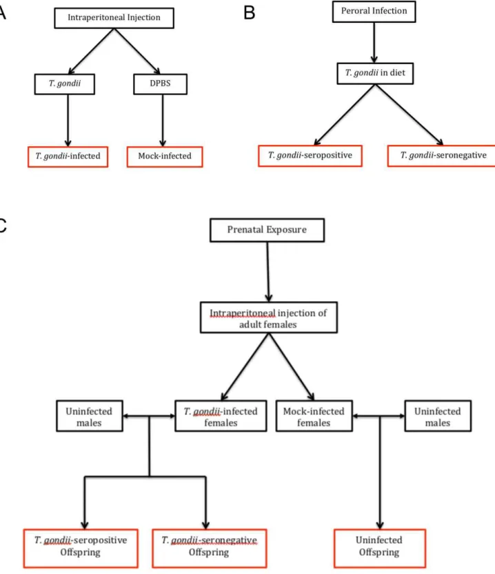
Exposure of mice toT. gondiiwas achieved using three routes of infection: intraperitoneal (IP), peroral (PO), and prenatal. An overview of how the experimental groups were generated via each type ofT. gondiiinfection is depicted in Figure 1.
IP Infection
Twenty-four 8-week old Balb/C mice were injected into the peritoneal cavity with either 400 tachyzoites of the Type II
T. gondiistrain, Prugniaud (PRU; n = 6 male; n = 6 female), or with Dulbecco’s phosphate buffered saline (DPBS; mock-infected; n = 6 male; n = 6 female). Serum samples were collected from a tail clip at 0, 15 and 21 days post-infection (dpi). IgG levels toT. gondii, gluten and complement factor C1q were measured with enzyme-linked immunosorbent assays (ELISAs), as described below. Over the course of the study, four of the female IPT. gondii-infected mice died. These animals were not included in the analyses.
PO Infection
A PO-infected cohort was generated with the goal of providing a more natural means of T. gondii infection and limiting to the extent possible any stress introduced by the procedure itself. Thus, in this PO model, we developed a diet containing infectious
T. gondii mixed with a crushed rodent chow that contains as its primary protein ingredient ground wheat (2018SX Teklad Global 18% Protein Extruded Rodent Diet; Harlan Laboratories, Madison, WI, U.S.A.). Animals were then allowed to eat freely (i.e. they were not forced). This PO method of inoculation was expected to produce infection in only a subset of animals that received this diet.
To prepare infectious T. gondii material, 5- and 8-week old BALB/c and mixed background BALB/c crossed with C57BL6, F4 donor mice were injected IP with 400 tachyzoites of the
T. gondii PRU strain. At five weeks post-infection, mice were sacrificed, their brains harvested, and serum obtained from a tail clip. Infectious brain homogenates were then mixed with crushed rodent chow.
Recipient mice were composed of two cohorts of BALB/c mice. Cohort 1 included twelve 12-week old mice (n = 6 male; n = 6 female); cohort 2 included ten 4-week old (n = 5 male; n = 5 female) and ten 6-week old mice (n = 5 male; n = 5 female). Recipient mice were food-deprived for 19 hours prior to infection. The food mixture of brain homogenate and crushed rodent chow was placed in the cages of all recipient mice, and mice were allowed to freely eat for three hours. Serum was collected at 4–5
Prenatal Infection
Female mice were injected IP with 400 tachyzoites of the
T. gondiiPRU strain.T. gondii-seropositive dams were mated with uninfected males. Eleven offspring were produced from this mating. The control group was composed of DPBS mock-infected dams mated with uninfected males. Four offspring resulted from this mating. Serum was taken from all offspring from a tail clipping at postnatal day 7. Antibodies toT. gondii, gluten and C1q were measured as described below.
Laboratory ELISA Procedures
In serum samples, IgG antibodies toT. gondii, gluten and C1q were measured by ELISAs. Commercially available ELISA kits for measuring serological T. gondii IgG were purchased from IBL America (Minneapolis, MN, U.S.A.) and used as previously described with a 1:50 serum dilution and an anti-mouse IgG secondary antibody [41]. IgG antibodies to wheat gluten and C1q were measured by ELISAs using previously described methods with some modification [11,42]. Whole gluten was extracted from the wheat cultivar Cheyenne, also as previously described [14]. C1q was purchased from Sigma-Aldrich (St. Louis, MO, U.S.A.) Wells of 96-well microtiter plates were incubated with one hundred ng of protein in 50ml carbonate buffer overnight at 4uC. The plates were incubated with serum samples diluted 1:50 in PBST for 2 h at 37uC. Plates were washed and incubated with peroxidase-conjugated goat-anti-mouse IgG secondary antibody for 30 min at 37uC (Southern Biotech, Birmingham, AL, U.S.A.). 2,29-azino-di-(3-ethylbenzthiazoline-6-sulfonate)(ABTS)/hydrogen peroxide solution (KPL Protein Research Products, Gaithersburg, MD, U.S.A.) was added for color development, and absorbance was measured at 405 nm, with a reference wavelength of 490 nm, in an automated microtiter plate reader (Molecular Devices, Menlo Park, CA, U.S.A.).
Using the above ELISA methods, standard curves of antibody levels (mg/ml) were generated forT. gondiiusing the calibrators available in the commercial kit and for gluten using a rabbit anti-gliadin polyclonal antibody (Sigma-Aldrich, St. Louis, MO, U.S.A.). The concentration of antibodies specific to T. gondii or to gluten was then calculated. For theT. gondiicalibrators provided in the commercial kit, conversion of International Units to milligrams per milliliter was performed according to Humphrey and Batty [43]. Standard curves for determination of serum IgG estimates are found in Figure 2.
Statistical Analyses
T. gondiiseropositivity was established based on values from the ELISA kit control standards run with each assay. Anti-T. gondii, -gluten and -C1q IgG levels were compared between: (1) IP
T. gondii-infected and IP mock-infected animals, (2) PO-infected
Figure 1. Overview of modes ofT. gondiiinfection and comparison groups.Panel A: Adult mice received intraperitoneal injections as shown to generate aT. gondii-infected and a mock-infected group. Experimental groups are boxed in red. Panel B: Adult mice received diets containingT. gondii, which resulted in mice that were successfully infected withT. gondii(T. gondii-seropositive) and those that were exposed to but did not develop an infection (T. gondii-seronegative). Panel C: IP- and mock-infected females were mated to uninfected males to produce offspring that were (1)T. gondii-exposed andT. gondii-seropositive, (2)T. gondii-exposed andT. gondii-seronegative, or (3) uninfected.
Results
Intraperitoneal (IP) Infection
We first measured anti-T. gondiiIgG levels to establishT. gondii -seropositive and T. gondii-seronegative groups. Following IP infection, highly statistically distinct T. gondii-seropositive and
T. gondii-seronegative groups were generated. The IP route of infection resulted inT. gondiiseropositivity in 100% of the animals that were inoculated with the parasite (n = 6 males; n = 6 females). Animals injected with a DPBS vehicle (mock-infected; n = 6 males;
Anti-T. gondiiIgG levels increased over time inT. gondii-inoculated animals compared to those that were mock-infected (Figure 3, panel A; repetitive measures ANOVA, between groups p#0.00001; interaction of infection group and time since infection, p#0.00001). There were no differences in T. gondii
IgG between sexes in either theT. gondii-infected or mock-infected groups.
In IP-inoculated mice, anti-gluten IgG increased over time in animals infected withT. gondii(n = 8) compared to those that were mock-infected (n = 12; Figure 3, panel B; repetitive measures
Figure 2. Standard curves from ELISAs for estimation of serological levels of anti-T. gondiiand anti-gluten IgG.Panel A: The amount of T. gondiicalibrator antibody is plotted according to absorbance. The standard curve shown is defined by the equation, y = 0.5342ln(x)+1.0478. The range of detection ofT. gondiiantibody for this assay is 100 ng-20 mg/ml. IP and PO inoculations producedT. gondiiantibodies in excess of 20 mg/ ml. Prenatal exposure toT. gondiiresulted in IgG levels of around 1 mg/ml in offspring. Panel B: The amount of gluten antibody is plotted according to absorbance. The gluten antibody curve is defined by the equation, y = 0.235ln(x)+0.8572. The range of detection for this assay is 20 ng–2 ug/ml. IP,
PO and prenatal exposures resulted in IgG levels of 50–90 ng/ml serological anti-gluten IgG. T1, T2, T3 refer to replicates of the assays run to construct these standard curves.
doi:10.1371/journal.pone.0050991.g002
and time remained significant for the males (n = 12; between infection groups, p#0.0002; interaction p#0.00001), but not females (n = 8; infection groups, p#0.49; interaction p#0.20). This difference between sexes in gluten antibody levels over time was attributable to the mock-infected group (Figure 3, panel B; sex p#0.008; interaction sex and time, p#0.04), whereas no difference in anti-gluten IgG levels over time was observed between sexes in theT. gondii-infected animals (Figure 3, panel B; sex p#0.69; interaction sex and time, p#0.72).
Complement C1q activation in IP-inoculated mice was significantly elevated compared to mock-infected mice and this response increased over time since infection (Figure 3, panel C; between infection groups p#0.00001; interaction p#0.00001; n = 7 T. gondii-infected, n = 11 mock-infected). Levels of C1q activation were not significantly different between sexes in the
T. gondii- or mock-infected groups. Peroral (PO) Exposure
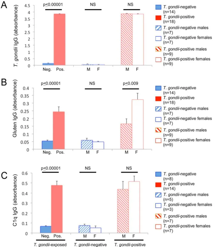
In animals exposed toT. gondiiin their diet (PO), ten of 12 mice from the first cohort (12 week-old mice tested 4–5 wpi) and eight of 20 mice from the second cohort (4- and 6-week old mice tested 3 wpi) tested seropositive forT. gondii. The PO rate of infection using this method was thus 56.25%. Seropositive mice had significantly higher levels of anti-T. gondiiIgG than those that were seronegative (Figure 4, panel A; two-tailed t-test, t =21.7e02, p#0.00001). There were no significant differences in anti-T. gondiiIgG between sexes (seropositive, n = 9 male, n = 9 female; seronegative, n = 7 male, n = 7 female).
In animals orally exposed toT. gondii, mean levels of anti-gluten IgG were significantly greater in T. gondii-seropositive (n = 18) vs
T. gondii-seronegative (n = 14) mice (Figure 4, panel B; two-tailed t-test, t =25.16, p#0.00001). In the T. gondii-seropositive group, anti-gluten IgG levels were significantly elevated in females compared to males (Figure 4, panel B; two-tailed t-test t =23.00, p#0.009). Gluten antibody levels were not significantly different between sexes in the mock-infected group (Figure 4, panel B; two-tailed t-test, t = 0.74, p#0.48).
C1q antibody levels were significantly increased in T. gondii -seropositive mice (n = 14) compared to those that were seroneg-ative (n = 8; Figure 4, panel C; two-tailed t-test t =26.44, p#0.00001). No sex-specific differences were detected.
Prenatal Exposure
Sexes were not determined for the 7-day old pups born in the prenatal cohort. Of the 11 mice born to the infected mothers, 100% were T. gondii-seropositive. The four mice born to uninfected mothers were T. gondii-seronegative. T. gondii IgG levels were significantly increased in the seropositive group (Figure 5, panel A; two-tailed t-test t =28.91, p#0.00001).
Mean gluten IgG levels of pups (n = 11) born to T. gondii
-C1q antibodies were significantly increased in offspring of
T. gondii-seropositive dams compared to offspring from seronega-tive dams (Figure 5, panel C; two-tailed t-test, t =23.7, p#0.002).
Discussion
Our findings convincingly indicate a direct relationship between infection withT. gondiiand the generation of antibodies to gluten. A standard laboratory rodent chow contained enough wheat to precipitate the anti-gluten immune response in infected animals. In particular, female mice infected perorally withT. gondii had a significantly stronger anti-gluten immune response compared to infected males or uninfected control animals. Female-specific effects were also found in IP-inoculated animals; however, this method of infection was associated with a high mortality rate and is itself a less natural means of infection by T. gondii than the peroral route. The anti-gluten immune response and activation of the complement system following prenatal exposure toT. gondii
infection coincides with a critical time period of postnatal neuronal development. These results, therefore, offer important milestones for future experiments to evaluate how the combined exposure to infection and dietary antigens, both known risk factors for the development of schizophrenia, might contribute to neurodevelop-mental hypotheses of psychiatric disease etiology.
Results from both the PO and IP portions of our study provide support that the female sex is more severely affected followingT. gondiiinfection. Four out of the six females inoculated IP withT. gondiidied, compared to100% viability in males infected using the same method. Indeed, others have shown that female mice are more susceptible to and more likely to die fromT. gondiiinfection than males [44,45]. Therefore, perhaps it is not surprising that PO
T. gondii launched a stronger anti-gluten immune activation in female mice compared to similarly infected males. Even in the IP mock vehicle group of our study, females exhibited significant elevations of gluten antibodies compared to pre-injection levels; males in this group showed no significant anti-gluten immune activation. This sex bias following a seemingly innocuous laboratory procedure is in keeping with reports from several groups regarding differential immune responses by female vs. male BALB/c mice [46,47,48]. Drude and colleagues (2011) reported that BALB/c mice undergoing standard laboratory injections with non-toxic substances such as saline and cyclodextrin exhibited sex-specific differences in the stress response, which in turn resulted in stress-induced lymphocytopenia [46]. Our results confirm that routine laboratory methods such as the IP route of delivery are not benign, and that stress-induced immune activation might, in fact, be compounded by exposure to dietary food antigens. Gluten antibody elevations in the IP mock-infected group of female mice further illustrate that it is the integrity of the GI environment that contributes to this food antigen response, a condition that can be brought on by stress or as modeled here byT. gondiiinfection. It is
Figure 3. IgG antibody levels following intraperitoneal (IP)T. gondiiinoculation.Panel A: Levels of anti-T. gondiiIgG in mice infected IP withT. gondii(red) varied significantly over time compared to mock-infected control mice (blue).T. gondiiantibody levels were not different between sexes (hashed bar – males; open bar – females). Panel B: Levels of anti-gluten IgG in mice infected IP withT. gondiivaried significantly over time compared to mock-infected control mice. Between mock-infected male and female mice, anti-gluten IgG levels varied significantly over time. In IPT. gondii-infected mice, anti-gluten IgG levels over time were not significantly (ns) different between sexes. Panel C: Levels of anti-C1q IgG in IP-infected mice varied significantly over time compared to mock-infected control mice. Time, infection and sex refer to interaction variables in repetitive measures ANOVAs. Repetitive measures ANOVAs were used to generate listed p-values. Error bars indicate standard errors of the mean. NS refers to not significant. Dpi refers to days post-infection.
doi:10.1371/journal.pone.0050991.g003
Figure 4. IgG antibody levels following peroral (PO)T. gondiiexposure.Panel A: Levels of anti-T. gondiiIgG were significantly greater in mice that were seropositive forT. gondii(T. gondii-positive) compared to those that were seronegative (T. gondii-negative) following ingestion of rodent chow containingT. gondii. Panel B: Levels of anti-gluten IgG were significantly greater in mice that were seropositive forT. gondii(T. gondii-positive) compared to those that were seronegative (T. gondii-negative) following ingestion of rodent chow containingT. gondii. No significant differences (NS) in anti-gluten IgG levels were found between sexes in theT. gondii-negative group. In animals that wereT. gondii-positive, anti-gluten IgG levels were significantly higher in females compared to males. Panel C: C1q antibody levels were significantly greater inT. gondii-positive vsT. gondii-negative animals. No sex-specific differences were observed. Two-tailed t-test comparisons were used to generate listed p-values. Error bars indicate standard errors of the mean.
the ability of other intestinally-active pathogens to generate diet-related antibodies should be examined in animal models and in humans.
It is well-documented that stress leads to increased intestinal permeability [49,50,51] which, in turn, brings to question the inflammatory state of the GI tract brought on by T. gondii
infection. It is not known with certainty howT. gondiistrains gain access to systemic circulation, but a para-cellular route affecting epithelial tight junction proteins is suspected [52,53,54]. Following PO infection of mice withT. gondii, parasites can be detected in the lamina propria and in Peyer’s patches of the small intestine by one hour post-infection, and parasites are detected in circulation by 48 hours post-infection [53]. POT. gondiiinfection is used to generate ileitis and colonitis in mouse models, and therefore can promote an environment of inflammation that damages cells lining the GI tract [29,34,53,55,56]). Physical damage that compromises the GI barrier integrity in turn promotes systemic inflammation as intestinal bacteria are translocated into circulation [49]. In fact, Hand and colleagues recently established that PO infection with
T. gondii directly causes bacterial translocation of resident commensals, loss of immune tolerance to resident microbiota and activation of microbiota-specific T cells and inflammatory effector cells [34].
If T. gondii crosses into circulation without damaging barrier integrity, then we must speculate on other means by which the anti-gluten immune activation originates. The para- vs trans-cellular travel of gluten peptides across the GI barrier is itself not well-understood, but evidence for gluten association with both routes exists [57,58,59]. Sapone et al demonstrated that celiac disease and gluten sensitivity were, in fact, two separate clinical conditions based on standard GI permeability tests, which showed that only celiac disease was associated with GI leakage [60]. It may be that gluten directs its own passage as evident by interactions of gluten peptides with the intestinal permeability mediator, zonulin and other tight junction proteins; however, gluten-associated transcytosis has also been documented [57,58,59,61,62,63,64,65]. The anti-gluten immune activation following exposure to
T. gondii has especially interesting implications for exploring possible neurodevelopmental mechanisms by which gluten sensi-tivity might be involved with diseases such as schizophrenia and autism. In these neurodevelopmental disorders, a food antigen pathology has long been suspected for a subset of individuals, yet direct evidence for prenatal gluten exposure and subsequent gluten involvement in the central nervous system has been elusive. In a
large birth registry-based study in Sweden, elevated levels of maternal anti-gluten IgG were recently found to increase the risk of non-affective psychosis in offspring [66]. Our experiments demonstrate that exposure to T. gondii infection during fetal development results in an anti-gluten immune response in offspring. Furthermore, we document that the complement pathway is also activated during this time period. C1q was used here as a gauge for generalized systemic immune activation following infection. Of interest to psychiatric diseases, however, is the role of C1q in synaptic pruning during development [67,68]. In the early postnatal days of mouse CNS development, cortical C1q mRNA expression is extensive compared to activity at postnatal day 30 [67,68]. In our study, serological C1q was significantly elevated in postnatal day 7 offspring exposed toT. gondii compared to unexposed offspring. Future studies are planned to examine how such prenatal exposures might affect subsequent synaptic organization.
In summary, the models described in this paper provide appropriate experimental tools to examine the impacts of gluten peptides, T. gondii and associated immune activation on brain physiology. As we accumulate more information from analyses of clinical biomarkers, we can adapt these animal models to test the effects of dietary modifications and other types of infections on behavioral endpoints, the pharmacological outcomes of specific antipsychotics on immune system parameters, and the autoim-mune responses triggered by T. gondii infection. Ultimately, we envision a translational system by which we can fully evaluate the interface of environmental perturbation and genetic predisposition as it relates to serious neurodevelopmental disorders such as schizophrenia, bipolar disorder and autism.
Acknowledgments
We thank Dr. Lorraine Jones-Brando for a critical reading of this manuscript. We thank Dr. Donald D. Kasarda of the U.S. Department of Agriculture for providing us with the wheat flour used for gluten extraction. We also thank Chunxia Yang and Maleeha Syed for technical assistance.
Author Contributions
Conceived and designed the experiments: EGS GK MVP RHY. Performed the experiments: GK KLG JX. Analyzed the data: EGS. Contributed reagents/materials/analysis tools: EGS AA MVP RHY. Wrote the paper: EGS AA MVP RHY.
References
1. Alaedini A, Green PH (2005) Narrative review: celiac disease: understanding a complex autoimmune disorder. Annals of Internal Medicine 142: 289–298. 2. Dickerson F, Stallings C, Origoni A, Vaughan C, Khushalani S, et al. (2011)
Markers of gluten sensitivity and celiac disease in bipolar disorder. Bipolar Disord13: 52–58.
3. Dickerson F, Stallings C, Origoni A, Vaughan C, Khushalani S, et al. (2010) Markers of gluten sensitivity and celiac disease in recent-onset psychosis and multi-episode schizophrenia. Biol Psychiatry 68: 100–104.
4. Dohan FC (1979) Schizophrenia and neuroactive peptides from food. Lancet 1: 1031.
5. Dohan FC (1970) Coeliac disease and schizophrenia. Lancet 1: 897–898. 6. Dohan FC (1980) Hypothesis: genes and neuroactive peptides from food as cause
of schizophrenia. Adv Biochem Psychopharmacol 22: 535–548.
7. Dohan FC (1981) Schizophrenia, celiac disease, gluten antibodies, and the importance of beta. Biol Psychiatry 16: 1115–1117.
8. Reichelt KL, Seim AR, Reichelt WH (1996) Could schizophrenia be reasonably explained by Dohan’s hypothesis on genetic interaction with a dietary peptide overload? Progress in Neuro-Psychopharmacology & Biological Psychiatry 20: 1083–1114.
9. Whiteley P, Haracopos D, Knivsberg AM, Reichelt KL, Parlar S, et al. (2010) The ScanBrit randomised, controlled, single-blind study of a gluten- and casein-free dietary intervention for children with autism spectrum disorders. Nutr Neurosci 13: 87–100.
10. Reichelt KL (1994) Exorphins in Schizophrenia and Autism. Journal of Neurochemistry 63: S86–S86.
Figure 5. IgG antibody levels following prenatal exposure toT. gondii.Panel A: In 7-day old pups, anti-T. gondiiIgG levels were significantly elevated in those whose mothers were seropositive forT. gondiicompared to those whose mothers were uninfected. Panel B: 7-day old offspring exposed prenatally toT. gondiihad significantly greater IgG to gluten than offspring from uninfected mothers. Panel C: 7-day old offspring of mothers who were seropositive forT. gondiihad significantly stronger complement factor C1q activation than offspring from uninfected mothers. Two-tailed t-test comparisons were used to generate listed significant p-values. Error bars indicate standard errors of the mean.
11. Severance EG, Alaedini A, Yang S, Halling M, Gressitt KL, et al. (2012) Gastrointestinal inflammation and associated immune activation in schizophre-nia. Schizophr Res 138: 48–53.
12. Severance EG, Dickerson FB, Halling M, Krivogorsky B, Haile L, et al. (2010) Subunit and whole molecule specificity of the anti-bovine casein immune response in recent onset psychosis and schizophrenia. Schizophr Res 118: 240– 247.
13. Severance EG, Dupont D, Dickerson FB, Stallings CR, Origoni AE, et al. (2010) Immune activation by casein dietary antigens in bipolar disorder. Bipolar Disord 12: 834–842.
14. Samaroo D, Dickerson F, Kasarda DD, Green PH, Briani C, et al. (2010) Novel immune response to gluten in individuals with schizophrenia. Schizophr Res 118: 248–255.
15. Dohan FC (1988) Genetic hypothesis of idiopathic schizophrenia: its exorphin connection. Schizophr Bull 14: 489–494.
16. Reichelt KL (1991) Peptides in Schizophrenia. Biol Psychiatry 29: 515–516. 17. Reichelt KL, Reichelt WH, Stensrud M (1995) The role of peptides in
schizophrenia. Journal of Neurochemistry 65: S43–S43.
18. Reichelt KL, Stensrud M (1998) Increase in urinary peptides prior to the diagnosis of schizophrenia. Schizophr Res 34: 211–213.
19. Flegr J, Lenochova P, Hodny Z, Vondrova M (2011) Fatal attraction phenomenon in humans: cat odour attractiveness increased for toxoplasma-infected men while decreased for toxoplasma-infected women. PLoS Neglected Tropical Diseases 5: e1389.
20. Gatkowska J, Wieczorek M, Dziadek B, Dzitko K, Dlugonska H (2012) Behavioral changes in mice caused by Toxoplasma gondii invasion of brain. Parasitology Research 111: 53–8.
21. Goodwin DG, Hrubec TC, Klein BG, Strobl JS, Werre SR, et al. (2012) Congenital infection of mice withToxoplasma gondiiinduces minimal change in behavior and no change in neurotransmitter concentrations. The Journal of Parasitology 98: 706–12.
22. Kannan G, Moldovan K, Xiao JC, Yolken RH, Jones-Brando L, et al. (2010)
Toxoplasma gondii strain-dependent effects on mouse behaviour. Folia para-sitologica 57: 151–155.
23. Torrey EF, Bartko JJ, Yolken RH (2012)Toxoplasma gondiiand other risk factors for schizophrenia: An update. Schizophr Bull 38: 642–7.
24. Torrey EF, Bartko JJ, Lun ZR, Yolken RH (2007) Antibodies toToxoplasma gondii
in patients with schizophrenia: a meta-analysis. Schizophr Bull 33: 729–736. 25. Webster JP, McConkey GA (2010)Toxoplasma gondii-altered host behaviour: clues
as to mechanism of action. Folia parasitologica 57: 95–104.
26. Webster JP (2007) The effect ofToxoplasma gondiion animal behavior: playing cat and mouse. Schizophr Bull 33: 752–756.
27. Webster JP (2001) Rats, cats, people and parasites: the impact of latent toxoplasmosis on behaviour. Microbes and infection/Institut Pasteur 3: 1037– 1045.
28. Xiao J, Kannan G, Jones-Brando L, Brannock C, Krasnova IN, et al. (2012) Sex-specific changes in gene expression and behavior induced by chronic Toxoplasma infection in mice. Neuroscience 206: 39–48.
29. Bereswill S, Munoz M, Fischer A, Plickert R, Haag LM, et al. (2010) Anti-inflammatory effects of resveratrol, curcumin and simvastatin in acute small intestinal inflammation. PLoS One 5: e15099.
30. Erridge C, Duncan SH, Bereswill S, Heimesaat MM (2010) The induction of colitis and ileitis in mice is associated with marked increases in intestinal concentrations of stimulants of TLRs 2, 4, and 5. PloS One 5: e9125. 31. Munoz M, Heimesaat MM, Danker K, Struck D, Lohmann U, et al. (2009)
Interleukin (IL)-23 mediatesToxoplasma gondii-induced immunopathology in the gut via matrixmetalloproteinase-2 and IL-22 but independent of IL-17. J Exp Med 206: 3047–3059.
32. Schreiner M, Liesenfeld O (2009) Small intestinal inflammation following oral infection withToxoplasma gondiidoes not occur exclusively in C57BL/6 mice: review of 70 reports from the literature. Mem Inst Oswaldo Cruz 104: 221–233. 33. Craven M, Egan CE, Dowd SE, McDonough SP, Dogan B, et al. (2012) Inflammation drives dysbiosis and bacterial invasion in murine models of ileal Crohn’s disease. PloS One 7: e41594.
34. Hand TW, Dos Santos LM, Bouladoux N, Molloy MJ, Pagan AJ, et al. (2012) Acute gastrointestinal infection induces long-lived microbiota-specific T cell responses. Science 337: 1553–1556.
35. Lidar M, Lipschitz N, Langevitz P, Barzilai O, Ram M, et al. (2009) Infectious serologies and autoantibodies in Wegener’s granulomatosis and other vasculit-ides: novel associations disclosed using the Rad BioPlex 2200. Annals of the New York Academy of Sciences 1173: 649–657.
36. Rostami Nejad M, Rostami K, Cheraghipour K, Nazemalhosseini Mojarad E, Volta U, et al. (2011) Celiac disease increases the risk of Toxoplasma gondii infection in a large cohort of pregnant women. The American Journal of Gastroenterology 106: 548–549.
37. Frank MM, Fries LF (1991) The role of complement in inflammation and phagocytosis. Immunology Today 12: 322–326.
38. Barrington R, Zhang M, Fischer M, Carroll MC (2001) The role of complement in inflammation and adaptive immunity. Immunol Rev 180: 5–15.
41. Xiao J, Buka SL, Cannon TD, Suzuki Y, Viscidi RP, et al. (2009) Serological pattern consistent with infection with type IToxoplasma gondiiin mothers and risk of psychosis among adult offspring. Microbes Infect 11: 1011–1018. 42. Severance EG, Gressitt K, Halling M, Stallings CR, Origoni AE, et al. (2012)
Complement C1q formation of immune complexes with milk caseins and wheat glutens in schizophrenia. Neurobiology of Disease 48: 447–53.
43. Humphrey JH, Batty I (1976) Letter: International units and standards. Lancet 1: 800–801.
44. Roberts CW, Cruickshank SM, Alexander J (1995) Sex-determined resistance to
Toxoplasma gondiiis associated with temporal differences in cytokine production. Infection and immunity 63: 2549–2555.
45. Liesenfeld O, Nguyen TA, Pharke C, Suzuki Y (2001) Importance of gender and sex hormones in regulation of susceptibility of the small intestine to peroral infection withToxoplasma gondiitissue cysts. The Journal of Parasitology 87: 1491–1493.
46. Drude S, Geissler A, Olfe J, Starke A, Domanska G, et al. (2011) Side effects of control treatment can conceal experimental data when studying stress responses to injection and psychological stress in mice. Lab Animal 40: 119–128. 47. Silva IA, El Nabawi M, Hoover D, Silbergeld EK (2005) Prenatal HgCl2
exposure in BALB/c mice: gender-specific effects on the ontogeny of the immune system. Developmental and Comparative Immunology 29: 171–183. 48. Wynne O, Horvat JC, Osei-Kumah A, Smith R, Hansbro PM, et al. (2011)
Early life infection alters adult BALB/c hippocampal gene expression in a sex specific manner. Stress 14: 247–261.
49. Lambert GP (2009) Stress-induced gastrointestinal barrier dysfunction and its inflammatory effects. Journal of Animal Science 87: E101–108.
50. Collins SM, Bercik P (2009) The relationship between intestinal microbiota and the central nervous system in normal gastrointestinal function and disease. Gastroenterology 136: 2003–2014.
51. Soderholm JD, Perdue MH (2001) Stress and gastrointestinal tract. II. Stress and intestinal barrier function. American Journal of Physiology Gastrointestinal and Liver Physiology 280: G7–G13.
52. Munoz M, Liesenfeld O, Heimesaat MM (2011) Immunology ofToxoplasma gondii.Immunological Reviews 240: 269–285.
53. Liesenfeld O (1999) Immune responses to Toxoplasma gondii in the gut. Immunobiology 201: 229–239.
54. Weight CM, Carding SR (2012) The protozoan pathogenToxoplasma gondii
targets the paracellular pathway to invade the intestinal epithelium. Annals of the New York Academy of Sciences 1258: 135–142.
55. Liesenfeld O (2002) Oral infection of C57BL/6 mice withToxoplasma gondii: a new model of inflammatory bowel disease? J Infect Dis 185 Suppl 1: S96–101. 56. Egan CE, Cohen SB, Denkers EY (2011) Insights into inflammatory bowel disease usingToxoplasma gondiias an infectious trigger. Immunology and Cell Biology 90: 668–75.
57. Menard S, Lebreton C, Schumann M, Matysiak-Budnik T, Dugave C, et al. (2012) Paracellular versus transcellular intestinal permeability to gliadin peptides in active celiac disease. The American Journal of Pathology 180: 608–615. 58. Matysiak-Budnik T, Candalh C, Dugave C, Namane A, Cellier C, et al. (2003)
Alterations of the intestinal transport and processing of gliadin peptides in celiac disease. Gastroenterology 125: 696–707.
59. Heyman M, Abed J, Lebreton C, Cerf-Bensussan N (2011) Intestinal permeability in coeliac disease: insight into mechanisms and relevance to pathogenesis. Gut 61: 1355–64.
60. Sapone A, Lammers KM, Casolaro V, Cammarota M, Giuliano MT, et al. (2011) Divergence of gut permeability and mucosal immune gene expression in two gluten-associated conditions: celiac disease and gluten sensitivity. BMC Medicine 9: 23.
61. Lammers KM, Lu R, Brownley J, Lu B, Gerard C, et al. (2008) Gliadin induces an increase in intestinal permeability and zonulin release by binding to the chemokine receptor CXCR3. Gastroenterology 135: 194–204 e193. 62. Tripathi A, Lammers KM, Goldblum S, Shea-Donohue T, Netzel-Arnett S, et
al. (2009) Identification of human zonulin, a physiological modulator of tight junctions, as prehaptoglobin-2. Proceedings of the National Academy of Sciences of the United States of America 106: 16799–16804.
63. Drago S, El Asmar R, Di Pierro M, Grazia Clemente M, Tripathi A, et al. (2006) Gliadin, zonulin and gut permeability: Effects on celiac and non-celiac intestinal mucosa and intestinal cell lines. Scandinavian Journal of Gastroen-terology 41: 408–419.
64. Sander GR, Cummins AG, Henshall T, Powell BC (2005) Rapid disruption of intestinal barrier function by gliadin involves altered expression of apical junctional proteins. FEBS Letters 579: 4851–4855.
65. Rallabhandi P (2012) Gluten and celiac disease–an immunological perspective. Journal of AOAC International 95: 349–355.
66. Karlsson H, Blomstrom A, Wicks S, Yang S, Yolken RH, et al. (2012) Maternal Antibodies to Dietary Antigens and Risk for Nonaffective Psychosis in Offspring. The American Journal of Psychiatry 169: 625–32.
67. Boulanger LM (2009) Immune proteins in brain development and synaptic plasticity. Neuron 64: 93–109.